Granulocytic sarcoma was first described by the British [[physician]] A. Burns in 1811. A granulocytic sarcoma is a solid [[tumor]] composed of immature [[malignant]] [[white blood cell]]s called [[myeloblast]]s. Grossly the neoplastic tissue usually appears firm with a fish-flesh appearance. On microscopic histopathological analysis, diffuse monotonous infiltrate that may or may not destroy underlying normal structures, tingible body macrophages) that impart a starry sky appearance are characteristic findings of granulocytic sarcoma. Granulocytic sarcoma must be differentiated from other diseases that cause lymphoma such as non-Hodgkin lymphomas of the lymhoblastic type, [[Burkitt lymphoma]], large-cell lymphoma, and small round cell tumors. Symptoms of granulocytic sarcoma may include violaceous, raised, nontender plaques or nodules on skin and painful gums which bleed easily with tooth brushing and other minor trauma. Physical examination may be remarkable for gingival hypertrophyt, lymphadenopathy, and violaceous, raised, nontender plaques or nodules on skin. Findings on immunohistochemical stainings include CD20, CD43, CD68, and myeloperoxidase. Systemic [[chemotherapy]] against the leukemia is typically utilized as the first-line treatment.
Granulocytic sarcoma (GS, also known as chloroma) was first discovered by Allen Burns, a British physician, in 1811. The term chloroma was first used by King to address the greenish appearance of the tumor due to [[myeloperoxidase]]. The association of the GS with [[Acute myeloid leukemia|acute myeloid leukemia (AML)]] was first recognized by Dock in 1902. GS can be classified into two categories based on its co-occurence with other malignancies. Infiltration of the tumor with [[myeloblasts]] is the main characteristic of the tumor on [[H&E stain]]. GS rises from primitive precursors of [[granulocytes]]. The disease is an extramedullary manifestation of myeloid diseases, however, it can occur as a primary disease. Aggregation of [[myeloblasts]], [[promyelocytes]] and [[Myelocyte|myelocytes]] outside of the bone marrow presents itself as these solid tumors. Tumors can occur at any site and can appear as green, gray, white or brown masses. GS must be differentiated from other diseases that can present as extramedullary solid tumors. All patients with GS must be evaluated for concurrent or future malignancies as GS can occur in the course of or prior to other malignancies. The prevalence of GS is approximately 2 per 1,000,000 individuals worldwide. The most important risk factor for development of GS is genetic mutations and susceptibility. Symptoms of GS may include the following: Symptoms due to mass effect such as [[deafness]], [[ptosis]], altered vision, intestinal obstruction, headache, neck pain, abdominal pain, and constitutional symptoms. [[Chemotherapy]] is the main stain of treatment in patients with GS. Even patients with isolated GS must receive systemic treatment to better the prognosis.
==Historical Perspective==
==Historical Perspective==
* Granulocytic sarcoma was first described by the British [[physician]] A. Burns in 1811<ref>Burns A. Observations of surgical anatomy, in Head and Neck. London, England, Royce, 1811, p. 364.</ref>, although the term ''chloroma'' did not appear until 1853.<ref>King A. A case of chloroma. Monthly J Med 17:17, 1853.</ref> This name is derived from the [[Greek language|Greek]] word ''chloros'' (green), as these tumors often have a green tint due to the presence of [[myeloperoxidase]]. The link between chloroma and [[acute leukemia]] was first recognized in 1902 by Dock and Warthin.<ref>Dock G, Warthin AS. A new case of chloroma with leukemia. Trans Assoc Am Phys 19:64, 1904, p. 115.</ref> However, because up to 30% of these tumors can be white, gray, or brown rather than green, the more correct term ''granulocytic sarcoma'' was proposed by Rappaport in 1967<ref>Rappaport H. Tumors of the hematopoietic system, in Atlas of Tumor Pathology, Section III, Fascicle 8. Armed Forces Institute of Pathology, Washington DC, 1967, pp. 241-247.</ref> and has since become virtually synonymous with the term chloroma.
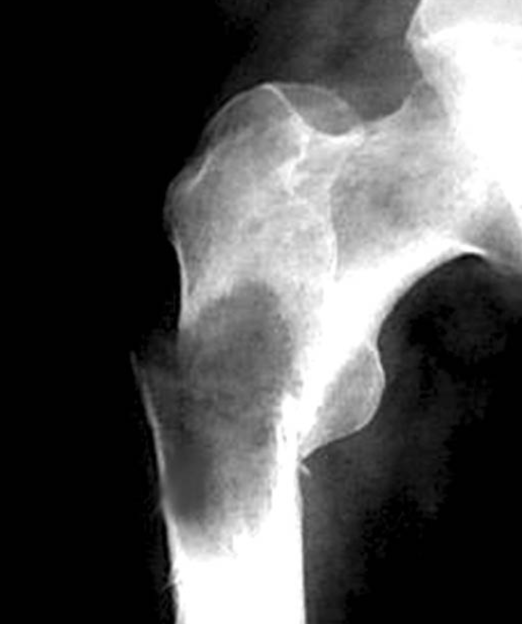
*[[File:Screenshot 2019-05-02 Myeloid sarcoma.png|thumb|Plain radiograph of femur in a patient with GS. Courtesy of image: pathology outlines (http://www.pathologyoutlines.com/topic/bonemyeloidsarcoma.html)]]Granulocytic sarcoma (GS, also known as chloroma) was first discovered by Allen Burns, a British physician, in 1811 <ref>{{Cite journal|last=Burns|first=Allen|date=|title=Observations of surgical anatomy, in Head andNeck.|url=|journal=London, England, Royce, 1811|volume=|pages=364-366|via=}}</ref>.
* The term chloroma was introduced by King in 1853 because of the green color of the lesion when exposed to air.
*The term chloroma was first used by King to address the greenish appearance of the tumor due to [[myeloperoxidase]].
*The association of the GS with [[Acute myeloid leukemia|acute myeloid leukemia (AML)]] was first recognized by Dock in 1902 <ref>{{Cite journal|last=Dock G, Warthin AS|first=|date=|title=A new case of chloroma withleukemia.|url=|journal=Trans Assoc Am Phys, 1904|volume=19:64|pages=115|via=}}</ref>.
*The term "granulocytic sarcoma" was suggested by Rappaport in 1967 to grant generalisability to it <ref>{{Cite book|title=Tumors of the hematopoietic system, inAtlas of Tumor Pathology, Section III|last=Rappaport H|first=|publisher=Fascicle 8. ArmedForces Institute of Pathology|year=1967|isbn=|location=Washington|pages=241-247}}</ref>.
==Classification==
* GS can be classified into two categories based on its co-occurence with other malignancies:
* A granulocytic sarcoma is a solid [[tumor]] composed of immature [[malignant]] [[white blood cell]]s called [[myeloblast]]s. A chloroma is an extramedullary manifestion of [[acute myeloid leukemia]]
* Infiltration of the tumor with [[myeloblasts]] is the main characteristic of the tumor on [[H&E stain]].
* Currently, any extramedullary manifestion of acute myeloid leukemia can be termed a granulocytic sarcoma or chloroma.
* GS rises from primitive precursors of [[granulocytes]].
* Specific terms which overlap with granulocytic sarcoma include:
* The disease is an extramedullary manifestation of myeloid diseases, however, it can occur as a primary disease.
:* Leukemia cutis, describing infiltration of the [[dermis]] (skin) by leukemic cells, which is also referred to as cutaneous granulocytic sarcoma
* Aggregation of [[myeloblasts]], [[Promyelocyte|promyelocytes]] and [[Myelocyte|myelocytes]] outside of the bone marrow presents itself as these solid tumors.
:* Meningeal leukemia, or invasion of the [[subarachnoid space]] by leukemic cells, is usually considered distinct from chloroma, although very rarely occurring solid [[central nervous system]] tumors composed of leukemic cells can be termed chloromas.
* Tumors can occur at any site and can appear as green, gray, white or brown masses.
===In acute leukemia===
==Differentiating GS from other Diseases==
* Chloromas are rare; exact estimates of their incidence are lacking, but they are uncommonly seen even by physicians specializing in the treatment of [[leukemia]]. Chloromas may be somewhat more common in patients with the following disease features:<ref name="review">Byrd JC, Edenfield JW, Shields DJ, et al: Extramedullary myeloid tumours in acute nonlymphocytic leukaemia: A clinical review. J Clin Oncol 13:1800, 1995.</ref>
GS must be differentiated from other diseases that can present as extramedullary solid tumors, such as:
:*FAB class M4 or M5
* [[Lymphoma|Large cell lymphoma]]
:*Those with specific [[cytogenetics|cytogenetic]] abnormalities (e.g. t(8;21) or inv(16))
* Non-Hodgkin lymphoma
:*Those whose [[myeloblast]]s express [[T-cell]] surface markers, [[CD13]], or [[CD14]]
* [[Thymoma]]
:*Those with high peripheral [[white blood cell]] counts
* [[Myeloma]]
:* However, even in patients with the above risk factors, chloroma remains an uncommon complication of acute myeloid leukemia.
* [[Sarcoma|Esosinophilic sarcoma]]
* Rarely, a chloroma can develop as the sole manifestation of relapse after apparently successful treatment of acute myeloid leukemia. In keeping with the general behavior of chloromas, such an event must be regarded as an early herald of a systemic relapse, rather than as a localized process. In one review of 24 patients who developed isolated chloromas after treatment for acute myeloid leukemia, the mean interval until bone marrow relapse was 7 months (range, 1 to 19 months).<ref>Byrd JC, Weiss RB. Recurrent granulocytic sarcoma: an unusual variation of acute myeloid leukemia associated with 8;21 chromosomal translocation and blast expression of the neural cell adhesion molecule. Cancer 73:2107-2112, 1994.</ref>
* [[Ewing sarcoma]]
* The most common localizations are skin, soft tissue, bone, and lymph nodes.1 Primary involvement of the gastrointestinal (GI) tract is rare.
* Extramedullary sites of [[hematopoiesis]]
===In myeloproliferative or myelodysplastic syndromes===
* [[Burkitt lymphoma]]
* Chloromas may occur in patients with a diagnosis of [[myelodysplastic syndrome]] (MDS) or [[myeloproliferative syndrome]]s (MPS) (e.g. [[chronic myelogenous leukemia]] (CML), [[polycythemia vera]], [[essential thrombocytosis]], or [[myelofibrosis]]). The detection of a chloroma is considered ''de facto'' evidence that these pre-malignant conditions have transformed into an acute leukemia requiring appropriate treatment. For example, presence of a chloroma is sufficient to indicate that chronic myelogenous leukemia has entered its ''blast crisis'' phase.
* [[Hypereosinophilic syndrome]]
===Primary chloroma===
* [[Polycythemia vera]]
* Very rarely, chloroma can occur without a known pre-existing or concomitant diagnosis of acute leukemia or MDS/MPS; this is known as primary chloroma. Diagnosis is particularly challenging in this situation (see below). In almost all reported cases of primary chloroma, acute leukemia has developed shortly afterward (median time to development of acute leukemia 7 months, range 1-25 months). Therefore, primary chloroma should probably be considered an initial manifestation of acute leukemia, rather than a localized process, and treated as such.
All patients with GS must be evaluated for concurrent or future malignancies as GS can occur in the course of or prior to other malignancies.
* Grossly the neoplastic tissue usually appears firm with a fish-flesh appearance.
*On microscopic histopathological analysis, diffuse monotonous infiltrate that may or may not destroy underlying normal structures, tingible body macrophages) that impart a starry sky appearance are characteristic findings of granulocytic sarcoma.
==Differentiating Granulocytic sarcoma from other Diseases==
*Granulocytic sarcoma must be differentiated from other diseases that cause lymphoma such as:
:*Non-Hodgkin lymphomas of the lymhoblastic type
:*Burkitt lymphoma
:*Large-cell lymphoma
:*Small round cell tumors
==Epidemiology and Demographics==
==Epidemiology and Demographics==
* The prevalence of GS is approximately 2 per 1,000,000 individuals worldwide.
* Most of the cases of GS are case reports and the disease is extremely rare.
===Age===
===Age===
*Granulocytic sarcoma is more commonly observed among older patients with a median age of 56 years.
* Patients of all age groups may develop GS.
* GS associated with [[AML]] occurs more commonly in children.
===Gender===
===Gender===
*Males are more commonly affected with granulocytic sarcoma than females with male-to-female ratio of 1:2.
* GS affects both men and women.
* Due to the rarity of the disease it is not clear whether there is a gender predilection for it.
===Race===
* There is no racial predilection for GS.
==Risk Factors==
* Risk factors for GS are usually chromosomal aberrations and include<ref name=":0">{{Cite journal|last=Daniela Dörfel et al.|first=|date=2016|title=Cardiac Myeloid Sarcoma: Multimodality Radiologic Imaging Features and Pathologic Correlation|url=https://www.amjmed.com/article/S0002-9343(16)30356-4/fulltext|journal=The American Journal of Medicine|volume=129|pages=e117-e120|via=}}</ref>:
** [[Trisomy 8]]
** Monosomy 7
** MLL gene rearrangement
** [[NPM1]] mutations
** [[FLT3]] mutations
== Natural History, Complications and Prognosis==
== Natural History, Complications and Prognosis==
* There is conflicting evidence on the [[prognosis|prognostic]] significance of chloromas in patients with acute myeloid leukemia. In general, they are felt to augur a poorer prognosis, with a poorer response to treatment and worse survival<ref>Tanravahi R, Qumsiyeh M, Patil S, et al: Extramedullary leukemia adversely affects hematologic complete remission and overall survival in patients with t(8;21)(q22;q22): Results from Cancer and Leukemia Group B 8461. J Clin Oncol 15:466, 1997.</ref>; however, others have reported that chloromas associate, as a biologic marker, with other poor prognostic factors, and therefore do not have independent prognostic significance.<ref>Bisschop MM, Revesz T, Bierings M, et al: Extramedullary infiltrates at diagnosis have no prognostic significance in children with acute myeloid leukemia. Leukemia 15:46, 2001.</ref>
* GS evolve over time, that depends on the co-occurence of the disease with other malignancies.
* GS may present before evidences of other malignancies manifest or after these malignancies are evident.
* Symptoms of the isolated GS depends on the location and the site of the tumor.
* Majority of cases are associated with [[AML]] or other [[myeloproliferative]]/[[Myelodysplastic syndrome|myelodysplastic syndromes]].
* Majority of GS tumors are found in the soft tissues such as the peritoneum, lymph nodes, CNS and skin. They are also found in bone and [[periosteum]].
* Early clinical features include weight loss, fatigue. Other manifestations of the tumor depend on its size and location.
* Prognosis of GS depends on its association with other malignacies. In cases of isolated GS the prognosis is good. However, GS associated with [[myeloproliferative disorders]] has poor prognosis.
* Prognosis of isolated GS with chromosome 8 abnormalities is worse than other cases of isolated GS.
== Diagnosis ==
== Diagnosis ==
===Diagnostic Criteria===
* There are no predefined criteria for diagnosis of granulocytic sarcoma.
* Granulocytic sarcoma must be suspected in patients with [[AML]] or [[Myelodysplastic syndrome|myelodysplastic syndromes]]. Diagnosis must be confirmed with histopathologic study of the specimen.
=== Symptoms ===
=== Symptoms ===
*Symptoms of granulocytic sarcoma may include the following:
Symptoms of GS may include the following:
:*Violaceous, raised, nontender plaques or nodules on skin
* Symptoms due to mass effect such as [[deafness]], [[ptosis]], altered vision, intestinal obstruction, etc.
:*Painful gums which bleed easily with tooth brushing and other minor trauma
* [[Headache]], neck pain, abdominal pain,etc. based on the site of the tumor
* Constitutional symptoms such as fever, fatigue, etc.
=== Physical Examination ===
=== Physical Examination ===
*Physical examination may be remarkable for:
Patients with GS can present with varying presentations.
:*Gingival hypertrophy
:*Lymphadenopathy
Physical examination may be remarkable for:
:*Violaceous, raised, nontender plaques or nodules on skin
* [[Lymphadenopathy]] (in cases associated with [[AML]] and other [[Myeloproliferative syndrome|myeloproliferative syndromes]])
* Skin lesions (of varying colors such as green, grey, brown, etc.)
* Organ enlargement such as [[hepatosplenomegaly]]
* [[Petechiae]] in patients with [[thrombocytopenia]]
* [[Hearing loss]]
* [[Heart murmurs]] in cases with heart chamber masses
* [[Crackles]] on lung auscultation
* [[Abdominal distension|Abdominal distention]]/tenderness in cases with intestinal obstruction
* Limb swelling due to different pathologies such as [[Deep vein thrombosis|deep vein thrombosis (DVT)]]
=== Laboratory Findings ===
=== Laboratory Findings ===
*There are no specific laboratory findings associated with granulocytic sarcoma.
* In cases associated with [[AML]]/[[CML]], [[anemia]], [[thrombocytopenia]] with normal, low or high white blood cells can be present.
==Imaging Findings===
* In cases associated with [[Polycythemia vera CT|polycythemia vera]], [[thrombocytosis]] and high levels of hemoglobin is present in complete blood count (CBC).
*There are no imaging study findings associated with granulocytic sarcoma.
* High [[eosinophil]] levels can be present in CBC.
===Imaging Findings===
* In cases of CNS involvement, magnetic resonance imaging (MRI) or CT scan of the CNS can reveal extra-axial masses.
* In cases with soft tissue involvement, sonogram of the tissue can reveal the mass.
GS appears as<ref>{{Cite journal|last=Guermazi et al.|first=|date=2002|title=Granulocytic sarcoma (chloroma): imaging findings in adults and children|url=https://www.ajronline.org/doi/full/10.2214/ajr.178.2.1780319|journal=American Journal of Roentgenology|volume=178|pages=319-25|via=}}</ref>:
* Hyperdense/isodence to brain/muscle in CT scan without enhancement
* Isointense/hyperintense on T2-weighted MRI
* Isointemse/hypointense on T1-weighted MRI
[[File:Chloroma histopathology micrograph (H&E stain).jpeg|thumb|Histopathologic micrograph of GS. Infiltration with myeloblasts which can be seen in the course of AML. Courtesy of image: Wikipedia (By Nephron - Own work, CC BY-SA 3.0, <nowiki>https://commons.wikimedia.org/w/index.php?curid=15893726</nowiki>)]]Abdominal plain radiogram can reveal obstruction, [[intussusception]], etc.
Echocardiogram may reveal mobile masses in any heart chamber in cases of heart involvement.
Chest radiographs can show lymph node enlargement and consolidation.
=== Other Diagnostic Studies ===
=== Other Diagnostic Studies ===
*Granulocytic sarcoma may also be diagnosed using immunohistochemical stainings for expression of myeloid associated enzymes.
* Peripheral blood smear can reveal circulating [[Blast|blasts]].
*Findings on immunohistochemical stainings include CD20, CD43, CD68, and myeloperoxidase.
* [[Flow cytometry]] can help differentiate [[AML]] from [[Acute lymphoblastic leukemia|acute lymphoblastic leukemia (ALL)]].
* Definitive diagnosis of a chloroma usually requires a [[biopsy]] of the lesion in question.
* Histopathologic analysis of biopsy specimen retrieved by excision or [[fine needle aspiration]] shows abundant [[myeloblasts]].
* Histopathologic analysis of GS lesions reveals high infiltration with [[myeloblasts]].
== Treatment ==
== Treatment ==
=== Medical Therapy ===
=== Medical Therapy ===
* Systemic chemotherapy against the leukemia is typically utilized as the first-line treatment, unless there is an emergent indication for local treatment of the chloroma (e.g. compromise of the [[spinal cord]]). Chloromas are typically quite sensitive to standard anti-leukemic [[chemotherapy]].
* [[Chemotherapy]] is the main stain of treatment in patients with GS. Even patients with isolated GS must receive systemic treatment to better the prognosis.
* If the chloroma is persistent after completion of induction chemotherapy, local treatment such as [[surgery]] or [[radiation therapy]] is often considered.
* Patients receiving [[cytarabine]] have better prognosis<ref name=":0" />.
* Patients presenting with a primary chloroma typically receive systemic chemotherapy, as development of acute leukemia is nearly universal in the short term after detection of the chloroma.
* Patients with chemotherpeutic regimens accepted for [[AML]] had longer period of progression to [[AML]].
* Patients treated for acute leukemia who relapse with an isolated chloroma are typically treated with systemic therapy for relapsed leukemia. However, as with any relapsed leukemia, outcomes are unfortunately poor.
Treatment includes different regimens<ref>{{Cite journal|last=Yilmaz, A. F., Saydam, G., Sahin, F., & Baran, Y.|first=|date=2013|title=Granulocytic sarcoma: a systematic review|url=https://www.ncbi.nlm.nih.gov/pmc/articles/PMC3875275/|journal=American Journal of Blood Research|volume=|pages=|via=}}</ref>:
* Patients with "pre-leukemic" conditions such as [[myelodysplastic syndrome]]s or myeloproliferative syndromes who develop a chloroma are often treated as if they have transformed to acute leukemia.
* Idarubicine and [[cytarabine]]
* [[Fludarabine]] and [[cytarabine]]
* Idarubicine and [[G-CSF]]
* [[Cyclophosphamide]], [[cytarabine]], [[topotecan]] and [[G-CSF]]
All of these therapeutic agents act through DNA damage and interferes with DNA synthesis.
===Bone marrow transplantation===
* [[Hematopoietic stem cell transplantation]] can be considered as a treatment option following induction [[chemotherapy]] in patients with [[AML]].<ref name=":1">{{Cite journal|last=Richard L. Bakst, Martin S. Tallman, Dan Douer and Joachim Yahalom|first=|date=2011|title=How I treat extramedullary acute myeloid leukemia|url=http://www.bloodjournal.org/content/118/14/3785?sso-checked=true|journal=Blood|volume=118|pages=3785-3793|via=}}</ref>
===Radiation===
* Radiation can be considered as an adjunctive therapy<ref name=":1" />.
* Combination of chemotherapy and radiotherapy can be considered in patients with CNS involvement or when rapid regression of symptoms is required.
=== Surgery ===
* Surgery alone is not a good treatment strategy for GS.
* Surgery can be considered prior to chemotherapy in patients where debulking can better the prognosis and help with symptom relief.<ref>{{Cite journal|last=Antic D et al.|first=|date=2013|title=Is there a "gold" standard treatment for patients with isolated myeloid sarcoma?|url=https://www.sciencedirect.com/science/article/abs/pii/S0753332212001060?via%3Dihub|journal=Biomed Pharmacother|volume=67|pages=72-77|via=}}</ref>
* Surgery can also have a diagnostic role in cases where excision of the mass provides specimen for histopatjologic diagnosis.
=== Prevention ===
=== Prevention ===
*There are no primary preventive measures available for granulocytic sarcoma.
* There are no primary preventive measures available for GS.
Granulocytic sarcoma (GS, also known as chloroma) was first discovered by Allen Burns, a British physician, in 1811. The term chloroma was first used by King to address the greenish appearance of the tumor due to myeloperoxidase. The association of the GS with acute myeloid leukemia (AML) was first recognized by Dock in 1902. GS can be classified into two categories based on its co-occurence with other malignancies. Infiltration of the tumor with myeloblasts is the main characteristic of the tumor on H&E stain. GS rises from primitive precursors of granulocytes. The disease is an extramedullary manifestation of myeloid diseases, however, it can occur as a primary disease. Aggregation of myeloblasts, promyelocytes and myelocytes outside of the bone marrow presents itself as these solid tumors. Tumors can occur at any site and can appear as green, gray, white or brown masses. GS must be differentiated from other diseases that can present as extramedullary solid tumors. All patients with GS must be evaluated for concurrent or future malignancies as GS can occur in the course of or prior to other malignancies. The prevalence of GS is approximately 2 per 1,000,000 individuals worldwide. The most important risk factor for development of GS is genetic mutations and susceptibility. Symptoms of GS may include the following: Symptoms due to mass effect such as deafness, ptosis, altered vision, intestinal obstruction, headache, neck pain, abdominal pain, and constitutional symptoms. Chemotherapy is the main stain of treatment in patients with GS. Even patients with isolated GS must receive systemic treatment to better the prognosis.
Historical Perspective
Plain radiograph of femur in a patient with GS. Courtesy of image: pathology outlines (http://www.pathologyoutlines.com/topic/bonemyeloidsarcoma.html)Granulocytic sarcoma (GS, also known as chloroma) was first discovered by Allen Burns, a British physician, in 1811 [1].
The term chloroma was first used by King to address the greenish appearance of the tumor due to myeloperoxidase.
Majority of GS tumors are found in the soft tissues such as the peritoneum, lymph nodes, CNS and skin. They are also found in bone and periosteum.
Early clinical features include weight loss, fatigue. Other manifestations of the tumor depend on its size and location.
Prognosis of GS depends on its association with other malignacies. In cases of isolated GS the prognosis is good. However, GS associated with myeloproliferative disorders has poor prognosis.
Prognosis of isolated GS with chromosome 8 abnormalities is worse than other cases of isolated GS.
Diagnosis
Diagnostic Criteria
There are no predefined criteria for diagnosis of granulocytic sarcoma.
Granulocytic sarcoma must be suspected in patients with AML or myelodysplastic syndromes. Diagnosis must be confirmed with histopathologic study of the specimen.
Symptoms
Symptoms of GS may include the following:
Symptoms due to mass effect such as deafness, ptosis, altered vision, intestinal obstruction, etc.
Headache, neck pain, abdominal pain,etc. based on the site of the tumor
Constitutional symptoms such as fever, fatigue, etc.
Physical Examination
Patients with GS can present with varying presentations.
Hyperdense/isodence to brain/muscle in CT scan without enhancement
Isointense/hyperintense on T2-weighted MRI
Isointemse/hypointense on T1-weighted MRI
Histopathologic micrograph of GS. Infiltration with myeloblasts which can be seen in the course of AML. Courtesy of image: Wikipedia (By Nephron - Own work, CC BY-SA 3.0, https://commons.wikimedia.org/w/index.php?curid=15893726)
Abdominal plain radiogram can reveal obstruction, intussusception, etc.
Echocardiogram may reveal mobile masses in any heart chamber in cases of heart involvement.
Chest radiographs can show lymph node enlargement and consolidation.
Other Diagnostic Studies
Peripheral blood smear can reveal circulating blasts.
Histopathologic analysis of GS lesions reveals high infiltration with myeloblasts.
Treatment
Medical Therapy
Chemotherapy is the main stain of treatment in patients with GS. Even patients with isolated GS must receive systemic treatment to better the prognosis.
Patients receiving cytarabine have better prognosis[4].
Patients with chemotherpeutic regimens accepted for AML had longer period of progression to AML.
Radiation can be considered as an adjunctive therapy[7].
Combination of chemotherapy and radiotherapy can be considered in patients with CNS involvement or when rapid regression of symptoms is required.
Surgery
Surgery alone is not a good treatment strategy for GS.
Surgery can be considered prior to chemotherapy in patients where debulking can better the prognosis and help with symptom relief.[8]
Surgery can also have a diagnostic role in cases where excision of the mass provides specimen for histopatjologic diagnosis.
Prevention
There are no primary preventive measures available for GS.
References
↑Burns, Allen. "Observations of surgical anatomy, in Head andNeck". London, England, Royce, 1811: 364–366.
↑Dock G, Warthin AS. "A new case of chloroma withleukemia". Trans Assoc Am Phys, 1904. 19:64: 115.
↑Rappaport H (1967). Tumors of the hematopoietic system, inAtlas of Tumor Pathology, Section III. Washington: Fascicle 8. ArmedForces Institute of Pathology. pp. 241–247.

.jpeg)