Glycogen storage disease type II physical examination: Difference between revisions
No edit summary |
No edit summary |
||
| Line 52: | Line 52: | ||
* [[Tremors]] may be present | * [[Tremors]] may be present | ||
* [[Spasm]] of legs may be rarely present | * [[Spasm]] of legs may be rarely present | ||
==Image gallery== | |||
Images shown below are courtesy of Professor Peter Anderson DVM PhD and published with permission. [http://www.peir.net © PEIR, University of Alabama at Birmingham, Department of Pathology] | |||
<div align="left"> | |||
<gallery heights="175" widths="175"> | |||
Image:218243.jpg|Pompe's Disease, Glycogen Storage Disease Type II. Child in crib | |||
Image:227286.jpg|Pompe's Disease, Glycogen Storage Disease Type II | |||
Image:227289.jpg|Pompe's Disease, Glycogen Storage Disease Type II | |||
</gallery> | |||
</div> | |||
<div align="left"> | |||
<gallery heights="175" widths="175"> | |||
Image:227292.jpg|Pompe's Disease, Glycogen Storage Disease Type II, 9 years old patient | |||
Image:227295.jpg|Pompe's Disease, Glycogen Storage Disease Type II, 9 years old patient | |||
</gallery> | |||
</div> | |||
<div align="left"> | |||
<gallery heights="175" widths="175"> | |||
Image:227298.jpg|Pompe's Disease, Glycogen Storage Disease Type II | |||
Image:227313.jpg|Pompe's Disease, Glycogen Storage Disease Type II | |||
</gallery> | |||
</div> | |||
==References== | ==References== | ||
{{reflist|2}} | {{reflist|2}} | ||
Latest revision as of 18:55, 30 January 2018
|
Glycogen storage disease type II Microchapters |
|
Differentiating Glycogen storage disease type II from other Diseases |
|---|
|
Diagnosis |
|
Treatment |
|
Case Studies |
|
Glycogen storage disease type II physical examination On the Web |
|
American Roentgen Ray Society Images of Glycogen storage disease type II physical examination |
|
FDA on Glycogen storage disease type II physical examination |
|
CDC on Glycogen storage disease type II physical examination |
|
Glycogen storage disease type II physical examination in the news |
|
Blogs on Glycogen storage disease type II physical examination |
|
Directions to Hospitals Treating Glycogen storage disease type II |
|
Risk calculators and risk factors for Glycogen storage disease type II physical examination |
Editor-In-Chief: C. Michael Gibson, M.S., M.D. [1]; Associate Editor(s)-in-Chief: Anmol Pitliya, M.B.B.S. M.D.[2]
Overview
Physical examination of patients with glycogen storage disease type 2 (GSD type 2) is usually remarkable for muscular weakness, hypotonia, absent deep tendon reflex and paucity of movements. Patients with infantile GSD type 2 usually appear dyspneic, pale, and/or cyanotic.
Physical Examination
- Physical examination of patients with glycogen storage disease type 2 (GSD type 2) is usually remarkable for muscular weakness, hypotonia, absent deep tendon reflexes and paucity of movements.[1][2][3]
Appearance of the Patient
- Patients with infantile GSD type 2 usually appear dyspneic, pale, and/or cyanotic
- Sometimes the appearance is also called as "Floppy baby appearance"
Vital Signs
Skin
HEENT
- Head lag
- Laxity of facial muscles
- Enlarged tongue may be present.
- Tougue fibrillation and/or absent tongue movements may be present
Neck
- Neck examination of patients with GSD type 2 is usually normal.
Lungs
Heart
Abdomen
- Hepatomegaly may be present
- Splenomegaly may be present
Back
- Scoliosis may be present
Genitourinary
- Genitourinary examination of patients with GSD type 2 is usually normal.
Neuromuscular
- Positive Gower's sign
- Hypotonia
- Absent deep tendon reflex
Extremities
- Calf muscles feel firm on palpation
- Decreased deep tendon reflexes
- Tremors may be present
- Spasm of legs may be rarely present
Image gallery
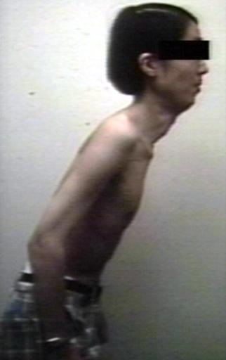
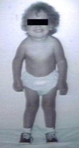
Images shown below are courtesy of Professor Peter Anderson DVM PhD and published with permission. © PEIR, University of Alabama at Birmingham, Department of Pathology
-
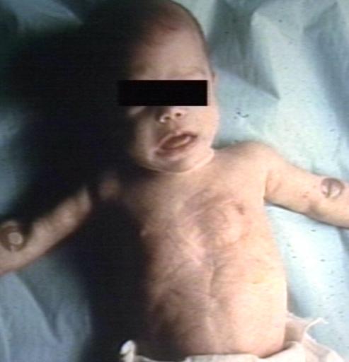
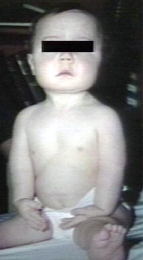
Pompe's Disease, Glycogen Storage Disease Type II. Child in crib
-
Pompe's Disease, Glycogen Storage Disease Type II
-
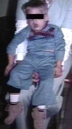
Pompe's Disease, Glycogen Storage Disease Type II


-
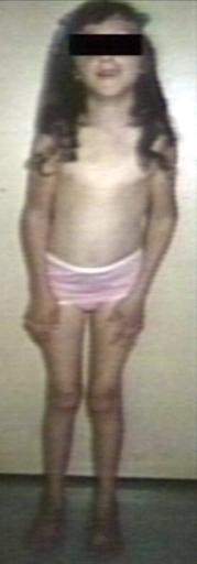
Pompe's Disease, Glycogen Storage Disease Type II, 9 years old patient
-
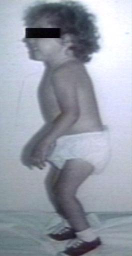
Pompe's Disease, Glycogen Storage Disease Type II, 9 years old patient


-
Pompe's Disease, Glycogen Storage Disease Type II
-
Pompe's Disease, Glycogen Storage Disease Type II


References
- ↑ van den Hout HM, Hop W, van Diggelen OP, Smeitink JA, Smit GP, Poll-The BT; et al. (2003). "The natural course of infantile Pompe's disease: 20 original cases compared with 133 cases from the literature". Pediatrics. 112 (2): 332–40. PMID 12897283.
- ↑ Winkel LP, Hagemans ML, van Doorn PA, Loonen MC, Hop WJ, Reuser AJ; et al. (2005). "The natural course of non-classic Pompe's disease; a review of 225 published cases". J Neurol. 252 (8): 875–84. doi:10.1007/s00415-005-0922-9. PMID 16133732.
- ↑ Kishnani PS, Howell RR (2004). "Pompe disease in infants and children". J Pediatr. 144 (5 Suppl): S35–43. doi:10.1016/j.jpeds.2004.01.053. PMID 15126982.