Glioblastoma multiforme pathophysiology
|
Glioblastoma multiforme Microchapters |
|
Diagnosis |
|---|
|
Treatment |
|
Case Studies |
|
Glioblastoma multiforme pathophysiology On the Web |
|
American Roentgen Ray Society Images of Glioblastoma multiforme pathophysiology |
|
Risk calculators and risk factors for Glioblastoma multiforme pathophysiology |
Editor-In-Chief: C. Michael Gibson, M.S., M.D. [1]
Overview
Pathophysiology
Genetics
- Development of glioblastoma is the result from multiple genetic mutations.
- Genes involved in the pathogenesis of primary glioblastoma include:[1]
- Genes involved in the pathogenesis of secondary glioblastoma include:[1]
- IDH1
- p53
- Chromosome 10q
- Chromosome 17p
- Chromosome 19q
Associated Conditions
Glioblastoma may be associated with:[1]
- Neurofibromatosis type 1
- Li-Fraumeni syndrome
- Turcot syndrome
- Ollier disease
- Maffucci syndrome
- Tuberous sclerosis
- Von Hippel-Lindau disease
Gross Pathology
On gross pathology, the characteristic findings of glioblastomas include:[1][2]
- Supratentorial white matter is the most common location
- Poorly-marginated, diffusely infiltrating mass with central necrotic core
- Ill-defined borders
- Tumor may be firm or gelatinous
- Variable appearance (firm and white, to soft and yellow, to cystic with hemorrhage)
- Midline shift due to tumor mass
- Presents as bihemispheric "butterfly glioma" in the corpus callosum

Microscopic Pathology
On microscopic histopathological analysis, the characteristic findings of glioblastomas include:[1][2]
- Pleomorphic astrocytes with marked atypia and mitosis
- Necrosis and microvascular proliferation
- +/-"Pseudopalisading necrosis": tumor cells lined-up like a picket fence around necrotic areas
Markers
Glioblastoma is demonstrated by positivity to tumor marker such as GFAP.
-
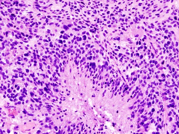
Glioblastoma (histology slide)
.jpg)
References
- ↑ 1.0 1.1 1.2 1.3 1.4 Pathology of glioblastoma multiforme. Dr Dylan Kurda and Dr Frank Gaillard et al. Radiopaedia 2015. http://radiopaedia.org/articles/glioblastoma
- ↑ 2.0 2.1 Pathology of glioblastoma multiforme. Libre Pathology. http://librepathology.org/wiki/index.php/Glioblastoma