Eosinophilic esophagitis other diagnostic studies: Difference between revisions
(Page Creation/Layout) |
Ahmed Younes (talk | contribs) (→Biopsy) |
||
| (14 intermediate revisions by 2 users not shown) | |||
| Line 1: | Line 1: | ||
__NOTOC__ | __NOTOC__ | ||
{{Eosinophilic esophagitis}} | {{Eosinophilic esophagitis}} | ||
{{CMG}} {{AE}} | {{CMG}} {{AE}} {{Ajay}} | ||
{{ | |||
==Overview== | ==Overview== | ||
[[Endoscopy|Endoscopic]] [[abnormalities]] in patients with EoE include fixed [[esophageal ring]] which is corrugated, white [[Exudate|exudates]] or [[plaques]], longitudinal furrows, [[Mucous membrane|mucosal]] [[pallor]], [[diffuse]] [[esophageal]] narrowing, [[Mucous membrane|mucosal]] fragility leading to [[esophageal]] [[lacerations]] during the [[endoscopy]]. | |||
==Other Diagnostic Studies== | ==Other Diagnostic Studies== | ||
===Endoscopy=== | |||
*[[Mucosal]] [[biopsies]] of the [[esophagus]] should be obtained in all patients in whom EoE is a clinical possibility regardless of the [[Endoscopy|endoscopic]] appearance. | |||
*[[Endoscopic]] [[abnormalities]] in patients with EoE are as follows:<ref name="urlTable 3: Proposed classification and grading system for the endoscopic assessment of the esophageal features of eosinophilic esophagitis (<a id=ref-link-section-1 title= href=/articles/#ref44">44</a>)">{{cite web |url=https://www.nature.com/articles/ajg201371/tables/3 |title=Table 3: Proposed classification and grading system for the endoscopic assessment of the esophageal features of eosinophilic esophagitis (<a id=ref-link-section-1 title="" href=/articles/#ref44>44</a>) |format= |work= |accessdate=}}</ref><ref name="urlVertical lines in distal esophageal mucosa (VLEM): a true endoscopic manifestation of esophagitis in children? - PubMed - NCBI">{{cite web |url=https://www.ncbi.nlm.nih.gov/pubmed/9199905?dopt=Abstract&holding=npg |title=Vertical lines in distal esophageal mucosa (VLEM): a true endoscopic manifestation of esophagitis in children? - PubMed - NCBI |format= |work= |accessdate=}}</ref><ref name="urlFragility of the esophageal mucosa: a pathognomonic endoscopic sign of primary eosinophilic esophagitis? - PubMed - NCBI">{{cite web |url=https://www.ncbi.nlm.nih.gov/pubmed/12612531?dopt=Abstract&holding=npg |title=Fragility of the esophageal mucosa: a pathognomonic endoscopic sign of primary eosinophilic esophagitis? - PubMed - NCBI |format= |work= |accessdate=}}</ref><ref name="urlEosinophilic esophagitis: red on microscopy, white on endoscopy. - PubMed - NCBI">{{cite web |url=https://www.ncbi.nlm.nih.gov/pubmed/15383737?dopt=Abstract&holding=npg |title=Eosinophilic esophagitis: red on microscopy, white on endoscopy. - PubMed - NCBI |format= |work= |accessdate=}}</ref><ref name="urlThe prevalence and diagnostic utility of endoscopic features of eosinophilic esophagitis: a meta-analysis. - PubMed - NCBI">{{cite web |url=https://www.ncbi.nlm.nih.gov/pubmed/22610003?dopt=Abstract&holding=npg |title=The prevalence and diagnostic utility of endoscopic features of eosinophilic esophagitis: a meta-analysis. - PubMed - NCBI |format= |work= |accessdate=}}</ref> | |||
**Fixed [[esophageal]] ring which is corrugated | |||
**White [[exudate]] | |||
**Longitudinal furrows | |||
**[[Mucous membrane|Mucosal]] [[pallor]] | |||
**[[Diffuse]] [[esophageal]] narrowing | |||
**[[Mucosal]] fragility leading to [[esophageal]] lacerations during the [[endoscopy]] | |||
*However, because these [[Endoscopy|endoscopic]] features have been described in other [[esophageal]] [[disorders]], none can be considered [[pathognomonic]] for EoE. | |||
<gallery> | |||
File:Eosinophilic esophagitis endo.jpg|Endoscopy of the esophagus: Eosinophilic esophagitis <br> Source: Wikimedia | |||
</gallery> | |||
===Biopsy=== | |||
*Characteristic features are as follows: | |||
**> 20 eosinophils/0.24 mm2. | |||
**[[Papillae]] are elongated | |||
**[[Papillae]] reach into the top 1/3 of the epithelial layer | |||
**Basal cell hyperplasia; > 3 cells thick or >15% of epithelial thickness | |||
[[Image:Eosinophilic esophagiits path.jpg|thumb|center|150px|[[H&E]] stain of [[esophagus]] [[biopsy]] showing eosinophilic esophagitis, manifested by an infiltration of [[eosinophils]] in the [[lamina propria]]]] | |||
==References== | ==References== | ||
Latest revision as of 23:42, 19 December 2017
|
Eosinophilic Esophagitis Microchapters |
|
Differentiating Eosinophilic Esophagitis from other Diseases |
|---|
|
Diagnosis |
|
Treatment |
|
Case Studies |
|
Eosinophilic esophagitis other diagnostic studies On the Web |
|
American Roentgen Ray Society Images of Eosinophilic esophagitis other diagnostic studies |
|
Eosinophilic esophagitis other diagnostic studies in the news |
|
Risk calculators and risk factors for Eosinophilic esophagitis other diagnostic studies |
Editor-In-Chief: C. Michael Gibson, M.S., M.D. [1] Associate Editor(s)-in-Chief: Ajay Gade MD[2]]
Overview
Endoscopic abnormalities in patients with EoE include fixed esophageal ring which is corrugated, white exudates or plaques, longitudinal furrows, mucosal pallor, diffuse esophageal narrowing, mucosal fragility leading to esophageal lacerations during the endoscopy.
Other Diagnostic Studies
Endoscopy
- Mucosal biopsies of the esophagus should be obtained in all patients in whom EoE is a clinical possibility regardless of the endoscopic appearance.
- Endoscopic abnormalities in patients with EoE are as follows:[1][2][3][4][5]
- Fixed esophageal ring which is corrugated
- White exudate
- Longitudinal furrows
- Mucosal pallor
- Diffuse esophageal narrowing
- Mucosal fragility leading to esophageal lacerations during the endoscopy
- However, because these endoscopic features have been described in other esophageal disorders, none can be considered pathognomonic for EoE.
-
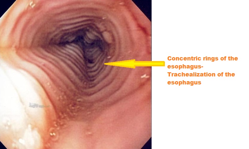
Endoscopy of the esophagus: Eosinophilic esophagitis
Source: Wikimedia

Biopsy
- Characteristic features are as follows:

References
- ↑ 44</a>)">"Table 3: Proposed classification and grading system for the endoscopic assessment of the esophageal features of eosinophilic esophagitis (<a id=ref-link-section-1 title="" href=/articles/#ref44>44</a>)".
- ↑ "Vertical lines in distal esophageal mucosa (VLEM): a true endoscopic manifestation of esophagitis in children? - PubMed - NCBI".
- ↑ "Fragility of the esophageal mucosa: a pathognomonic endoscopic sign of primary eosinophilic esophagitis? - PubMed - NCBI".
- ↑ "Eosinophilic esophagitis: red on microscopy, white on endoscopy. - PubMed - NCBI".
- ↑ "The prevalence and diagnostic utility of endoscopic features of eosinophilic esophagitis: a meta-analysis. - PubMed - NCBI".