Congenital adrenal hyperplasia pathophysiology
|
Congenital adrenal hyperplasia main page |
Pathophisiology
Gross Pathology
Gross pathology findings in patients with congenital adrenal hyperplasia are:[1][2]
- Enlarged adrenal glands
- Wrinkled surface adrenal glands
- Cerebriform pattern adrenal glands (pathognomonic sign)
- Normal ultrasound appearances may also be seen
- Testicular masses may be identified representing adrenal rest tissue
Microscopic Pathology
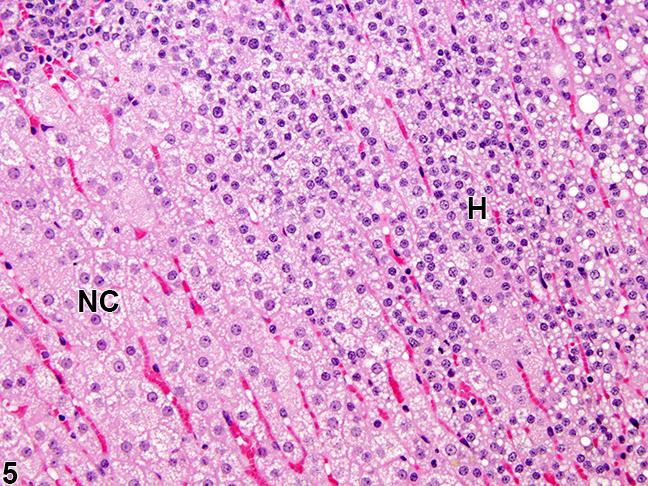
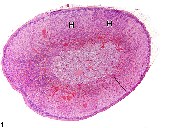
In congenital adrenal hyperplasia, hyperplastic cells are usually but not always smaller, with cytoplasm that can be vacuolated also often more basophilic. Rare mitotic figures may be present, but the hyperplastic cells typically lack features of cellular atypia.[3]
 |
 |
References
- ↑ Congenital adrenal hyperplasia. Dr Henry Knipe and Dr M Venkatesh . Radiopaedia.org 2015.http://radiopaedia.org/articles/congenital-adrenal-hyperplasia
- ↑ Teixeira SR, Elias PC, Andrade MT, Melo AF, Elias Junior J (2014). "The role of imaging in congenital adrenal hyperplasia". Arq Bras Endocrinol Metabol. 58 (7): 701–8. PMID 25372578.
- ↑ 3.0 3.1 3.2 "Adrenal Gland - Hyperplasia - Nonneoplastic Lesion Atlas".
