Chlamydia trachomatis: Difference between revisions
Becca Cohen (talk | contribs) No edit summary |
Becca Cohen (talk | contribs) No edit summary |
||
| Line 51: | Line 51: | ||
Image: Chlamydia10.jpeg| McCoy cell monolayers with Chlamydia trachomatis inclusion bodies (50X mag). <SMALL><SMALL>''[http://phil.cdc.gov/phil/home.asp From Public Health Image Library (PHIL).] ''<ref name=PHIL> {{Cite web | title = Public Health Image Library (PHIL) | url = http://phil.cdc.gov/phil/home.asp}}</ref></SMALL></SMALL> | Image: Chlamydia10.jpeg| McCoy cell monolayers with Chlamydia trachomatis inclusion bodies (50X mag). <SMALL><SMALL>''[http://phil.cdc.gov/phil/home.asp From Public Health Image Library (PHIL).] ''<ref name=PHIL> {{Cite web | title = Public Health Image Library (PHIL) | url = http://phil.cdc.gov/phil/home.asp}}</ref></SMALL></SMALL> | ||
Image: Chlamydia09.jpeg| Photomicrograph depicts HeLa cells infected with Type-A Chlamydia trachomatis (400X mag). <SMALL><SMALL>''[http://phil.cdc.gov/phil/home.asp From Public Health Image Library (PHIL).] ''<ref name=PHIL> {{Cite web | title = Public Health Image Library (PHIL) | url = http://phil.cdc.gov/phil/home.asp}}</ref></SMALL></SMALL> | Image: Chlamydia09.jpeg| Photomicrograph depicts HeLa cells infected with Type-A Chlamydia trachomatis (400X mag). <SMALL><SMALL>''[http://phil.cdc.gov/phil/home.asp From Public Health Image Library (PHIL).] ''<ref name=PHIL> {{Cite web | title = Public Health Image Library (PHIL) | url = http://phil.cdc.gov/phil/home.asp}}</ref></SMALL></SMALL> | ||
Image: Chlamydia03.jpeg| Patient’s left eye with the upper lid retracted in order to reveal the inflamed conjunctival membrane lining the inside of both the upper and lower lids, due to what was determined to be a case of inclusion conjunctivitis caused by the bacterium, Chlamydia trachomatis. <SMALL><SMALL>''[http://phil.cdc.gov/phil/home.asp From Public Health Image Library (PHIL).] ''<ref name=PHIL> {{Cite web | title = Public Health Image Library (PHIL) | url = http://phil.cdc.gov/phil/home.asp}}</ref></SMALL></SMALL> | |||
Revision as of 18:16, 12 June 2015
| Chlamydia trachomatis | ||||||||||||
|---|---|---|---|---|---|---|---|---|---|---|---|---|
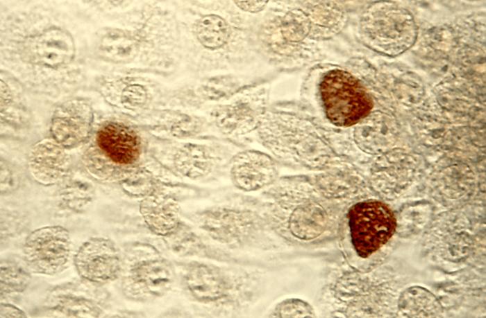
 C. trachomatis inclusion bodies (brown) in a McCoy cell culture.
| ||||||||||||
| Scientific classification | ||||||||||||
| ||||||||||||
| Binomial name | ||||||||||||
| Chlamydia trachomatis Busacca, 1935 |
Chlamydia trachomatis is one of three bacterial species in the genus Chlamydia, family Chlamydiaceae, class Chlamydiae, phylum Chlamydiae, domain Bacteria. C. trachomatis has only been found living inside the cells of humans, causing the following conditions:
In men
In women
- Cervicitis
- Pelvic inflammatory disease (PID)
- Premature birth
- Ectopic pregnancy
- Pelvic pain, chronic or acute
- Newborn eye (trachoma) or lung infection
In both sexes
- Lymphogranuloma venereum
- Urethritis
- Infertility
- Proctitis (rectal disease and bleeding)
- Reactive arthritis
- Trachoma
C. trachomatis has also been detected in some patients with temporomandibular joint (TMJ) disease. It may be treated with any of several antibiotics: azithromycin, erythromycin or doxycycline/tetracycline.
C. trachomatis was the first chlamydial agent discovered in humans. It comprises two human biovars: trachoma and lymphogranuloma venereum (LGV). Many, but not all, C. trachomatis strains have an extrachromosomal plasmid. Chlamydia species are readily identified and distinguished from other chlamydial species using DNA-based tests. Most strains of C. trachomatis are recognized by monoclonal antibodies [mAbs] to epitopes in the VS4 region of MOMP. However, these mAbs may also crossreact with the other two Chlamydia species, Chlamydia suis and Chlamydia muridarum.
Gallery
-
Photomicrograph of Chlamydia trachomatis taken from a urethral scrape. From Public Health Image Library (PHIL). [1]
-
McCoy cell monolayers with Chlamydia trachomatis inclusion bodies (200X mag). From Public Health Image Library (PHIL). [1]
-
McCoy cell monolayers with Chlamydia trachomatis inclusion bodies (50X mag). From Public Health Image Library (PHIL). [1]
-
Photomicrograph depicts HeLa cells infected with Type-A Chlamydia trachomatis (400X mag). From Public Health Image Library (PHIL). [1]
-
Patient’s left eye with the upper lid retracted in order to reveal the inflamed conjunctival membrane lining the inside of both the upper and lower lids, due to what was determined to be a case of inclusion conjunctivitis caused by the bacterium, Chlamydia trachomatis. From Public Health Image Library (PHIL). [1]
External links
- Chlamydiae.com [1]
- Template:GPnotebook
- article at reuters.com
ar:تراخوما da:Klamydia de:Chlamydia trachomatis nl:Chlamydia trachomatis no:Chlamydia trachomatis uk:Chlamydia trachomatis
![Photomicrograph of Chlamydia trachomatis taken from a urethral scrape. From Public Health Image Library (PHIL). [1]](/images/2/21/Chlamydia15.jpeg)
![McCoy cell monolayers with Chlamydia trachomatis inclusion bodies (200X mag). From Public Health Image Library (PHIL). [1]](/images/8/88/Chlamydia11.jpeg)
![McCoy cell monolayers with Chlamydia trachomatis inclusion bodies (50X mag). From Public Health Image Library (PHIL). [1]](/images/e/e2/Chlamydia10.jpeg)
![Photomicrograph depicts HeLa cells infected with Type-A Chlamydia trachomatis (400X mag). From Public Health Image Library (PHIL). [1]](/images/2/29/Chlamydia09.jpeg)
![Patient’s left eye with the upper lid retracted in order to reveal the inflamed conjunctival membrane lining the inside of both the upper and lower lids, due to what was determined to be a case of inclusion conjunctivitis caused by the bacterium, Chlamydia trachomatis. From Public Health Image Library (PHIL). [1]](/images/3/3c/Chlamydia03.jpeg)