Chagas disease
| Chagas disease | |
 | |
|---|---|
| Photomicrograph of Giemsa-stained Trypanosoma cruzi crithidia (CDC) |
|
Chagas disease Microchapters |
|
Diagnosis |
|---|
|
Treatment |
|
Case Studies |
|
Chagas disease On the Web |
|
American Roentgen Ray Society Images of Chagas disease |
For patient information click here
Editor-In-Chief: C. Michael Gibson, M.S., M.D. [1]; Associate Editor(s)-in-Chief: Tamar Sifri [2]
Synonyms and keywords: American trypanosomiasis; Trypanosoma cruzi infection
Overview
Historical Perspective
Pathophysiology
Causes
Differentiating Chagas disease from other Diseases
Epidemiology and Demographics
Risk Factors
Natural History, Complications and Prognosis
Diagnosis
History and Symptoms | Physical Examination | Laboratory Findings | Electrocardiogram | Other Diagnostic Studies
Treatment
Medical Therapy | Surgery | Primary Prevention | Secondary Prevention | Cost-Effectiveness of Therapy | Future or Investigational Therapies
Case Studies
Life Cycle
An infected triatomine insect vector (or "kissing" bug) takes a blood meal and releases trypomastigotes in its feces near the site of the bite wound. Trypomastigotes enter the host through the wound or through intact mucosal membranes, such as the conjunctiva (1). Common triatomine vector species for trypanosomiasis belong to the genera Triatoma, Rhodnius, and Panstrongylus. Inside the host, the trypomastigotes invade cells near the site of inoculation, where they differentiate into intracellular amastigotes (2). The amastigotes multiply by binary fission (3) and differentiate into trypomastigotes, and then are released into the circulation as bloodstream trypomastigotes (4). Trypomastigotes infect cells from a variety of tissues and transform into intracellular amastigotes in new infection sites. Clinical manifestations can result from this infective cycle. The bloodstream trypomastigotes do not replicate (different from the African trypanosomes). Replication resumes only when the parasites enter another cell or are ingested by another vector. The "kissing" bug becomes infected by feeding on human or animal blood that contains circulating parasites (5). The ingested trypomastigotes transform into epimastigotes in the vector's midgut (6). The parasites multiply and differentiate in the midgut (7) and differentiate into infective metacyclic trypomastigotes in the hindgut (8). Trypanosoma cruzi can also be transmitted through blood transfusions, organ transplantation, transplacentally, and in laboratory accidents.
-
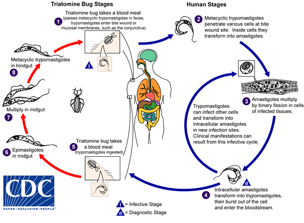
Life cycle of Trypanosoma cruzi
Adapted from CDC

Related Chapters
- Tropical disease
- Drugs for Neglected Diseases Initiative
- Distinguish from: Chaga mushroom
Template:Protozoal diseases Template:Link FA ar:شاجاس ca:Malaltia de Chagas de:Chagas-Krankheit it:Malattia di Chagas lt:Čagaso liga ms:Penyakit Cagas nl:Ziekte van Chagas sv:Chagas sjukdom