Bowen’s disease
Template:DiseaseDisorder infobox Editor-In-Chief: C. Michael Gibson, M.S., M.D. [1]
Associate Editor-In-Chief: Cafer Zorkun, M.D., Ph.D. [2]
Overview
Bowen's disease (BD) is a neoplastic skin disease, considered either as an early stage or intraepidermal form of squamous cell carcinoma. It was named after Dr John T. Bowen, the doctor who first described it in 1912.
Causes
Causes of BD include solar damage, arsenic, immunosuppression (including AIDS), viral infection (human papillomavirus or HPV) and chronic skin injury and dermatoses.

Signs and symptoms
Bowen's disease typically presents as a gradually enlarging, well demarcated erythematous plaque with an irregular border and surface crusting or scaling. BD may occur at any age in adults but is rare before the age of 30 years - most patients are aged over 60. Any site may be affected, although involvement of palms or soles is uncommon. BD occurs predominantly in women (70-85% of cases); about three-quarters of patients have lesions on the lower leg (60-85%), usually in previously or presently sun-exposed areas of skin. A persistent progressive non-elevated red scaly or crusted plaque which is due to an intradermal carcinoma and is potentially malignant. Atypical squamous (resembling fish scales) cells proliferate through the whole thickness of the epidermis. The lesions may occur anywhere on the skin surface or on mucosal surfaces. The cause most frequently found is trivalent arsenic compounds. Freezing, cauterization or diathermy coagulation is often effective treatment.
Histology
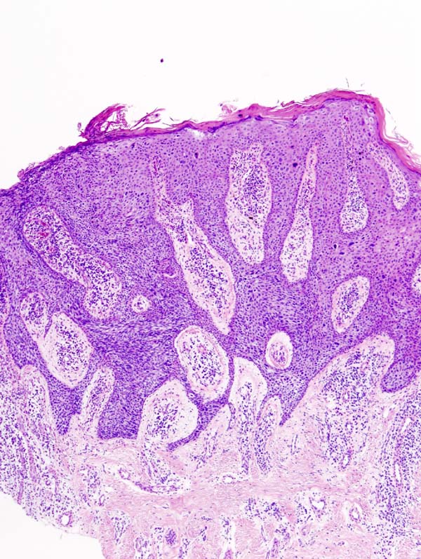
Bowen's is equivalent to squamous cell carcinoma in situ. The entire tumor is confined to the epidermis and does not invade into the dermis. The cells in Bowen's are extremely unusual or atypical under the microscope and in many cases look worse under the microscope than the cells of many outright and invading squamous-cell carcinomas. The degree of atypia (strangeness, unusualness) seen under the microscope best tells how cells may behave should they invade another portion of the body.
Treatment
Photodynamic therapy (PDT), Cryotherapy (freezing) or local chemotherapy (with 5-fluorouracil) are favored by some clinicians over excision. Because the cells of Bowen's disease have not invaded the dermis, it has a much better prognosis than invasive squamous cell carcinoma.