Autoimmune hepatitis physical examination: Difference between revisions
Jump to navigation
Jump to search
(→Skin) |
(→Skin) |
||
| Line 15: | Line 15: | ||
*[[Jaundice]] | *[[Jaundice]] | ||
*Spider angiomata | *Spider angiomata | ||
<gallery widths= | <gallery widths=250px> | ||
File:Spider nevus.jpg|[[spider angiomata]] |Source: | File:Spider nevus.jpg|[[spider angiomata]] |Source: | ||
UploadedImage-02.jpg | Description {{dermref}} | UploadedImage-02.jpg | Description {{dermref}} | ||
Revision as of 17:28, 28 December 2017
|
Autoimmune hepatitis Microchapters |
|
Diagnosis |
|---|
|
Treatment |
|
Case Studies |
|
Autoimmune hepatitis physical examination On the Web |
|
American Roentgen Ray Society Images of Autoimmune hepatitis physical examination |
|
Risk calculators and risk factors for Autoimmune hepatitis physical examination |
Editor-In-Chief: C. Michael Gibson, M.S., M.D. [1]; Associate Editor(s)-in-Chief: :Manpreet Kaur, MD [2]
Overview
Physical Examination
- Physical examination of patients with Autoimmune hepatitis is usually remarkable for:
- Jaundice
- Hepatomegaly
- Spider angiomata.
Appearance of the Patient
- Patients with Autoimmune hepatitis usually appear normal.
Skin
- Jaundice
- Spider angiomata
-
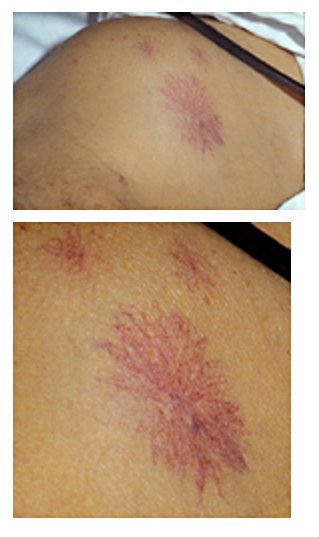
Source:
-
Description (Adapted from Dermatology Atlas)


HEENT
- Hirsutism
- Icteric sclera
- Acne
Abdomen
- Abdominal distention(Ascites)
- Hepatomegaly
- splenomegaly
Neuromuscular
- Patient present with hepatic encephalopathy
- Patient is disoriented to persons, place, and time
- Altered mental status