Aortic stenosis gross pathology: Difference between revisions
No edit summary |
No edit summary |
||
| Line 34: | Line 34: | ||
</div> | </div> | ||
== | ==[[Aortic stenosis other imaging findings autopsy|Aortic Stenosis Autopsy Report]]== | ||
==References== | ==References== | ||
Revision as of 19:11, 25 July 2011
|
Aortic Stenosis Microchapters |
|
Diagnosis |
|---|
|
Treatment |
|
Percutaneous Aortic Balloon Valvotomy (PABV) or Aortic Valvuloplasty |
|
Transcatheter Aortic Valve Replacement (TAVR) |
|
Case Studies |
|
Aortic stenosis gross pathology On the Web |
|
American Roentgen Ray Society Images of Aortic stenosis gross pathology |
|
Directions to Hospitals Treating Aortic stenosis gross pathology |
|
Risk calculators and risk factors for Aortic stenosis gross pathology |
Editor-In-Chief: C. Michael Gibson, M.S., M.D. [1]
Pathological Findings
Images shown below are courtesy of Professor Peter Anderson DVM PhD and published with permission. © PEIR, University of Alabama at Birmingham, Department of Pathology
-
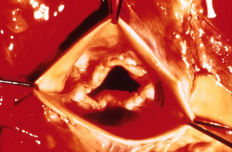
Aortic Stenosis, Bicuspid valve: Gross; excellent image of bicuspid and calcific valve showing a false raphe.
-
Aortic Stenosis, Bicuspid valve: Gross; good example of bicuspid valve
-
Aortic Stenosis, Bicuspid valve: Gross; image of bicuspid aortic valve, an excellent example
-
Aortic Stenosis, Bicuspid valve: Gross; close-up image of bicuspid aortic valve.
-
Aortic Stenosis, Bicuspid valve: Gross; close-up image of bicuspid aortic valve.
-
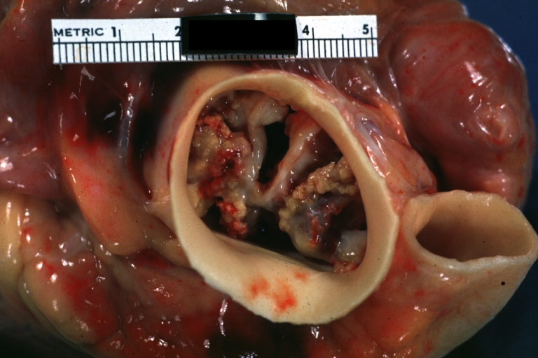
Bicuspid aortic valve
-
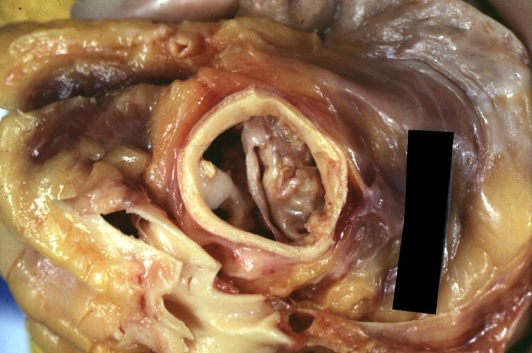
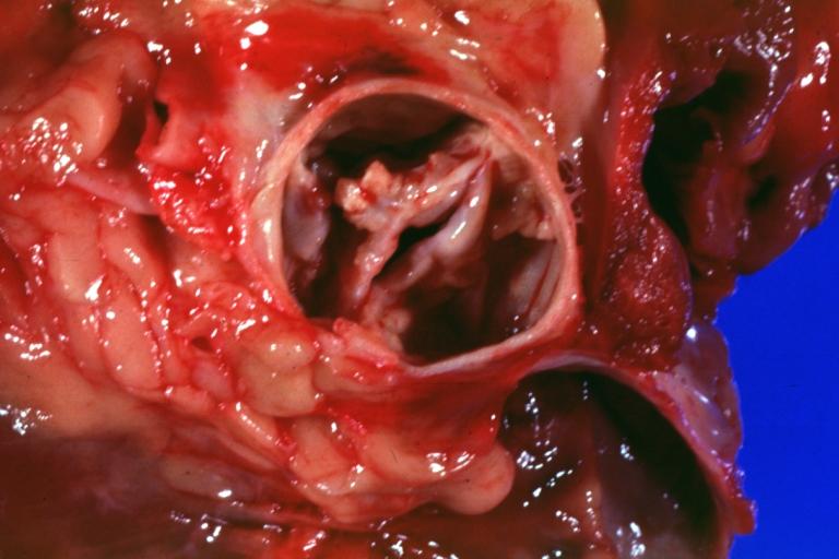
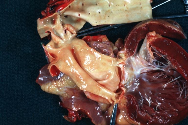
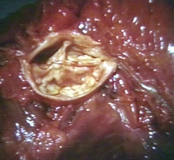
Gross natural color opened first portion aortic arch with bicuspid aortic valve shows stenosis and aortic root is dilated
-
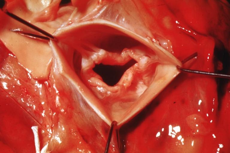
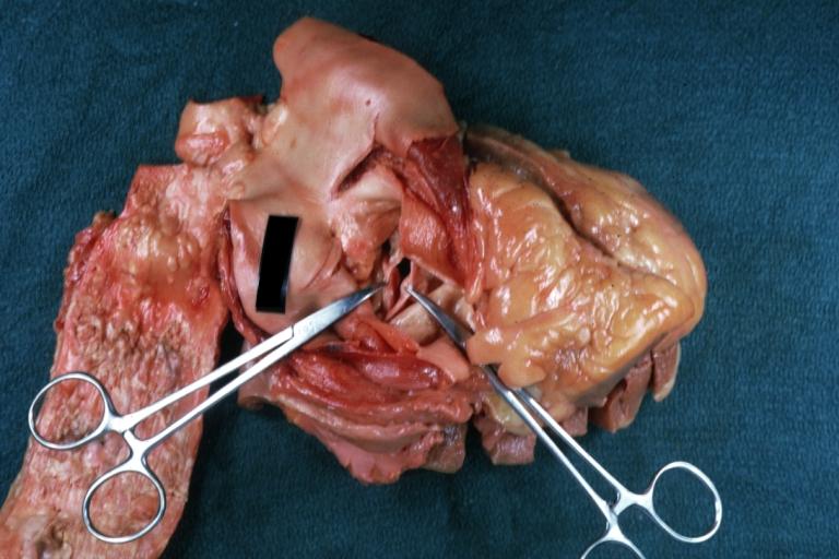
Aortic Stenosis Bicuspid: Gross; natural color opened left ventricular outflow tract with calcific masses on valve as well as anterior leaflet mitral valve probably did not cause significant stenosis
-
Bicuspid Aortic Valve with Repaired Aorta Coarctation: Gross natural color opened left ventricular outflow tract with uncomplicated bicuspid aortic valve repaired coarctation barely visible ruptured postoperative young female with ovaries Turner mosaic not ruled out
-
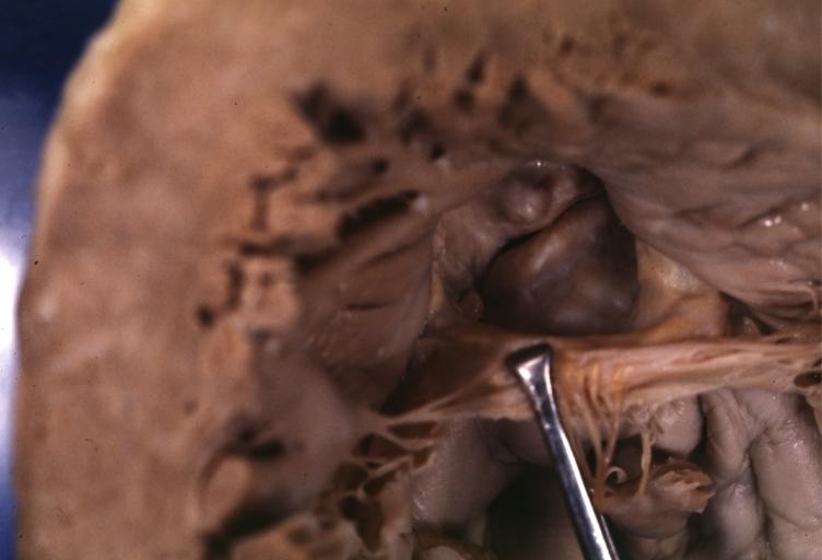
Bicuspid Aortic Stenosis: Gross; fixed tissue
-
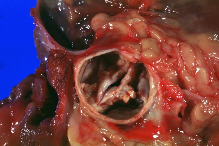
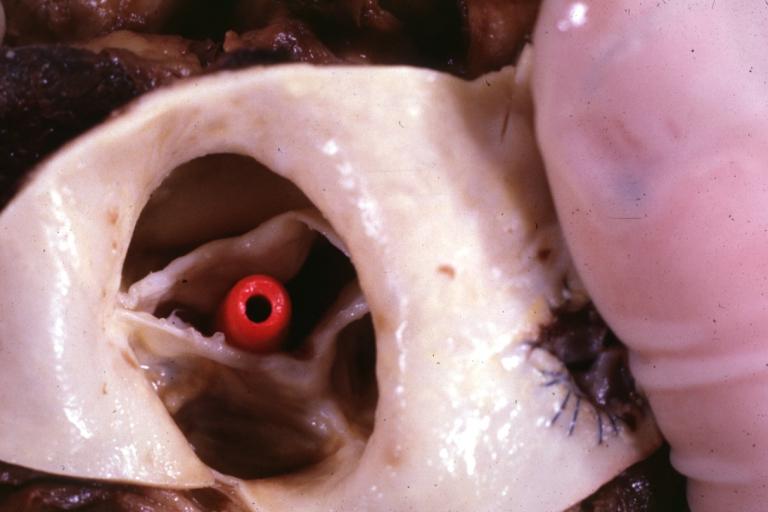
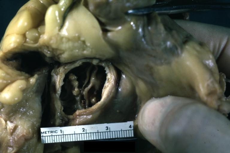
Aortic Stenosis, Bicuspid: Gross; fixed tissue view of stenotic valve through ventricular outlet track
-
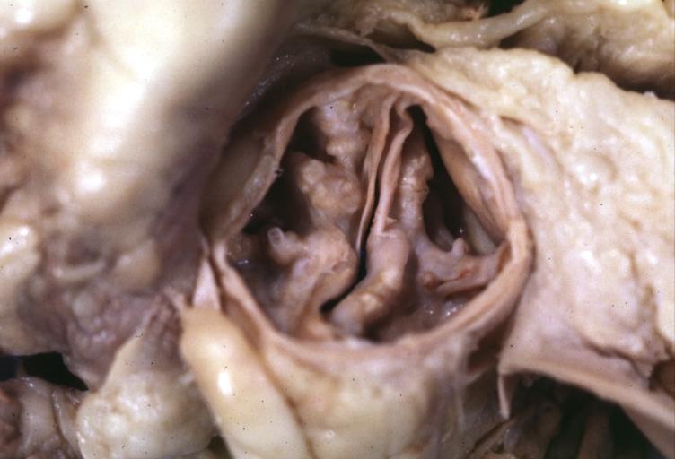
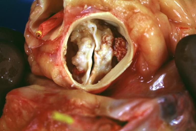
Aortic Stenosis Bicuspid: Gross; fixed tissue. Bicuspid valve and false raphe classical
-
Bicuspid aortic valve
-
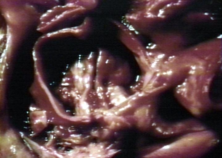
Bicuspid aortic valve
-
Bicuspid aortic valve
-
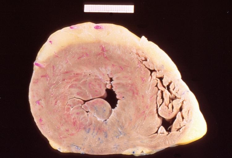
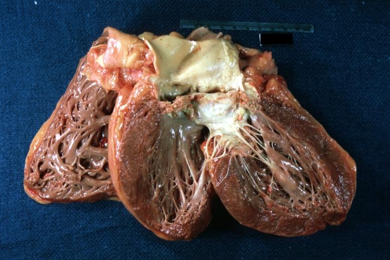
Left ventricular hypertrophy due to bicuspid aortic valve
-
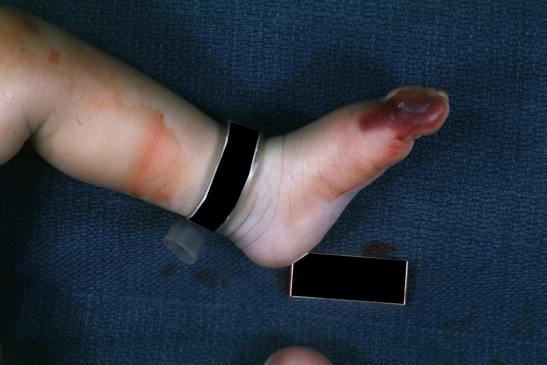
Congenital aortic stenosis: Gangrene toe In Infant: Gross, natural color, 1 month old child with congenital aortic stenosis
-
Unicuspid aortic stenosis


