Aortic stenosis gross pathology: Difference between revisions
No edit summary |
|||
| Line 1: | Line 1: | ||
{{ | {{Aortic stenosis}} | ||
{{CMG}} | {{CMG}} | ||
==Pathological Findings== | ==Pathological Findings== | ||
Images shown below are courtesy of Professor Peter Anderson DVM PhD and published with permission. [http://www.peir.net © PEIR, University of Alabama at Birmingham, Department of Pathology] | Images shown below are courtesy of Professor Peter Anderson DVM PhD and published with permission. [http://www.peir.net © PEIR, University of Alabama at Birmingham, Department of Pathology] | ||
<gallery> | |||
<gallery | |||
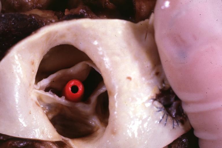
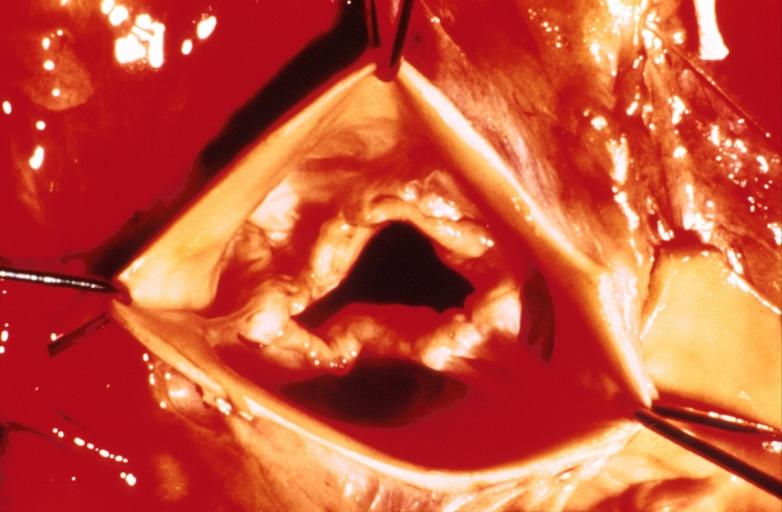
Image:Bicuspid aortic valve1.jpg|Aortic Stenosis, Bicuspid valve: Gross; excellent image of bicuspid and calcific valve showing a false raphe. | Image:Bicuspid aortic valve1.jpg|Aortic Stenosis, Bicuspid valve: Gross; excellent image of bicuspid and calcific valve showing a false raphe. | ||
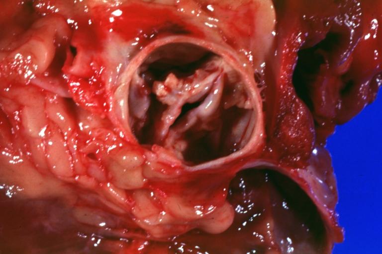
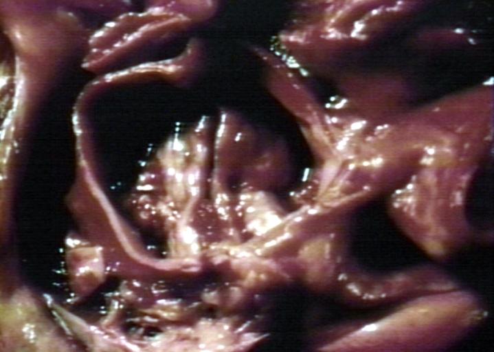
Image:Bicuspid aortic valve2.jpg|Aortic Stenosis, Bicuspid valve: Gross; good example of bicuspid valve | Image:Bicuspid aortic valve2.jpg|Aortic Stenosis, Bicuspid valve: Gross; good example of bicuspid valve | ||
Image:Bicuspid aortic valve3.jpg|Aortic Stenosis, Bicuspid valve: Gross; image of bicuspid aortic valve, an excellent example | Image:Bicuspid aortic valve3.jpg|Aortic Stenosis, Bicuspid valve: Gross; image of bicuspid aortic valve, an excellent example | ||
Image:Bicuspid aortic valve4.jpg|Aortic Stenosis, Bicuspid valve: Gross; close-up image of bicuspid aortic valve. | Image:Bicuspid aortic valve4.jpg|Aortic Stenosis, Bicuspid valve: Gross; close-up image of bicuspid aortic valve. | ||
Image:Bicuspid aortic valve5.jpg|Aortic Stenosis, Bicuspid valve: Gross; close-up image of bicuspid aortic valve. | Image:Bicuspid aortic valve5.jpg|Aortic Stenosis, Bicuspid valve: Gross; close-up image of bicuspid aortic valve. | ||
Image:Bicuspid aortic valve6.jpg|Bicuspid aortic valve | Image:Bicuspid aortic valve6.jpg|Bicuspid aortic valve | ||
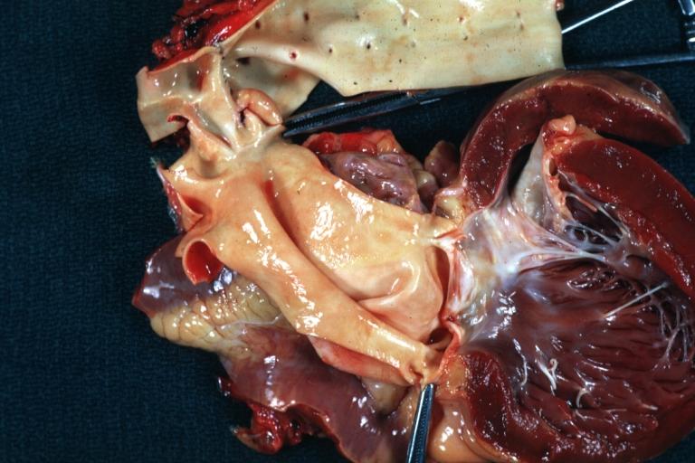
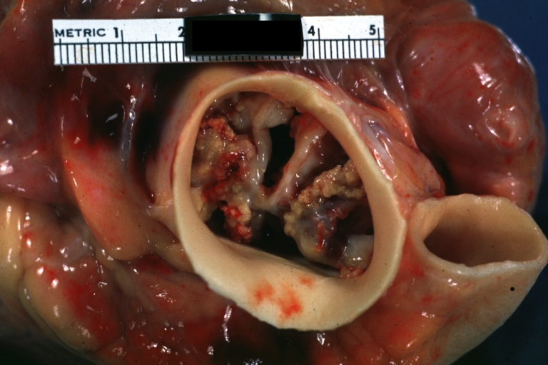
Image:Bicuspid aortic valve7.jpg|Gross natural color opened first portion aortic arch with bicuspid aortic valve shows stenosis and aortic root is dilated | Image:Bicuspid aortic valve7.jpg|Gross natural color opened first portion aortic arch with bicuspid aortic valve shows stenosis and aortic root is dilated | ||
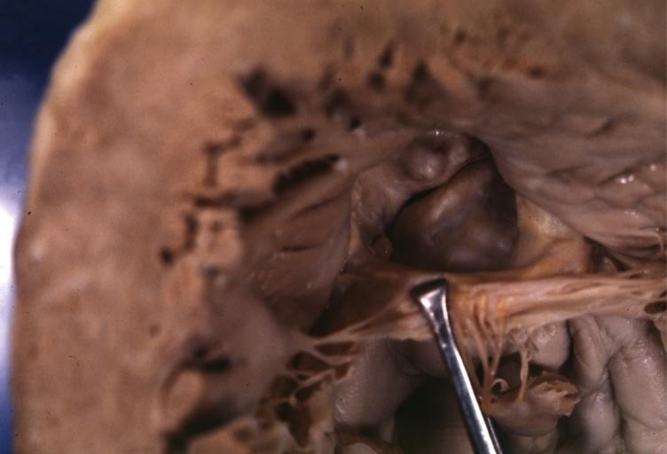
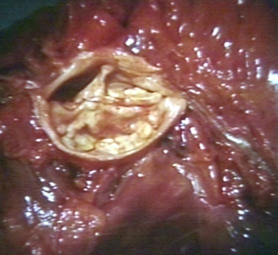
Image:Bicuspid aortic valve8.jpg|Aortic Stenosis Bicuspid: Gross; natural color opened left ventricular outflow tract with calcific masses on valve as well as anterior leaflet mitral valve probably did not cause significant stenosis | Image:Bicuspid aortic valve8.jpg|Aortic Stenosis Bicuspid: Gross; natural color opened left ventricular outflow tract with calcific masses on valve as well as anterior leaflet mitral valve probably did not cause significant stenosis | ||
Image:Bicuspid aortic valve9.jpg|Bicuspid Aortic Valve with Repaired Aorta Coarctation: Gross natural color opened left ventricular outflow tract with uncomplicated bicuspid aortic valve repaired coarctation barely visible ruptured postoperative young female with ovaries Turner mosaic not ruled out | Image:Bicuspid aortic valve9.jpg|Bicuspid Aortic Valve with Repaired Aorta Coarctation: Gross natural color opened left ventricular outflow tract with uncomplicated bicuspid aortic valve repaired coarctation barely visible ruptured postoperative young female with ovaries Turner mosaic not ruled out | ||
Image:Bicuspid aortic valve10.jpg|Bicuspid Aortic Stenosis: Gross; fixed tissue | Image:Bicuspid aortic valve10.jpg|Bicuspid Aortic Stenosis: Gross; fixed tissue | ||
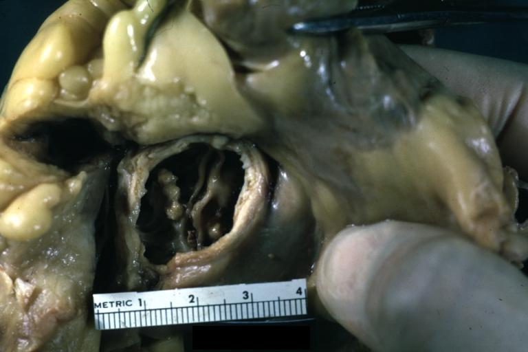
Image:Bicuspid aortic valve11.jpg|Aortic Stenosis, Bicuspid: Gross; fixed tissue view of stenotic valve through ventricular outlet track | Image:Bicuspid aortic valve11.jpg|Aortic Stenosis, Bicuspid: Gross; fixed tissue view of stenotic valve through ventricular outlet track | ||
Image:Bicuspid aortic valve12.jpg|Aortic Stenosis Bicuspid: Gross; fixed tissue. Bicuspid valve and false raphe classical | Image:Bicuspid aortic valve12.jpg|Aortic Stenosis Bicuspid: Gross; fixed tissue. Bicuspid valve and false raphe classical | ||
Image:Bicuspid aortic valve13.jpg|Bicuspid aortic valve | Image:Bicuspid aortic valve13.jpg|Bicuspid aortic valve | ||
Image:Bicuspid aortic valve14.jpg|Bicuspid aortic valve | Image:Bicuspid aortic valve14.jpg|Bicuspid aortic valve | ||
Image:Bicuspid aortic valve15.jpg|Bicuspid aortic valve | Image:Bicuspid aortic valve15.jpg|Bicuspid aortic valve | ||
Image:Bicuspid aortic valve16.jpg|Left ventricular hypertrophy due to bicuspid aortic valve | Image:Bicuspid aortic valve16.jpg|Left ventricular hypertrophy due to bicuspid aortic valve | ||
Image:Congenital aortic stenosis.jpg|Congenital aortic stenosis: Gangrene toe In Infant: Gross, natural color, 1 month old child with congenital aortic stenosis | Image:Congenital aortic stenosis.jpg|Congenital aortic stenosis: Gangrene toe In Infant: Gross, natural color, 1 month old child with congenital aortic stenosis | ||
Image:Unicuspid aortic stenosis.jpg|Unicuspid aortic stenosis | Image:Unicuspid aortic stenosis.jpg|Unicuspid aortic stenosis | ||
| Line 77: | Line 35: | ||
==An Autopsy Report== | ==An Autopsy Report== | ||
A 68-year-old man initially sought medical advice five years prior to his death. His symptoms at that time were [[exercise intolerance]] and occasional [[peripheral edema]]. He gave a history of a "[[heart murmur]]" that was diagnosed 25 years ago during an employment physical. No follow up care had been given for this murmur. | A 68-year-old man initially sought medical advice five years prior to his death. His symptoms at that time were [[exercise intolerance]] and occasional [[peripheral edema]]. He gave a history of a "[[heart murmur]]" that was diagnosed 25 years ago during an employment physical. No follow up care had been given for this murmur. | ||
| Line 83: | Line 40: | ||
===Autopsy Findings=== | ===Autopsy Findings=== | ||
Autopsy disclosed a markedly enlarged heart weighing 650 grams and having dilated chambers. The aortic valve was calcified and showed evidence of stenosis and insufficiency. The coronary arteries were narrowed 60 to 70% by atherosclerosis. No acute coronary occlusions were found and there was no evidence of [[myocardial infarction]]. | Autopsy disclosed a markedly enlarged heart weighing 650 grams and having dilated chambers. The aortic valve was calcified and showed evidence of stenosis and insufficiency. The coronary arteries were narrowed 60 to 70% by atherosclerosis. No acute coronary occlusions were found and there was no evidence of [[myocardial infarction]]. | ||
<gallery> | |||
<gallery | |||
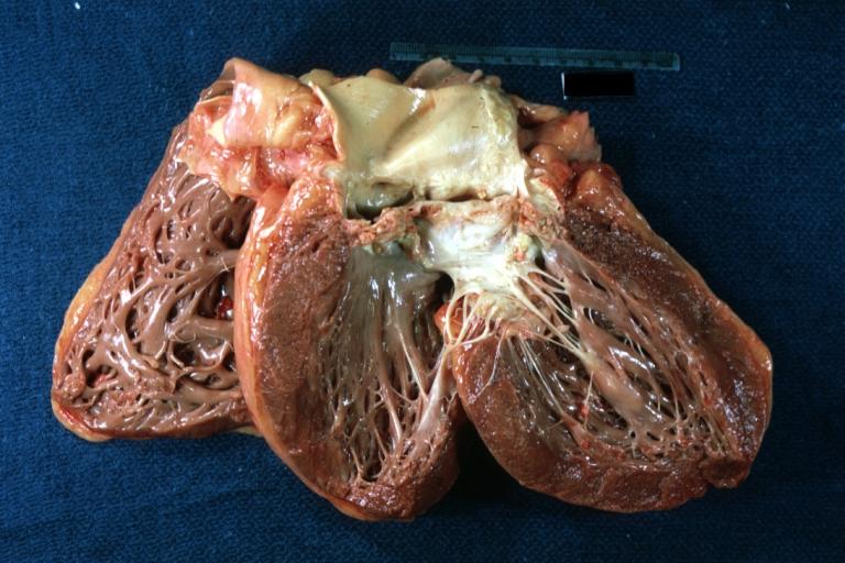
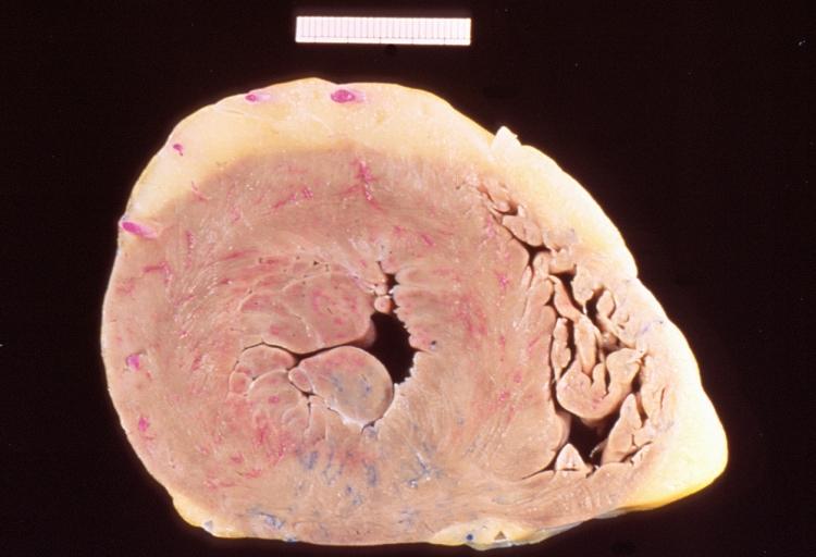
Image:Comparison of hypertrophic myocardium and normal Gross.JPG|This is a gross photograph of a cross section of a normal human heart taken at autopsy (right) and the heart from this case, which demonstrates concentric hypertrophy of the left ventricular wall. Note the marked thickening of the left ventricular wall. There is also moderate thickening of the right ventricular wall. | Image:Comparison of hypertrophic myocardium and normal Gross.JPG|This is a gross photograph of a cross section of a normal human heart taken at autopsy (right) and the heart from this case, which demonstrates concentric hypertrophy of the left ventricular wall. Note the marked thickening of the left ventricular wall. There is also moderate thickening of the right ventricular wall. | ||
Image:Comparison of hypertrophy and normal myocardial micro 1.JPG|This low-power photomicrograph shows normal myocardium (left) compared to hypertrophied myocardium (right). | Image:Comparison of hypertrophy and normal myocardial micro 1.JPG|This low-power photomicrograph shows normal myocardium (left) compared to hypertrophied myocardium (right). | ||
Image:Comparison of hypertrophy and normal myocardial micro 2.JPG|Normal myocardium (left) is compared here to hypertrophied myocardium (right). The muscle fibers are thicker and the nuclei are larger and darker in the hypertrophied myocardium.The clear spaces between the muscle fibers are due to processing artifacts and are not present during life. | Image:Comparison of hypertrophy and normal myocardial micro 2.JPG|Normal myocardium (left) is compared here to hypertrophied myocardium (right). The muscle fibers are thicker and the nuclei are larger and darker in the hypertrophied myocardium.The clear spaces between the muscle fibers are due to processing artifacts and are not present during life. | ||
Image:Comparison of hypertrophy and normal myocardial micro 3.JPG|Normal myocardium (left) is compared to hypertrophied myocardium (right). This high power view demonstrates the large dark nuclei (arrow) found in hypertrophied cardiac muscle cells. Polyploidy is a common feature in cardiac hypertrophy. Also note the increased size (thickness) of the individual cardiac muscle cell on the right compared to normal cardiac myocytes (left). | Image:Comparison of hypertrophy and normal myocardial micro 3.JPG|Normal myocardium (left) is compared to hypertrophied myocardium (right). This high power view demonstrates the large dark nuclei (arrow) found in hypertrophied cardiac muscle cells. Polyploidy is a common feature in cardiac hypertrophy. Also note the increased size (thickness) of the individual cardiac muscle cell on the right compared to normal cardiac myocytes (left). | ||
| Line 104: | Line 54: | ||
{{reflist|2}} | {{reflist|2}} | ||
[[Category:DiseaseState]] | [[Category:DiseaseState]] | ||
[[Category:Signs and symptoms]] | [[Category:Signs and symptoms]] | ||
| Line 113: | Line 61: | ||
[[Category:Congenital heart disease]] | [[Category:Congenital heart disease]] | ||
[[es:Estenosis aórtica]] | [[es:Estenosis aórtica]] | ||
[[fr:Rétrécissement aortique]] | [[fr:Rétrécissement aortique]] | ||
[[pl:Stenoza Aortalnej]] | [[pl:Stenoza Aortalnej]] | ||
[[pt:Estenose aórtica]] | [[pt:Estenose aórtica]] | ||
[[ro:Stenoza Aortică]] | [[ro:Stenoza Aortică]] | ||
[[tr:Aort darlığı]] | [[tr:Aort darlığı]] | ||
{{WH}} | {{WH}} | ||
{{WS}} | {{WS}} | ||
Revision as of 19:06, 25 July 2011
|
Aortic Stenosis Microchapters |
|
Diagnosis |
|---|
|
Treatment |
|
Percutaneous Aortic Balloon Valvotomy (PABV) or Aortic Valvuloplasty |
|
Transcatheter Aortic Valve Replacement (TAVR) |
|
Case Studies |
|
Aortic stenosis gross pathology On the Web |
|
American Roentgen Ray Society Images of Aortic stenosis gross pathology |
|
Directions to Hospitals Treating Aortic stenosis gross pathology |
|
Risk calculators and risk factors for Aortic stenosis gross pathology |
Editor-In-Chief: C. Michael Gibson, M.S., M.D. [1]
Pathological Findings
Images shown below are courtesy of Professor Peter Anderson DVM PhD and published with permission. © PEIR, University of Alabama at Birmingham, Department of Pathology
-
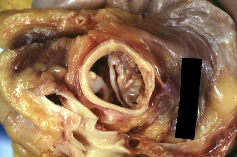
Aortic Stenosis, Bicuspid valve: Gross; excellent image of bicuspid and calcific valve showing a false raphe.
-
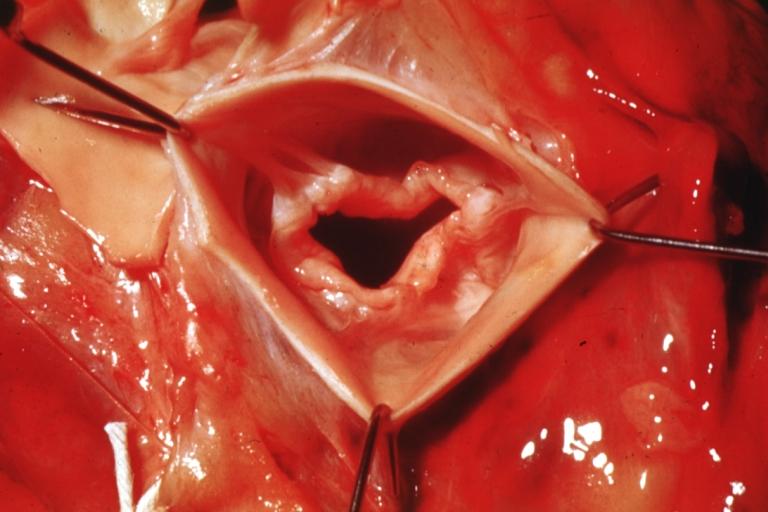
Aortic Stenosis, Bicuspid valve: Gross; good example of bicuspid valve
-
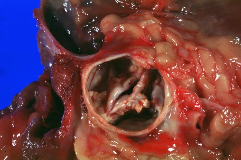
Aortic Stenosis, Bicuspid valve: Gross; image of bicuspid aortic valve, an excellent example
-
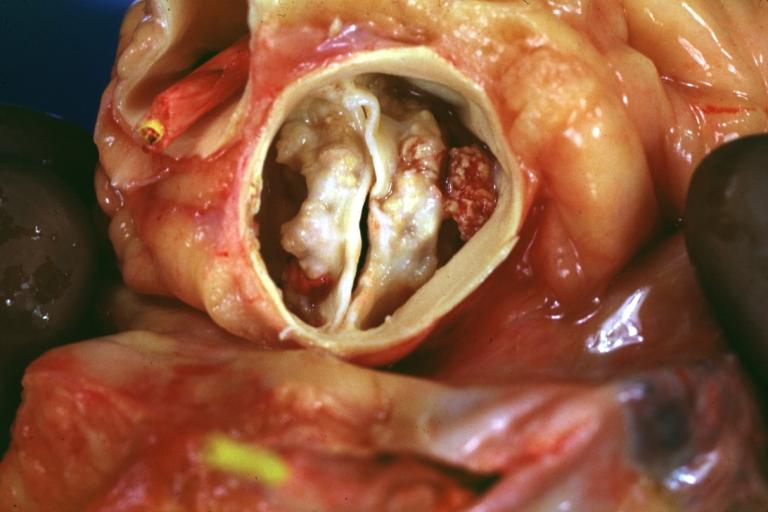
Aortic Stenosis, Bicuspid valve: Gross; close-up image of bicuspid aortic valve.
-
Aortic Stenosis, Bicuspid valve: Gross; close-up image of bicuspid aortic valve.
-
Bicuspid aortic valve
-
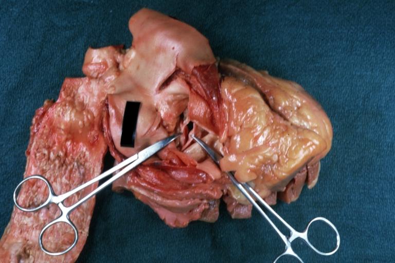
Gross natural color opened first portion aortic arch with bicuspid aortic valve shows stenosis and aortic root is dilated
-
Aortic Stenosis Bicuspid: Gross; natural color opened left ventricular outflow tract with calcific masses on valve as well as anterior leaflet mitral valve probably did not cause significant stenosis
-
Bicuspid Aortic Valve with Repaired Aorta Coarctation: Gross natural color opened left ventricular outflow tract with uncomplicated bicuspid aortic valve repaired coarctation barely visible ruptured postoperative young female with ovaries Turner mosaic not ruled out
-
Bicuspid Aortic Stenosis: Gross; fixed tissue
-
Aortic Stenosis, Bicuspid: Gross; fixed tissue view of stenotic valve through ventricular outlet track
-
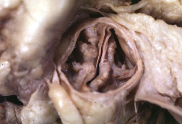
Aortic Stenosis Bicuspid: Gross; fixed tissue. Bicuspid valve and false raphe classical
-
Bicuspid aortic valve
-
Bicuspid aortic valve
-
Bicuspid aortic valve
-
Left ventricular hypertrophy due to bicuspid aortic valve
-
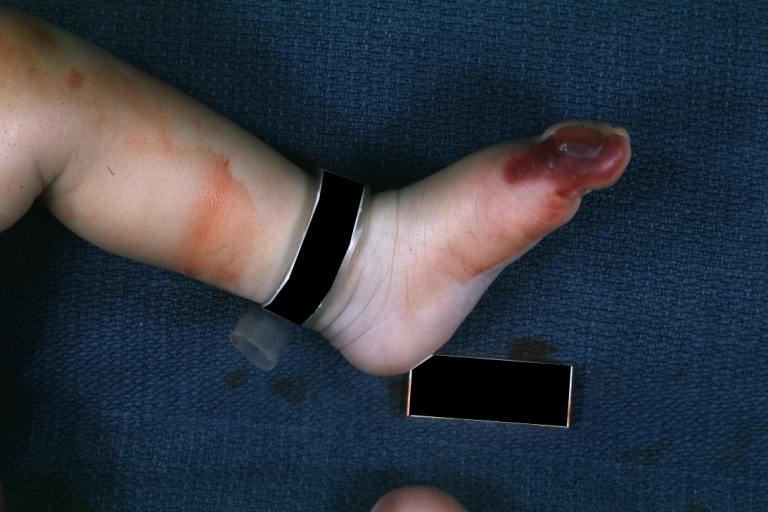
Congenital aortic stenosis: Gangrene toe In Infant: Gross, natural color, 1 month old child with congenital aortic stenosis
-
Unicuspid aortic stenosis


An Autopsy Report
A 68-year-old man initially sought medical advice five years prior to his death. His symptoms at that time were exercise intolerance and occasional peripheral edema. He gave a history of a "heart murmur" that was diagnosed 25 years ago during an employment physical. No follow up care had been given for this murmur.
The patient's terminal admission was for signs of severe heart failure--the patient had marked peripheral edema and shortness of breath and chest x-ray revealed significant cardiac enlargement and pulmonary edema with bilateral pleural effusions. He sustained a cardiac arrest shortly after admission and could not be resuscitated.
Autopsy Findings
Autopsy disclosed a markedly enlarged heart weighing 650 grams and having dilated chambers. The aortic valve was calcified and showed evidence of stenosis and insufficiency. The coronary arteries were narrowed 60 to 70% by atherosclerosis. No acute coronary occlusions were found and there was no evidence of myocardial infarction.
-
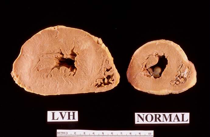
This is a gross photograph of a cross section of a normal human heart taken at autopsy (right) and the heart from this case, which demonstrates concentric hypertrophy of the left ventricular wall. Note the marked thickening of the left ventricular wall. There is also moderate thickening of the right ventricular wall.
-
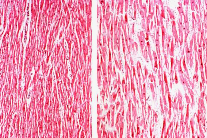
This low-power photomicrograph shows normal myocardium (left) compared to hypertrophied myocardium (right).
-
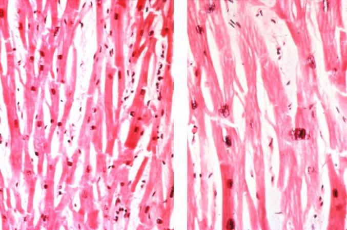
Normal myocardium (left) is compared here to hypertrophied myocardium (right). The muscle fibers are thicker and the nuclei are larger and darker in the hypertrophied myocardium.The clear spaces between the muscle fibers are due to processing artifacts and are not present during life.
-
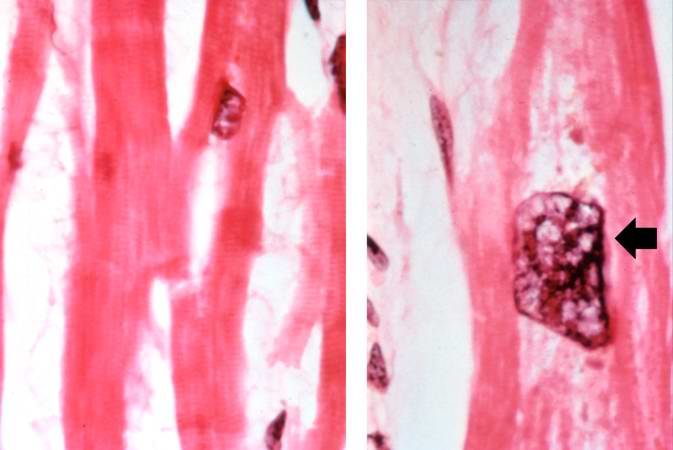
Normal myocardium (left) is compared to hypertrophied myocardium (right). This high power view demonstrates the large dark nuclei (arrow) found in hypertrophied cardiac muscle cells. Polyploidy is a common feature in cardiac hypertrophy. Also note the increased size (thickness) of the individual cardiac muscle cell on the right compared to normal cardiac myocytes (left).


