Aortic coarctation: Difference between revisions
No edit summary |
No edit summary |
||
| Line 29: | Line 29: | ||
==Diagnosis== | ==Diagnosis== | ||
[[Aortic coarctation symptoms | Symptoms]] | [[Aortic coarctation physical examination | Physical examination]] | [[Aortic coarctation electrocardiogram | Electrocardiogram]] | [[Aortic coarctation chest x-ray | Chest x-ray]] | [[Aortic coarctation echocardiography | Echocardiography]] | [[Aortic coarctation symptoms | Symptoms]] | [[Aortic coarctation physical examination | Physical examination]] | [[Aortic coarctation electrocardiogram | Electrocardiogram]] | [[Aortic coarctation chest x-ray | Chest x-ray]] | [[Aortic coarctation echocardiography | Echocardiography]] | [[Aortic coarctation angiography | Angiography]] | [[Aortic coarctation MRI | MRI]] | [[Aortic coarctation CT | CT]] | ||
===Angiography=== | ===Angiography=== | ||
Revision as of 17:21, 24 June 2011
For patient information click here'
|
Aortic coarctation Microchapters |
|
Diagnosis |
|---|
|
Treatment |
|
Case Studies |
|
Aortic coarctation On the Web |
|
American Roentgen Ray Society Images of Aortic coarctation |
Editor-In-Chief: C. Michael Gibson, M.S., M.D. [1]
Associate Editor-In-Chief: Cafer Zorkun, M.D., Ph.D. [2]
Overview
History
Epidemiology and demographics
Classification
Anatomy
Pathophysiology
Natural History
Genetics
Complications
Associated conditions
Diagnosis
Symptoms | Physical examination | Electrocardiogram | Chest x-ray | Echocardiography | Angiography | MRI | CT
Angiography
<youtube v=yvwCL3D8MFM />
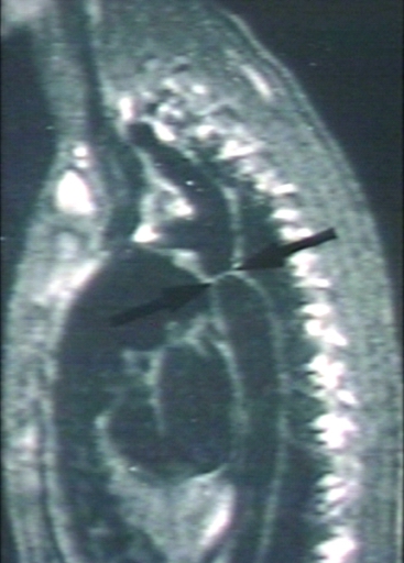
MRI
Magnetic resonance imaging (MRI) can define the location and severity of a coarctation. MRI can also detect associated cardiac abnormalities and is used for serial follow-up after surgical repair or balloon angioplasty. MR angiography has almost completely replaced invasive catheter based techniques for evaluating re coarctation. In adults with untreated coarctation blood often reaches the lower body through collaterals, eg. internal thoracic arteries via. the subclavian arteries. Those can be seen on MR or angiography.

-
![[Courtesy of RadsWiki and copylefted]](/images/7/72/Coarctation-of-the-aorta-MRI-003.jpg)
[Courtesy of RadsWiki and copylefted]
-
![[Courtesy of RadsWiki and copylefted]](/images/c/c6/Coarctation-of-the-aorta-MRI-002.jpg)
[Courtesy of RadsWiki and copylefted]
-
![[Courtesy of RadsWiki and copylefted]](/index.php/File:Coarctation-of-the-aorta-MRI-003.jpg)
![[Courtesy of RadsWiki and copylefted]](/index.php/File:Coarctation-of-the-aorta-MRI-002.jpg)

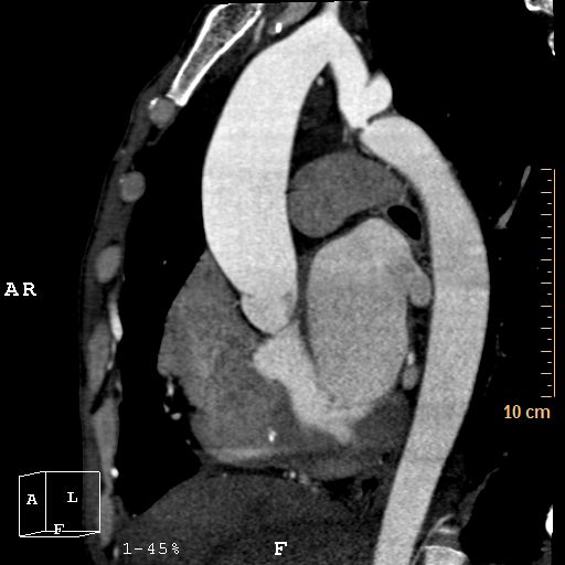
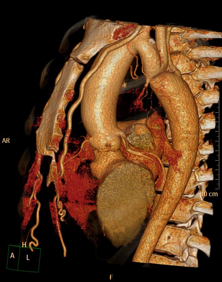
CT
CT images shown below are courtesy of Cafer Zorkun and copylefted
-

Aortic Coarctation
-
Aortic Coarctation
-

Aortic Coarctation
-
Aortic Coarctation


Therapy
Therapy is conservative if the patient is asymptomatic. If symptoms or hypertension are present, treatment for coarctation may be surgical or catheter based. The treatment choice depends on the patients age, the location of the coarctation and other associated anomalies. Recoartctaion after previous surgery is treated percutaneously with either balloon dilation and/or stenting.
References
External links
- A case of coarctation of the aorta was published in the New England Journal of Medicine in 2007 showing chest X-Rays and MRT Images.[1]
- Aortic Coarctation information from Seattle Children's Hospital Heart Center
- Overview and diagram at childrenscentralcal.org
- Diagram at kumc.edu
- Overview and diagram at umich.edu
- [3]
Template:Link FA de:Aortenisthmusstenose it:Coartazione dell'aorta no:Koarktasjon av aorta nn:Koarktasjon sr:Коарктација аорте uk:Коарктація аорти
- ↑ Quiros-Lopez R, Garcia-Alegria J (2007). "A medical mystery -- high blood pressure". N Engl J Med. 356 (25): 2630. PMID 17582073.