Alopecia pathophysiology: Difference between revisions
Jose Loyola (talk | contribs) |
No edit summary |
||
| (32 intermediate revisions by 2 users not shown) | |||
| Line 2: | Line 2: | ||
{{Alopecia}} | {{Alopecia}} | ||
{{CMG}}; {{AE}} {{ | {{CMG}}; {{AE}} {{Jose}} | ||
==Overview== | ==Overview== | ||
Since [[alopecia]] has many different causes, the pathophysiologic mechanism for its development varies widely according to the cause. [[Alopecia areata]], for example, is related with [[CD8+]] [[T-cell]] [[autoimmunity]], while [[androgenetic alopecia]] is related with androgen hormones effects' on the hair follicle which leads to its miniaturization and hair loss. [[Tinea capitis]] on the other hand is an infectious disease that can damage the hair follicle and lead to definitive hair loss. | |||
==Pathophysiology== | ==Pathophysiology== | ||
The most common causes for [[alopecia]] and its pathophysiology mechanism are briefly discussed below: | |||
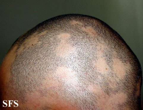
== | ===Alopecia Areata=== | ||
[[File:Alopecia_areata_12.jpeg|200px|thumb|left|Alopecia areata - from atlasdermatologico.com.br]] | |||
* The exact pathogenesis of [[alopecia areata]] is not fully understood. | |||
* It has been theorized that [[T-cell-mediated autoimmunity]] must be involved in its development. | |||
* The [[hair follicle]] typically has low levels of [[major histocompatibility complex expression]], which provides protection from the immune system. It is believed that in [[alopecia Areata]] that protection is lost, resulting in a [[CD8+]] [[T lymphocyte]] attack to the bulb of the hair, generating an [[inflammatory infiltrate]] in the [[peribulbar]] region of the [[hair follicle]].<ref name="pmid14582671">{{cite journal| author=Paus R, Ito N, Takigawa M, Ito T| title=The hair follicle and immune privilege. | journal=J Investig Dermatol Symp Proc | year= 2003 | volume= 8 | issue= 2 | pages= 188-94 | pmid=14582671 | doi=10.1046/j.1087-0024.2003.00807.x | pmc= | url=https://www.ncbi.nlm.nih.gov/entrez/eutils/elink.fcgi?dbfrom=pubmed&tool=sumsearch.org/cite&retmode=ref&cmd=prlinks&id=14582671 }} </ref><ref name="pmid24326544">{{cite journal| author=Paus R, Bertolini M| title=The role of hair follicle immune privilege collapse in alopecia areata: status and perspectives. | journal=J Investig Dermatol Symp Proc | year= 2013 | volume= 16 | issue= 1 | pages= S25-7 | pmid=24326544 | doi=10.1038/jidsymp.2013.7 | pmc= | url=https://www.ncbi.nlm.nih.gov/entrez/eutils/elink.fcgi?dbfrom=pubmed&tool=sumsearch.org/cite&retmode=ref&cmd=prlinks&id=24326544 }} </ref> | |||
*Genes involved in the pathogenesis of [[alopecia areata]] include [[MCHR2]] and [[MCHR2-AS1]] which are related to the [[MHC]] pathway ([[melanin concentrating hormone]]).<ref name="pmid27306922">{{cite journal| author=Fischer J, Degenhardt F, Hofmann A, Redler S, Basmanav FB, Heilmann-Heimbach S | display-authors=etal| title=Genomewide analysis of copy number variants in alopecia areata in a Central European cohort reveals association with MCHR2. | journal=Exp Dermatol | year= 2017 | volume= 26 | issue= 6 | pages= 536-541 | pmid=27306922 | doi=10.1111/exd.13123 | pmc= | url=https://www.ncbi.nlm.nih.gov/entrez/eutils/elink.fcgi?dbfrom=pubmed&tool=sumsearch.org/cite&retmode=ref&cmd=prlinks&id=27306922 }} </ref> | |||
* There is an association of [[alopecia Areata]] with other autoimmune diseases such as [[vitiligo]], [[autoimmune thyroid disease]], [[celiac disease]], [[systemic erythematous]], [[chronic atrophic gastritis]] which further reinforces its relationship with autoimmunity.<ref name="pmid28717940">{{cite journal| author=Trüeb RM, Dias MFRG| title=Alopecia Areata: a Comprehensive Review of Pathogenesis and Management. | journal=Clin Rev Allergy Immunol | year= 2018 | volume= 54 | issue= 1 | pages= 68-87 | pmid=28717940 | doi=10.1007/s12016-017-8620-9 | pmc= | url=https://www.ncbi.nlm.nih.gov/entrez/eutils/elink.fcgi?dbfrom=pubmed&tool=sumsearch.org/cite&retmode=ref&cmd=prlinks&id=28717940 }} </ref> | |||
<br> | |||
==== | ===Telogen effluvium=== | ||
* [[Telogen effluvium]] is not a disease per se, but it is a common cause of hair loss due to a triggering event that increases the number of hair follicles that are in the catagen or telogen phase (shedding and resting phase) of the hair development cycle. | |||
* | * It is of non-scarring type. | ||
*It is | * There are many different events that can act as triggers for this condition, such as [[febrile diseases]] ([[malaria]], [[HIV]], [[tuberculosis]]), [[drugs]] ([[oral contraceptives]], [[anticonvulsants]], [[beta blockers]], [[captopril]] [[antithyroid drugs]] and [[hypolipidemic drugs]]), [[thyroid diseases]], organ dysfunction, nutritional ([[iron]] or [[zinc]] deficiency) or local factors such as hair dye.<ref name="pmid26500992">{{cite journal| author=Malkud S| title=Telogen Effluvium: A Review. | journal=J Clin Diagn Res | year= 2015 | volume= 9 | issue= 9 | pages= WE01-3 | pmid=26500992 | doi=10.7860/JCDR/2015/15219.6492 | pmc=4606321 | url=https://www.ncbi.nlm.nih.gov/entrez/eutils/elink.fcgi?dbfrom=pubmed&tool=sumsearch.org/cite&retmode=ref&cmd=prlinks&id=26500992 }} </ref> | ||
* Telogen hairs are usually at least at 25% for the diagnosis of telogen effluvium to be made.<ref name="pmid28613598">{{cite journal| author=| title=StatPearls | journal= | year= 2020 | volume= | issue= | pages= | pmid=28613598 | doi= | pmc= | url= }} </ref> | |||
== | ===Traumatic alopecia=== | ||
In androgenetic alopecia | * Usually seen on children that pull their hair, the same mechanism as [[traction alopecia]]. | ||
* May be associated with [[trichotillomania]] - a psychiatric condition in which the patient repeatedly pulls their hair.<ref name="pmid30844205">{{cite journal| author=| title=StatPearls | journal= | year= 2020 | volume= | issue= | pages= | pmid=30844205 | doi= | pmc= | url= }} </ref> | |||
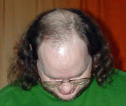
===Androgenetic alopecia=== | |||
[[File:Alopecia.jpg|200px|thumb|left|Androgenetic alopecia - from atlasdermatologico.com.br]] | |||
* In [[androgenetic alopecia]] there is marked miniaturization of the hair follicle and disruption of the hair cycle. | |||
* It is thought that in [[androgenetic alopecia]] the hair loss is the result of the shortening of the anagen phases of hair development and enlongation of the telogen phase that gradually takes place until the hair eventually doesn't even leave the skin surface.<ref name="pmid14585162">{{cite journal| author=Ellis JA, Sinclair R, Harrap SB| title=Androgenetic alopecia: pathogenesis and potential for therapy. | journal=Expert Rev Mol Med | year= 2002 | volume= 4 | issue= 22 | pages= 1-11 | pmid=14585162 | doi=10.1017/S1462399402005112 | pmc= | url=https://www.ncbi.nlm.nih.gov/entrez/eutils/elink.fcgi?dbfrom=pubmed&tool=sumsearch.org/cite&retmode=ref&cmd=prlinks&id=14585162 }} </ref> | |||
* The is also an increase in the period from the hair shedding to its regrowth.<ref name="pmid14585162">{{cite journal| author=Ellis JA, Sinclair R, Harrap SB| title=Androgenetic alopecia: pathogenesis and potential for therapy. | journal=Expert Rev Mol Med | year= 2002 | volume= 4 | issue= 22 | pages= 1-11 | pmid=14585162 | doi=10.1017/S1462399402005112 | pmc= | url=https://www.ncbi.nlm.nih.gov/entrez/eutils/elink.fcgi?dbfrom=pubmed&tool=sumsearch.org/cite&retmode=ref&cmd=prlinks&id=14585162 }} </ref> | |||
* The miniaturization affects the hair follicle globally, including the dermal papilla which is essential for its maintenance.<ref name="pmid14585162">{{cite journal| author=Ellis JA, Sinclair R, Harrap SB| title=Androgenetic alopecia: pathogenesis and potential for therapy. | journal=Expert Rev Mol Med | year= 2002 | volume= 4 | issue= 22 | pages= 1-11 | pmid=14585162 | doi=10.1017/S1462399402005112 | pmc= | url=https://www.ncbi.nlm.nih.gov/entrez/eutils/elink.fcgi?dbfrom=pubmed&tool=sumsearch.org/cite&retmode=ref&cmd=prlinks&id=14585162 }} </ref> | |||
* It is mediated by the presence of [[androgens]], which is further reinforced by the fact that eunuchs do not bald. | |||
* The molecular mechanism of action for the [[androgens]] such as [[testosterone]] and [[5α-dihydrotestosterone]] ([[DHT]]) to act on the hair follicle is not fully understood. | |||
* It is theorized that some genes that regulate the follicle cycling may be regulated by the presence of androgens and that the expression of such genes are related to the concentrations of [[androgen]] and [[androgen receptor]]s in the follicle.<ref name="pmid14585162">{{cite journal| author=Ellis JA, Sinclair R, Harrap SB| title=Androgenetic alopecia: pathogenesis and potential for therapy. | journal=Expert Rev Mol Med | year= 2002 | volume= 4 | issue= 22 | pages= 1-11 | pmid=14585162 | doi=10.1017/S1462399402005112 | pmc= | url=https://www.ncbi.nlm.nih.gov/entrez/eutils/elink.fcgi?dbfrom=pubmed&tool=sumsearch.org/cite&retmode=ref&cmd=prlinks&id=14585162 }} </ref> | |||
* It is also theorized through observation of families with [[androgenetic alopecia]] that these genes related to the disease may act in an [[autosomal dominant]] manner in men and [[autosomal recessive]] manner in women, though there is strong evidence for a [[polygenic]] mode of inheritance. | |||
* [[Finasteride]] is used to treat [[androgenetic alopecia]] being a potent [[5-alpha-reductase]] type-2 [[inhibitor]], inhibiting the conversion of [[testosterone]] to [[DHT]]. | |||
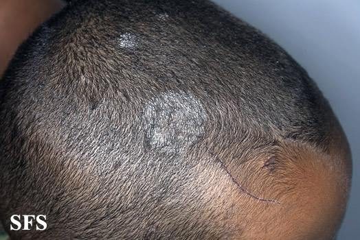
===Tinea capitis=== | |||
[[File:Tinea capitis47.jpg|200px|thumb|left|Tinea capitis - from atlasdermatologico.com.br]] | |||
* [[Tinea capitais]] is caused by [[dermatophyte]] species that are able to invade keratinized tissues like the hair. | |||
* It is usually transmitted via direct contact with organisms from other humans, animals, soil, or [[fomites]]. | |||
* The [[dermatophyte]] infects the hair and grows towards the [[stratum corneum]]. It then affects the hair which becomes brittle and break.<ref name="pmid30725594">{{cite journal| author=| title=StatPearls | journal= | year= 2020 | volume= | issue= | pages= | pmid=30725594 | doi= | pmc= | url= }} </ref> | |||
* It may present with black dots, which is the non-inflammatory form of the disease, causing fracture of the hair. It also may present with intense inflammation which leads to follicular destruction, and may complicate with [[kerion]], an abscess in the [[scalp]], or [[favus]], another inflammatory form in which there is a honeycomb destruction of the hair shaft. Both are severe forms of the disease.<ref name="pmid30725594">{{cite journal| author=| title=StatPearls | journal= | year= 2020 | volume= | issue= | pages= | pmid=30725594 | doi= | pmc= | url= }} </ref> | |||
<br> | |||
<br> | |||
<br> | |||
===Anagen Effluvium=== | |||
* [[Anagen effluvium]] occurs mostly due to chemotherapy. | |||
* Chemotherapeutic agents cause cessation of the hair growth during anagen phase, due to disruption of the cell cycle caused by the medication.<ref name="pmid30725594">{{cite journal| author=| title=StatPearls | journal= | year= 2020 | volume= | issue= | pages= | pmid=30725594 | doi= | pmc= | url= }} </ref> | |||
==References== | ==References== | ||
{{Reflist|2}} | {{Reflist|2}} | ||
[[Category:Disease]] | [[Category:Disease]] | ||
[[Category:Dermatology]] | [[Category:Dermatology]] | ||
Latest revision as of 17:39, 24 December 2020
|
Alopecia Microchapters |
|
Diagnosis |
|---|
|
Treatment |
|
Case Studies |
|
Alopecia pathophysiology On the Web |
|
American Roentgen Ray Society Images of Alopecia pathophysiology |
|
Risk calculators and risk factors for Alopecia pathophysiology |
Editor-In-Chief: C. Michael Gibson, M.S., M.D. [1]; Associate Editor(s)-in-Chief: José Eduardo Riceto Loyola Junior, M.D.[2]
Overview
Since alopecia has many different causes, the pathophysiologic mechanism for its development varies widely according to the cause. Alopecia areata, for example, is related with CD8+ T-cell autoimmunity, while androgenetic alopecia is related with androgen hormones effects' on the hair follicle which leads to its miniaturization and hair loss. Tinea capitis on the other hand is an infectious disease that can damage the hair follicle and lead to definitive hair loss.
Pathophysiology
The most common causes for alopecia and its pathophysiology mechanism are briefly discussed below:
Alopecia Areata

- The exact pathogenesis of alopecia areata is not fully understood.
- It has been theorized that T-cell-mediated autoimmunity must be involved in its development.
- The hair follicle typically has low levels of major histocompatibility complex expression, which provides protection from the immune system. It is believed that in alopecia Areata that protection is lost, resulting in a CD8+ T lymphocyte attack to the bulb of the hair, generating an inflammatory infiltrate in the peribulbar region of the hair follicle.[1][2]
- Genes involved in the pathogenesis of alopecia areata include MCHR2 and MCHR2-AS1 which are related to the MHC pathway (melanin concentrating hormone).[3]
- There is an association of alopecia Areata with other autoimmune diseases such as vitiligo, autoimmune thyroid disease, celiac disease, systemic erythematous, chronic atrophic gastritis which further reinforces its relationship with autoimmunity.[4]
Telogen effluvium
- Telogen effluvium is not a disease per se, but it is a common cause of hair loss due to a triggering event that increases the number of hair follicles that are in the catagen or telogen phase (shedding and resting phase) of the hair development cycle.
- It is of non-scarring type.
- There are many different events that can act as triggers for this condition, such as febrile diseases (malaria, HIV, tuberculosis), drugs (oral contraceptives, anticonvulsants, beta blockers, captopril antithyroid drugs and hypolipidemic drugs), thyroid diseases, organ dysfunction, nutritional (iron or zinc deficiency) or local factors such as hair dye.[5]
- Telogen hairs are usually at least at 25% for the diagnosis of telogen effluvium to be made.[6]
Traumatic alopecia
- Usually seen on children that pull their hair, the same mechanism as traction alopecia.
- May be associated with trichotillomania - a psychiatric condition in which the patient repeatedly pulls their hair.[7]
Androgenetic alopecia

- In androgenetic alopecia there is marked miniaturization of the hair follicle and disruption of the hair cycle.
- It is thought that in androgenetic alopecia the hair loss is the result of the shortening of the anagen phases of hair development and enlongation of the telogen phase that gradually takes place until the hair eventually doesn't even leave the skin surface.[8]
- The is also an increase in the period from the hair shedding to its regrowth.[8]
- The miniaturization affects the hair follicle globally, including the dermal papilla which is essential for its maintenance.[8]
- It is mediated by the presence of androgens, which is further reinforced by the fact that eunuchs do not bald.
- The molecular mechanism of action for the androgens such as testosterone and 5α-dihydrotestosterone (DHT) to act on the hair follicle is not fully understood.
- It is theorized that some genes that regulate the follicle cycling may be regulated by the presence of androgens and that the expression of such genes are related to the concentrations of androgen and androgen receptors in the follicle.[8]
- It is also theorized through observation of families with androgenetic alopecia that these genes related to the disease may act in an autosomal dominant manner in men and autosomal recessive manner in women, though there is strong evidence for a polygenic mode of inheritance.
- Finasteride is used to treat androgenetic alopecia being a potent 5-alpha-reductase type-2 inhibitor, inhibiting the conversion of testosterone to DHT.
Tinea capitis

- Tinea capitais is caused by dermatophyte species that are able to invade keratinized tissues like the hair.
- It is usually transmitted via direct contact with organisms from other humans, animals, soil, or fomites.
- The dermatophyte infects the hair and grows towards the stratum corneum. It then affects the hair which becomes brittle and break.[9]
- It may present with black dots, which is the non-inflammatory form of the disease, causing fracture of the hair. It also may present with intense inflammation which leads to follicular destruction, and may complicate with kerion, an abscess in the scalp, or favus, another inflammatory form in which there is a honeycomb destruction of the hair shaft. Both are severe forms of the disease.[9]
Anagen Effluvium
- Anagen effluvium occurs mostly due to chemotherapy.
- Chemotherapeutic agents cause cessation of the hair growth during anagen phase, due to disruption of the cell cycle caused by the medication.[9]
References
- ↑ Paus R, Ito N, Takigawa M, Ito T (2003). "The hair follicle and immune privilege". J Investig Dermatol Symp Proc. 8 (2): 188–94. doi:10.1046/j.1087-0024.2003.00807.x. PMID 14582671.
- ↑ Paus R, Bertolini M (2013). "The role of hair follicle immune privilege collapse in alopecia areata: status and perspectives". J Investig Dermatol Symp Proc. 16 (1): S25–7. doi:10.1038/jidsymp.2013.7. PMID 24326544.
- ↑ Fischer J, Degenhardt F, Hofmann A, Redler S, Basmanav FB, Heilmann-Heimbach S; et al. (2017). "Genomewide analysis of copy number variants in alopecia areata in a Central European cohort reveals association with MCHR2". Exp Dermatol. 26 (6): 536–541. doi:10.1111/exd.13123. PMID 27306922.
- ↑ Trüeb RM, Dias MFRG (2018). "Alopecia Areata: a Comprehensive Review of Pathogenesis and Management". Clin Rev Allergy Immunol. 54 (1): 68–87. doi:10.1007/s12016-017-8620-9. PMID 28717940.
- ↑ Malkud S (2015). "Telogen Effluvium: A Review". J Clin Diagn Res. 9 (9): WE01–3. doi:10.7860/JCDR/2015/15219.6492. PMC 4606321. PMID 26500992.
- ↑ "StatPearls". 2020. PMID 28613598.
- ↑ "StatPearls". 2020. PMID 30844205.
- ↑ 8.0 8.1 8.2 8.3 Ellis JA, Sinclair R, Harrap SB (2002). "Androgenetic alopecia: pathogenesis and potential for therapy". Expert Rev Mol Med. 4 (22): 1–11. doi:10.1017/S1462399402005112. PMID 14585162.
- ↑ 9.0 9.1 9.2 "StatPearls". 2020. PMID 30725594.