Cirrhosis physical examination
|
Cirrhosis Microchapters |
|
Diagnosis |
|---|
|
Treatment |
|
Case studies |
|
Cirrhosis physical examination On the Web |
|
American Roentgen Ray Society Images of Cirrhosis physical examination |
|
Risk calculators and risk factors for Cirrhosis physical examination |
Editor-In-Chief: C. Michael Gibson, M.S., M.D. [1] Associate Editor(s)-in-Chief: Aditya Govindavarjhulla, M.B.B.S. [2]
Overview
Many signs and symptoms may occur in the presence of cirrhosis or as a result of the complications or causes of cirrhosis. Many are nonspecific and may occur in other diseases and do not necessarily point to cirrhosis. Likewise, the absence of any sign or symptom does not rule out the possibility of cirrhosis.
Physical Examination
Skin
| RT-8OzD9j00}} | -fTGzcsygBI}} |

- Palmar erythema may be present. There are exaggerations of normal speckled mottling of the palm, due to altered sex hormone metabolism.

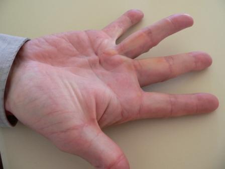
- Dupuytren's contracture may be present. There is thickening and shortening of palmar fascia that leads to flexion deformities of the fingers. It is thought to be due to fibroblastic proliferation and disorderly collagen deposition. It is relatively common (33% of patients).
 |
 |
Eyes
- Jaundice. It presents as a yellow discoloration of the skin, eyes, and mucus membranes due to increased bilirubin (at least 2-3 mg/dL or 30 mmol/L). Urine may also appear dark.
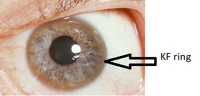
- Kayser-Fleischer rings may be present. They are dark rings that appear to encircle the iris of the eye.
Abdomen
- Liver size. It can be enlarged, normal, or shrunken.
- Splenomegaly. It is due to congestion of the red pulp as a result of portal hypertension.
- Ascites. It is an accumulation of fluid in the peritoneal cavity giving rise to flank dullness (needs about 1500 mL to detect flank dullness).
{{#ev:youtube|CHUBTgrU3Oc}}
- Caput medusa. In portal hypertension, the umbilical vein may open. Blood from the portal venous system may be shunted through the periumbilical veins into the umbilical vein and ultimately to the abdominal wall veins, manifesting as caput medusa.

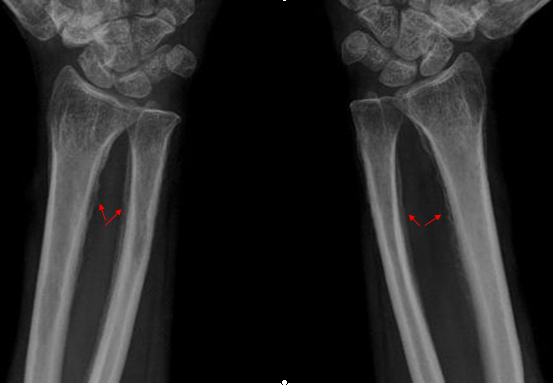
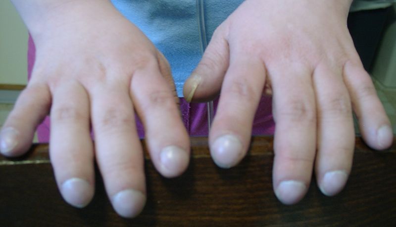
- Hypertrophic osteoarthropathy may be present. It consistis of chronic proliferative periostitis of the long bones that can cause considerable pain.
{{#ev:youtube|Or65nOrcz1A}}