Sickle-cell disease x ray
|
Sickle-cell disease Microchapters |
|
Diagnosis |
|---|
|
Treatment |
|
Case Studies |
|
Sickle-cell disease x ray On the Web |
|
American Roentgen Ray Society Images of Sickle-cell disease x ray |
|
Risk calculators and risk factors for Sickle-cell disease x ray |
Editor-In-Chief: C. Michael Gibson, M.S., M.D. [1]; Associate Editor(s)-in-Chief: Aarti Narayan, M.B.B.S [2], Shyam Patel [3]
Overview
X rays may be helpful in the diagnosis of specific pathological features that are found in sickle cell disease.
X Ray
X rays may be helpful in the diagnosis of specific pathological features that are found in sickle cell disease. X rays can show various pathological features of sickle cell disease, including pneumonia, acute chest syndrome, and osteonecrosis of the femoral head.[1] X-rays can also show osteoarticular involvement of bones. X-rays of the hands can show periarticular osteopenia and dactylitis (hand-foot syndrome) if the bones are involved.[1]
(Images shown below are courtesy of RadsWiki)
Patient #1: SCD patient with H shaped vertebrae
-
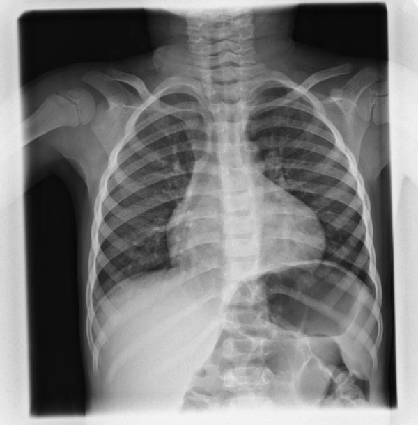
H shaped vertebral bodies
-
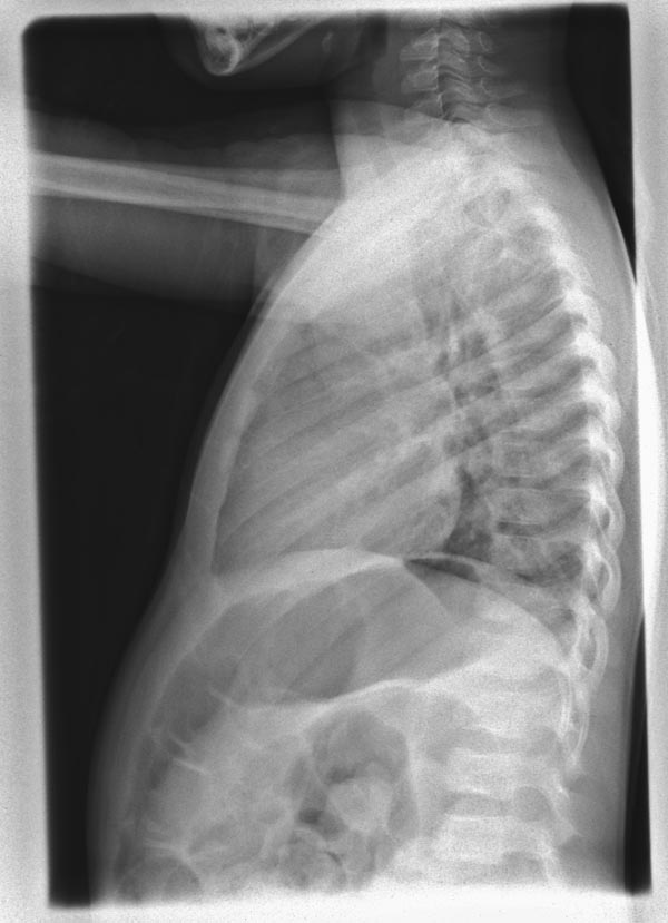
H shaped vertebral bodies
-
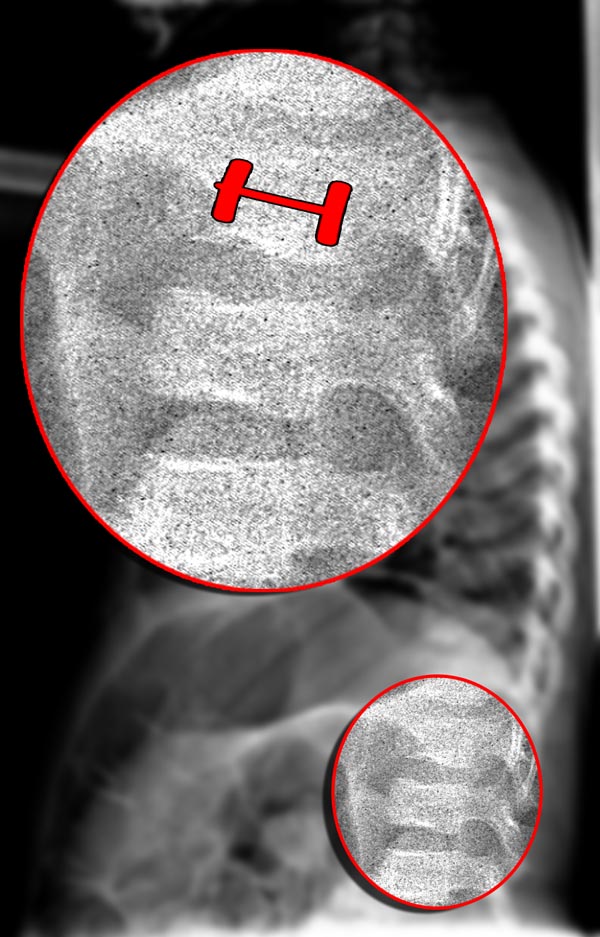
H shaped vertebral bodies


Patient #1: SCD patient with H shaped vertebrae


References
- ↑ 1.0 1.1 da Silva Junior GB, Daher Ede F, da Rocha FA (2012). "Osteoarticular involvement in sickle cell disease". Rev Bras Hematol Hemoter. 34 (2): 156–64. doi:10.5581/1516-8484.20120036. PMC 3459393. PMID 23049406.