Hepatic hemangioma MRI: Difference between revisions
Jump to navigation
Jump to search
No edit summary |
(→MRI) |
||
| Line 8: | Line 8: | ||
* Delayed enhancement: Lesion fills in the contrast | * Delayed enhancement: Lesion fills in the contrast | ||
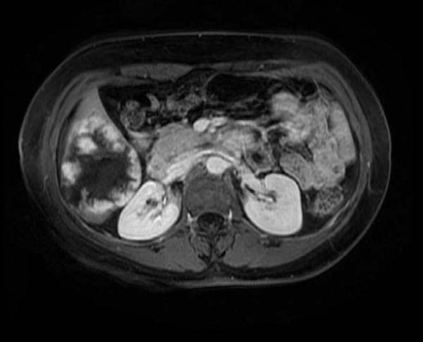
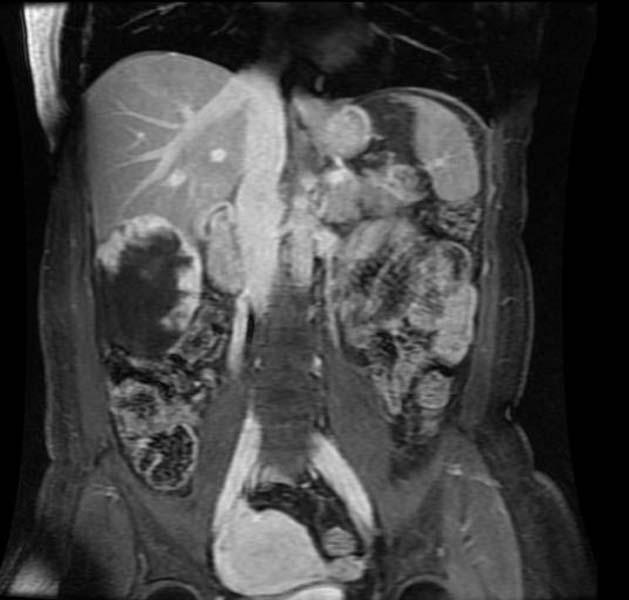
Shown below are the images of MRI in a patient of hepatic hemangioma. | Shown below are the images of MRI in a patient of hepatic hemangioma. | ||
<gallery perRow="3"> | <gallery perRow="3"> | ||
Image:Heptatic hemangioma 001.jpg|T2 FrFSE | Image:Heptatic hemangioma 001.jpg|T2 FrFSE | ||
| Line 17: | Line 17: | ||
Image:Heptatic hemangioma 006.jpg|T1 post Gad coronal | Image:Heptatic hemangioma 006.jpg|T1 post Gad coronal | ||
</gallery> | </gallery> | ||
[http://www.radswiki.net Images courtesy of RadsWiki] | |||
==References== | ==References== | ||
Revision as of 21:46, 7 March 2013
|
Hepatic hemangioma Microchapters |
|
Diagnosis |
|---|
|
Treatment |
|
Case Studies |
|
Hepatic hemangioma MRI On the Web |
|
American Roentgen Ray Society Images of Hepatic hemangioma MRI |
|
Risk calculators and risk factors for Hepatic hemangioma MRI |
Editor-In-Chief: C. Michael Gibson, M.S., M.D. [1]
MRI
- T2 hyperintense
- Portal venous enhancement: Peripheral nodular enhancement
- Delayed enhancement: Lesion fills in the contrast
Shown below are the images of MRI in a patient of hepatic hemangioma.
-
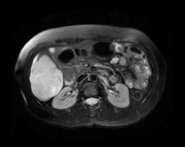
T2 FrFSE
-
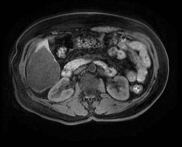
T1
-
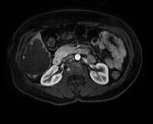
T1 post Gad
-
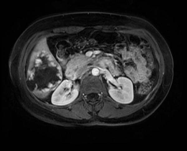
T1 post Gad
-
T1 post Gad
-
T1 post Gad coronal


