Sandbox:Maria: Difference between revisions
Jump to navigation
Jump to search
Sara Mohsin (talk | contribs) No edit summary |
Sara Mohsin (talk | contribs) No edit summary |
||
| Line 34: | Line 34: | ||
|} | |} | ||
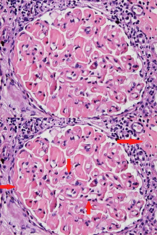
[[File:GM. kidneys gif.gif|thumb|200px|none| Light Microscopy. There is marked, global, homogeneous, eosinophilic thickening of the glomerular basement membrane with segmental accentuation. Homogeneous, eosinophilic globules are seen in the lumen of occasional capillary loops. The capillary lumina appear reduced in diameter but no inflammatory or proliferative changes are observed. The periglomerular interstitial space shows lymphocytic infiltration. The focal interstitial deposition of homogeneous eosinophilic material is present in the right upper corner of the picture (H&E × 400). [https://openi.nlm.nih.gov/detailedresult.php?img=PMC2600791_1757-1626-1-333-1&query=waldenstrom+macroglobulinaemia&it=xg&req=4&npos=31 Source: Castro H. et al, Department of Medicine, Division of General Internal Medicine, University of Miami/Jackson Memorial Medical Center, Miami, Florida, USA.]]] | |||
<ref name="pmid28701598">{{cite journal| author=Kiran U, Aggarwal S, Choudhary A, Uma B, Kapoor PM| title=The blalock and taussig shunt revisited. | journal=Ann Card Anaesth | year= 2017 | volume= 20 | issue= 3 | pages= 323-330 | pmid=28701598 | doi=10.4103/aca.ACA_80_17 | pmc=5535574 | url=https://www.ncbi.nlm.nih.gov/entrez/eutils/elink.fcgi?dbfrom=pubmed&tool=sumsearch.org/cite&retmode=ref&cmd=prlinks&id=28701598 }} </ref> | <ref name="pmid28701598">{{cite journal| author=Kiran U, Aggarwal S, Choudhary A, Uma B, Kapoor PM| title=The blalock and taussig shunt revisited. | journal=Ann Card Anaesth | year= 2017 | volume= 20 | issue= 3 | pages= 323-330 | pmid=28701598 | doi=10.4103/aca.ACA_80_17 | pmc=5535574 | url=https://www.ncbi.nlm.nih.gov/entrez/eutils/elink.fcgi?dbfrom=pubmed&tool=sumsearch.org/cite&retmode=ref&cmd=prlinks&id=28701598 }} </ref> | ||
Revision as of 17:27, 23 September 2020
Practice here
| Criteria | Symptomatic WM | Asymptomatic WM | IgM-Related Disorders | MGUS |
|---|---|---|---|---|
| IgM monoclonal protein | + | + | + | + |
| Bone marrow infiltration | + | + | - | - |
| Symptoms attributable to IgM | + | - | + | - |
| Symptoms attributable to tumor infiltration | + | - | - | - |

References
- ↑ Kiran U, Aggarwal S, Choudhary A, Uma B, Kapoor PM (2017). "The blalock and taussig shunt revisited". Ann Card Anaesth. 20 (3): 323–330. doi:10.4103/aca.ACA_80_17. PMC 5535574. PMID 28701598.