Sandbox:Sahar: Difference between revisions
Jump to navigation
Jump to search
No edit summary |
No edit summary |
||
| Line 170: | Line 170: | ||
* Second and third decades of life | * Second and third decades of life | ||
| style="padding: 5px 5px; background: #F5F5F5;" align="left" | | | style="padding: 5px 5px; background: #F5F5F5;" align="left" | | ||
* Female = male | |||
* Lipoma of joints affects men more frequently than women | |||
| style="padding: 5px 5px; background: #F5F5F5;" align="left" | | | style="padding: 5px 5px; background: #F5F5F5;" align="left" | | ||
* Most commonly seen in wrist and hand | |||
* May also affect ankle and foot | |||
* Bilateral and symmetric location in 50% of the cases | |||
| style="padding: 5px 5px; background: #F5F5F5;" align="left" | | | style="padding: 5px 5px; background: #F5F5F5;" align="left" | | ||
* May cause severe pain, trigger finger, or even symptoms of carpal tunnel syndrome | |||
| style="padding: 5px 5px; background: #F5F5F5;" align="left" | | | style="padding: 5px 5px; background: #F5F5F5;" align="left" | | ||
* May have 2 different shape: | |||
* A single adipose tissue extending along the tendon sheet | |||
* A lipomatous lesion composed mostly from hypertrophic synovial villi | |||
| style="padding: 5px 5px; background: #F5F5F5;" align="left" | | | style="padding: 5px 5px; background: #F5F5F5;" align="left" | | ||
* Radiologic imaging may show a lesion of less density than the surrounding tissue | |||
| | | | ||
|- | |- | ||
Revision as of 20:11, 15 November 2019
| Lipomatous tumor | Age of onset | Gender preponderance | Location | Clinical features | Pathologic appearance | Other features | Pathologic view |
|---|---|---|---|---|---|---|---|
| Angiolipoma |
|
|
|
|
|
|
 |
| Myolipoma |
|
|
|
|
|
|
 |
| Myelolipoma |
|
|
|
|
|
|
 |
| Spindle Cell/Pleomorphic Lipoma |
|
|
|
|
|
|
 |
| Chondroid Lipoma |
|
|
|
|
|
|
 |
| Hibernoma |
|
|
|
|
|
|
 |
| Lipomas of Tendon Sheaths and Joints |
|
|
|
|
|
|
|
| Intramuscular and Intermuscular Lipomas |  | ||||||
| Neural Fibrolipoma |
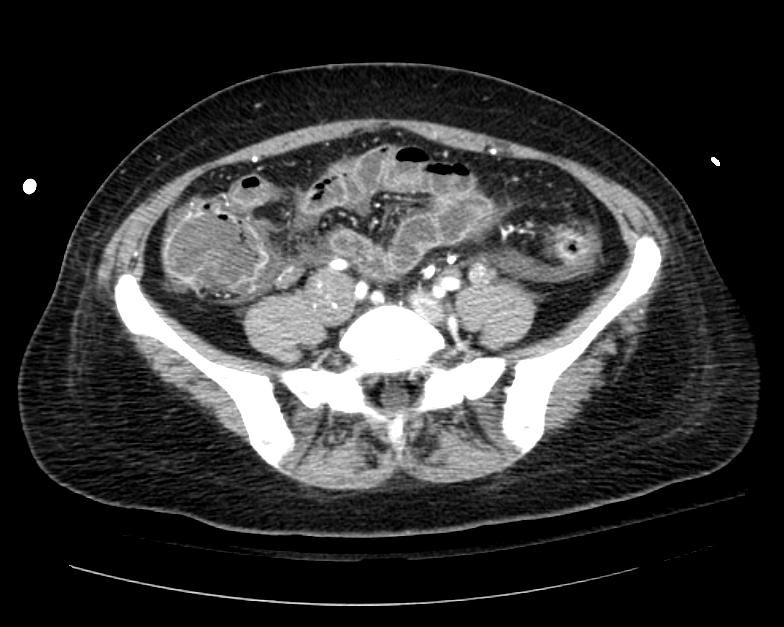
Example #1
The patient presented with S.O.B. one year after hysterectomy for a leiomyomatous uterus.
-
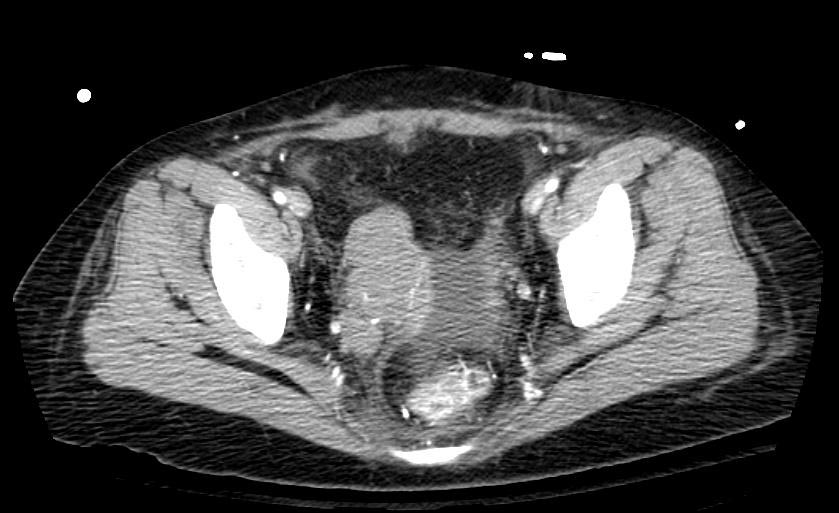
CT in Intravenous leiomyomatosis
-