Sandbox:Sahar: Difference between revisions
Jump to navigation
Jump to search
No edit summary |
No edit summary |
||
| Line 25: | Line 25: | ||
* Encapsulated, yellow nodules with a reddish tinge | * Encapsulated, yellow nodules with a reddish tinge | ||
* A combination of fatty tissue and vascular channels | * A combination of fatty tissue and vascular channels | ||
* Fibrin thrombi is present in vascular channels | * Fibrin thrombi is present in vascular channels (characteristic finding) | ||
| style="padding: 5px 5px; background: #F5F5F5;" align="left" | | | style="padding: 5px 5px; background: #F5F5F5;" align="left" | | ||
| Line 47: | Line 47: | ||
* A combination of mature adipocytes and sheets of well-differentiated smooth muscle | * A combination of mature adipocytes and sheets of well-differentiated smooth muscle | ||
* No nuclear atypia | * No nuclear atypia | ||
*Sieve-like appearance at low magnification (due to interspersed location of smooth muscle component) | |||
* | * | ||
* | * | ||
| style="padding: 5px 5px; background: #F5F5F5;" align="left" | | | style="padding: 5px 5px; background: #F5F5F5;" align="left" | | ||
* Benign | |||
* It is usually large and located in the deep soft tissues | |||
* | * | ||
|- | |- | ||
Revision as of 20:17, 13 November 2019
| Lipomatous tumor | Age of onset | Gender preponderance | Location | Clinical features | Diagnostic feature(s) | Other features |
|---|---|---|---|---|---|---|
| Angiolipoma |
|
|
|
|
|
|
| Myolipoma |
|
|
|
|
|
|
|
|
|
|
| |||
|
|
|
|
| |||
|
|
|
|
| |||
|
|
|
|
|
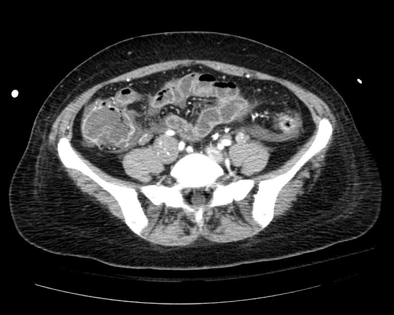
Example #1
The patient presented with S.O.B. one year after hysterectomy for a leiomyomatous uterus.
-
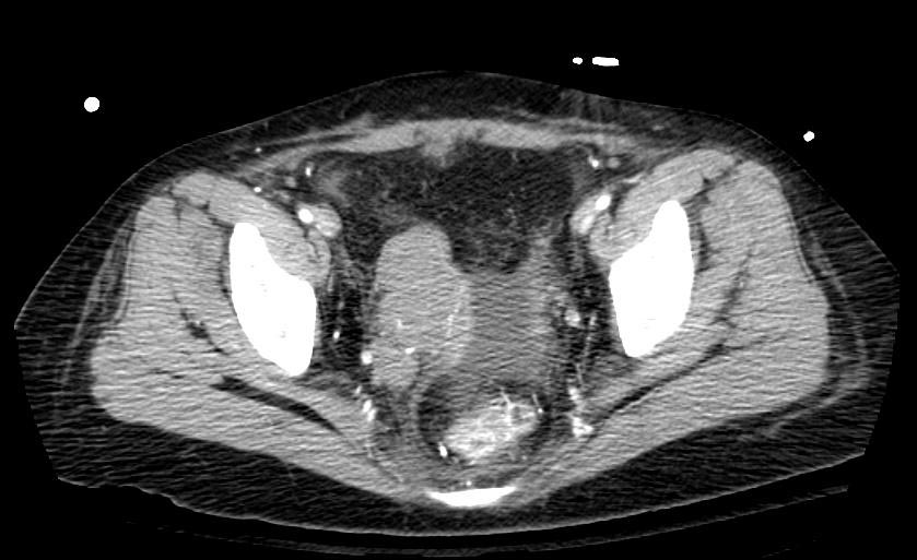
CT in Intravenous leiomyomatosis
-