Sickle-cell disease x ray: Difference between revisions
Shyam Patel (talk | contribs) No edit summary |
Shyam Patel (talk | contribs) No edit summary |
||
| Line 3: | Line 3: | ||
==Overview== | ==Overview== | ||
X-rays have utility in diagnosing specific pathological features that are found in sickle cell disease. | |||
==X ray findings== | ==X ray findings== | ||
X-rays can show various pathological features of sickle cell disease, including pneumonia, acute chest syndrome, and osteonecrosis of the femoral head.<ref name="pmid23049406">{{cite journal| author=da Silva Junior GB, Daher Ede F, da Rocha FA| title=Osteoarticular involvement in sickle cell disease. | journal=Rev Bras Hematol Hemoter | year= 2012 | volume= 34 | issue= 2 | pages= 156-64 | pmid=23049406 | doi=10.5581/1516-8484.20120036 | pmc=3459393 | url=https://www.ncbi.nlm.nih.gov/entrez/eutils/elink.fcgi?dbfrom=pubmed&tool=sumsearch.org/cite&retmode=ref&cmd=prlinks&id=23049406 }} </ref> | |||
(Images shown below are courtesy of RadsWiki) | (Images shown below are courtesy of RadsWiki) | ||
Revision as of 20:00, 14 October 2016
|
Sickle-cell disease Microchapters |
|
Diagnosis |
|---|
|
Treatment |
|
Case Studies |
|
Sickle-cell disease x ray On the Web |
|
American Roentgen Ray Society Images of Sickle-cell disease x ray |
|
Risk calculators and risk factors for Sickle-cell disease x ray |
Editor-In-Chief: C. Michael Gibson, M.S., M.D. [1]; Associate Editor(s)-in-Chief: Aarti Narayan, M.B.B.S [2] Shyam Patel [3]
Overview
X-rays have utility in diagnosing specific pathological features that are found in sickle cell disease.
X ray findings
X-rays can show various pathological features of sickle cell disease, including pneumonia, acute chest syndrome, and osteonecrosis of the femoral head.[1]
(Images shown below are courtesy of RadsWiki)
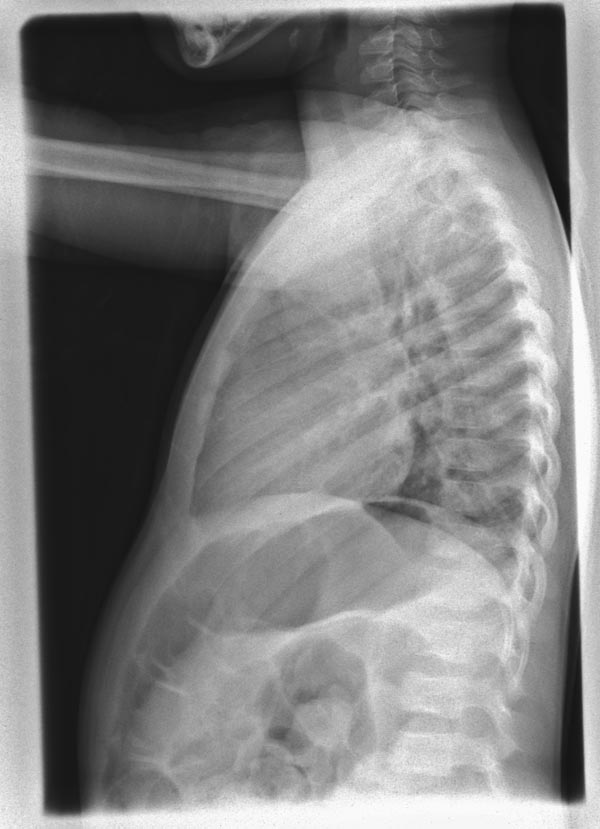
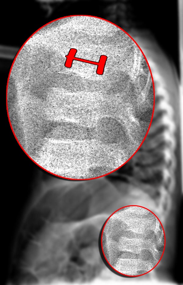
Patient #1: SCD patient with H shaped vertebrae
-
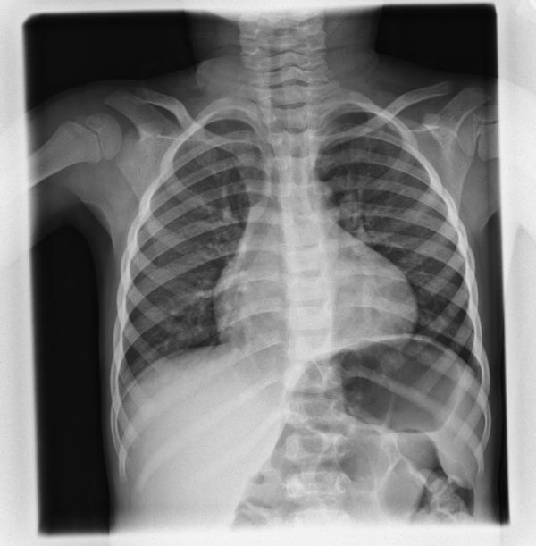
H shaped vertebral bodies
-
H shaped vertebral bodies
-
H shaped vertebral bodies


Patient #1: SCD patient with H shaped vertebrae


References
- ↑ da Silva Junior GB, Daher Ede F, da Rocha FA (2012). "Osteoarticular involvement in sickle cell disease". Rev Bras Hematol Hemoter. 34 (2): 156–64. doi:10.5581/1516-8484.20120036. PMC 3459393. PMID 23049406.