Ureter
|
WikiDoc Resources for Ureter |
|
Articles |
|---|
|
Media |
|
Evidence Based Medicine |
|
Clinical Trials |
|
Ongoing Trials on Ureter at Clinical Trials.gov Clinical Trials on Ureter at Google
|
|
Guidelines / Policies / Govt |
|
US National Guidelines Clearinghouse on Ureter
|
|
Books |
|
News |
|
Commentary |
|
Definitions |
|
Patient Resources / Community |
|
Directions to Hospitals Treating Ureter Risk calculators and risk factors for Ureter
|
|
Healthcare Provider Resources |
|
Continuing Medical Education (CME) |
|
International |
|
|
|
Business |
|
Experimental / Informatics |
Steven C. Campbell, M.D., Ph.D.
Overview
In human anatomy, the ureters are the ducts that carry urine from the kidneys to the urinary bladder, passing anterior to the psoas major. The ureters are muscular tubes that can propel urine along by the motions of peristalsis. In the adult, the ureters are usually 25-30cm long.
In humans, the ureters enter the bladder through the back, running within the wall of the bladder for a few centimetres. There are no valves in the ureters, backflow being prevented by pressure from the filling of the bladder, as well as the tone of the muscle in the bladder wall.
In the female, the ureters pass through the mesometrium on the way to the urinary bladder.
Histology

The ureter has a diameter of about 3 millimeters, and the lumen is star-shaped. Like the bladder, it is lined with transitional epithelium, and contains layers of smooth muscle.
The epithelial cells of the ureter are stratified (in many layers), are normally round in shape but become squamous (flat) when stretched. The lamina propria is thick and elastic (as it is important that it is impermeable).
There are two spiral layers of smooth muscle in the ureter wall, an inner loose spiral, and an outer tight spiral. The inner loose spiral is sometimes described as longitudinal, and the outer as circular, (this is the opposite to the situation in the gastrointestinal tract). The distal third of the ureter contains another layer of outer longitudinal muscle.
The adventitia of the ureter, like elsewhere is composed of fibrous connective tissue, that binds it to adjacent tissues.
Diseases and disorders
Medical problems that can affect the ureter include:
- Cancer of the ureter
- Passage of kidney stones
- Ureterocele
- Megaureter
- Vesico-ureteric reflux
- Anatomical abnormalities, such as duplexing and ectopia
External links
- Template:SUNYAnatomyLabs - "Posterior Abdominal Wall: Internal Structure of a Kidney"
- Template:SUNYAnatomyFigs - "Relationship of the ureter to the uterine artery."
- Template:SUNYAnatomyFigs - "Mid-sagittal section of male pelvis."
- Template:SUNYAnatomyImage
- Template:SUNYAnatomyImage
- Template:IowaHistologyInteractive
- Template:UCDavisOrganology - "Mammal, ureter (LM, Medium)"
- Histology at KUMC urinary-renal15 - "Ureter"
- Template:ViennaCrossSection
Additional images
-
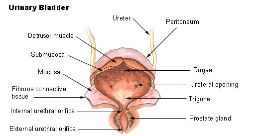 Bladder
 -
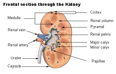 Frontal section through the kidney
 -
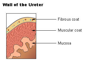 Wall of the ureter.
 -
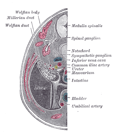 Transverse section of human embryo eight and a half to nine weeks old.
 -
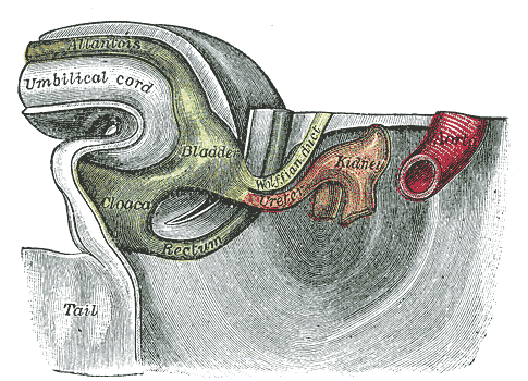 Tail end of human embryo thirty-two to thirty-three days old.
 -
 The relations of the viscera and large vessels of the abdomen. (Seen from behind, the last thoracic vertebra being well raised.)
 -
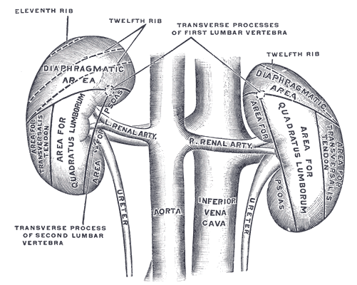 The posterior surfaces of the kidneys, showing areas of relation to the parietes.
 -
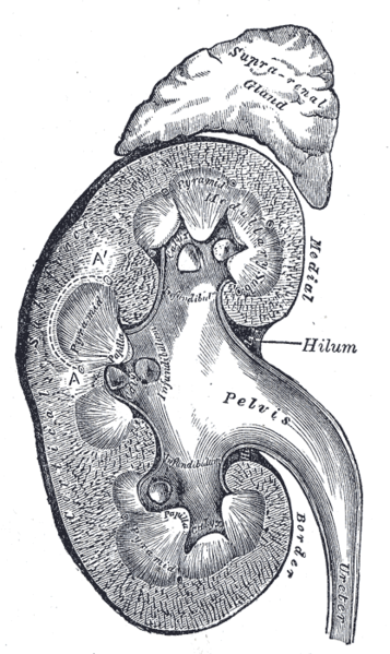 Vertical section of kidney.
 -
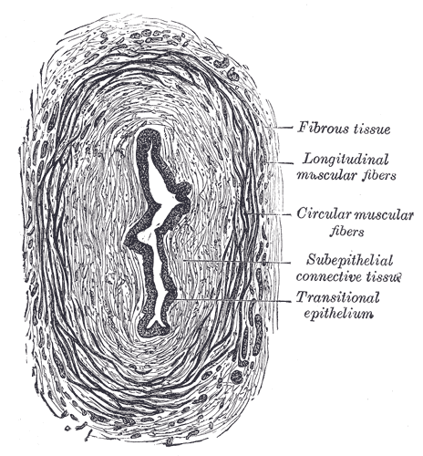 Transverse section of ureter.
 -
 The interior of bladder.
 -
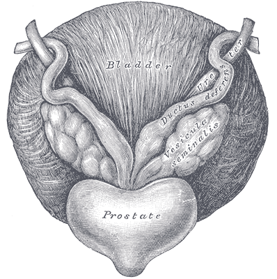 Fundus of the bladder with the vesiculæ seminales.
 -
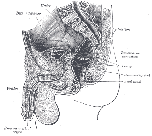 Median sagittal section of male pelvis.


ar:حالب cs:Močovod de:Harnleiter id:Ureter it:Uretere he:שופכן ku:Mîzlûl lt:Šlapimtakis nl:Urineleider no:Urinleder sk:Močovod sr:Мокраћовод sh:Mokraćovod fi:Virtsanjohdin