Spondylolysis
| Spondylolysis | |
| ICD-9 | 738.4 |
|---|---|
| DiseasesDB | 12322 |
| MeSH | D013169 |
Template:Search infobox Editor-In-Chief: C. Michael Gibson, M.S., M.D. [1]; Associate Editor-In-Chief: Cafer Zorkun, M.D., Ph.D. [2]
- Not to be confused with spondylosis or spondylolisthesis.
Overview
Spondylolysis is a defect in the pars interarticularis of a vertebra. The great majority of cases occur in the lowest of the lumbar vertebrae (L5), but spondylolysis may also occur in the other lumbar vertebrae, as well as in the thoracic vertebrae.
Spondylolysis occurs in three to six percent of the population.[1][2]
Causes
It is typically caused by stress fracture of the bone, and is associated with certain activities, such as weight lifting and gymnastics. It has been proposed that the pars interarticularis is especially vulnerable when the spine is in an extended position, and a force suddenly presses the vertebrae together, such as when landing on ones feet after a hop. This pressure acts like a nutcracker on the pars interarticularis and can fracture it in susceptible individuals. [3]
Spondylolysis also runs in families and is more prevalent in some populations, suggesting a hereditary component such as a tendency toward thin vertebral bone.
Spondylolysis is the most common cause of spondylolisthesis in pediatric patients. In the older population, degenerative disc disease commonly leads to spondylolisthesis without spondylolysis; in these instances, the spinal canal gets narrowed because the spinolaminar arch at one level slides forward on the lower level effectively flattening the canal. The hereditary factor mentioned above is quite notable, since the frequency of spondylolisthesis in Eskimos is 30–50%.
Diagnosis
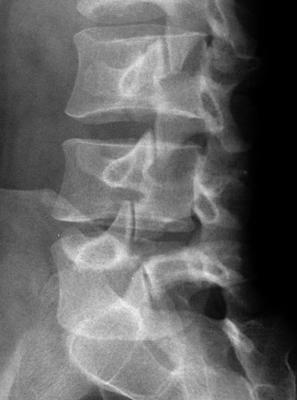
The defect is seen in the oblique lumbar radiograph. Image of Pars Defect<-- Picture of x-ray. An oblique x-ray of the lumbar spine shows what appears to be a "scotty dog" first described by Lachapelle. The nose of the dog is the costal/transverse process; the ear, the superior facet; the neck, the pars interarticularis; the collar, the pars defect (dark on x-ray); the eye, the pedicle seen end on; the body, the lamina; the hindefoot, the spinous process; the tail if pointing straight up=opp. superior articular facet and if pointing horizontally is the transverse process of the opposite side; and the forefoot, the inferior articular process.[3][4]
Treatment
According to a meta-analysis of randomized controlled trials by the Cochrane Collaboration, the role of surgery compared to for adults with degenerative lumbar spondylosis is unclear.[5] It is not clear if any randomized controlled trials have been performed of pediatric or adolescent patients.
References
- ↑ Standaert CJ, Herring SA (2000). "Spondylolysis: a critical review". British journal of sports medicine. 34 (6): 415–22. PMID 11131228.
- ↑ Bono CM (2004). "Low-back pain in athletes". The Journal of bone and joint surgery. American volume. 86-A (2): 382–96. PMID 14960688.
- ↑ "Glossary". Retrieved 2007-07-05.
- ↑ "Lumbar Spondylolisthesis as seen on X-Ray Films". Retrieved 2007-07-05.
- ↑ Gibson JN, Waddell G (2005). "Surgery for degenerative spondylosis". Cochrane database of systematic reviews (Online) (4): CD001352. doi:10.1002/14651858.CD001352.pub3. PMID 16235281.
External links
Template:Diseases of the musculoskeletal system and connective tissue