Uploads by Vishnu Vardhan Serla
Jump to navigation
Jump to search
This special page shows all uploaded files.
| Date | Name | Thumbnail | Size | Description | Versions |
|---|---|---|---|---|---|
| 19:42, 20 December 2012 | Trichomonas pap test.jpg (file) |  |
794 KB | Micrograph showing Trichomoniasis (infection with Trichomonas vaginalis). Pap test. Pap stain. Trichomoniasis is frequently associated with abundant inflammatory cells (esp. neutrophils) - not seen in this image. | 1 |
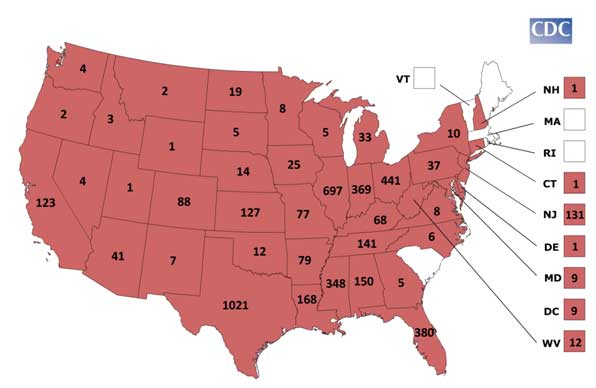
| 14:27, 21 November 2012 | Sle byCounty.jpg (file) |  |
17 KB | 1 | |
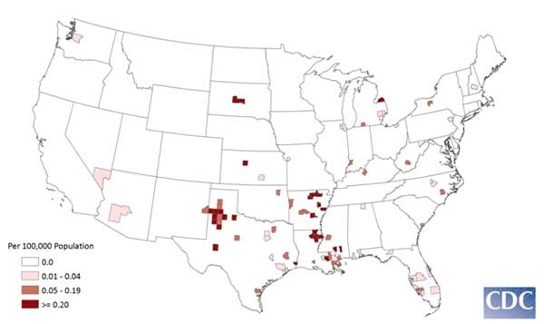
| 14:14, 21 November 2012 | Sle state map.jpg (file) |  |
22 KB | 1 | |
| 21:51, 20 November 2012 | Sle chart.jpg (file) |  |
14 KB | 1 | |
| 21:37, 20 November 2012 | 10228 lores.jpg (file) |  |
182 KB | 1 | |
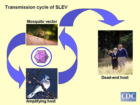
| 21:33, 20 November 2012 | Sle transmission cycle 450px.jpg (file) | 57 KB | 1 | ||
| 19:03, 20 November 2012 | Remove-b.jpg (file) |  |
25 KB | 1 | |
| 19:03, 20 November 2012 | Remove-a.jpg (file) |  |
21 KB | 1 | |
| 18:43, 20 November 2012 | Tickhandbook.pdf (file) | 6.82 MB | 1 | ||
| 16:39, 20 November 2012 | STARI.jpg (file) |  |
36 KB | 1 | |
| 16:29, 20 November 2012 | Lgmap-lone star tick.jpg.png (file) |  |
72 KB | 1 | |
| 15:44, 20 November 2012 | Tick sizes.jpg (file) |  |
153 KB | 1 | |
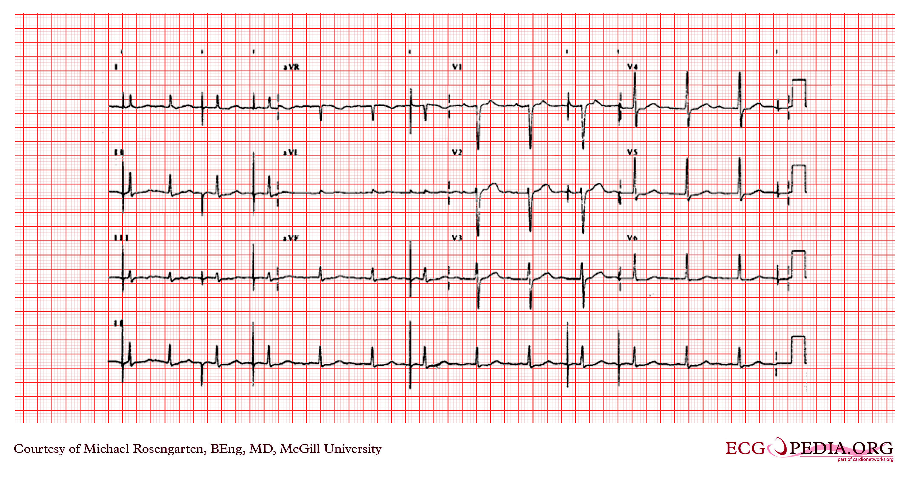
| 20:25, 16 October 2012 | Non Q MI.jpg (file) |  |
4.46 MB | 1 | |
| 19:27, 16 October 2012 | Paced atrial rhythm with a bipolar atrial pacemaker.jpg (file) |  |
4.46 MB | 1 | |
| 18:55, 16 October 2012 | Pacemaker with ventriculophasic effect..jpg (file) | 630 KB | 1 | ||
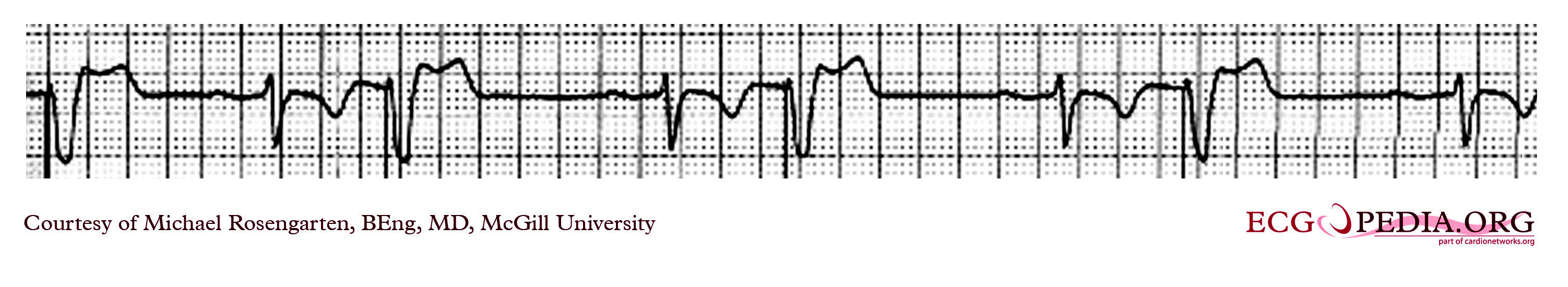
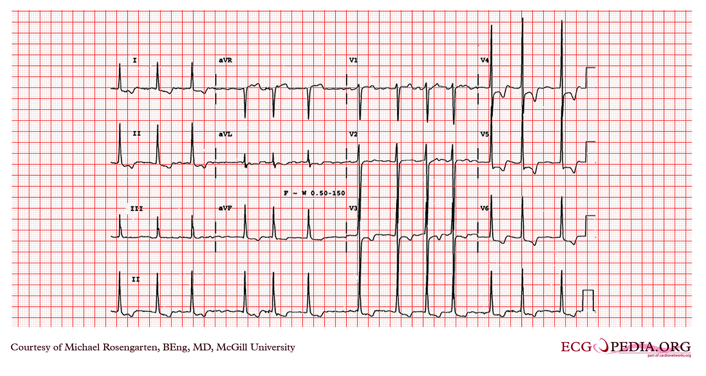
| 18:31, 16 October 2012 | Mobitz II AV block.jpg (file) |  |
17 KB | 1 | |
| 18:20, 16 October 2012 | Limb Lead Reversal 1.jpg (file) |  |
5.87 MB | 1 | |
| 18:11, 16 October 2012 | Limb Lead Reversal.jpg (file) |  |
5.87 MB | 1 | |
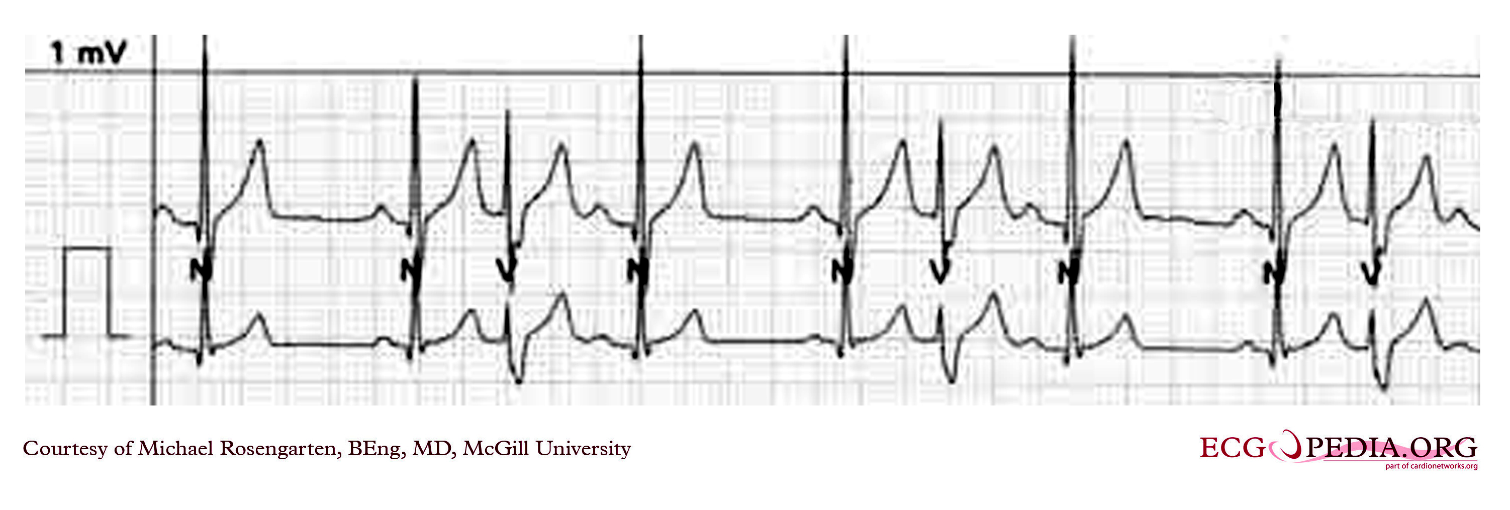
| 17:41, 16 October 2012 | Premature Ventricular Contractions with trigeminal rhythm.jpg (file) |  |
816 KB | 1 | |
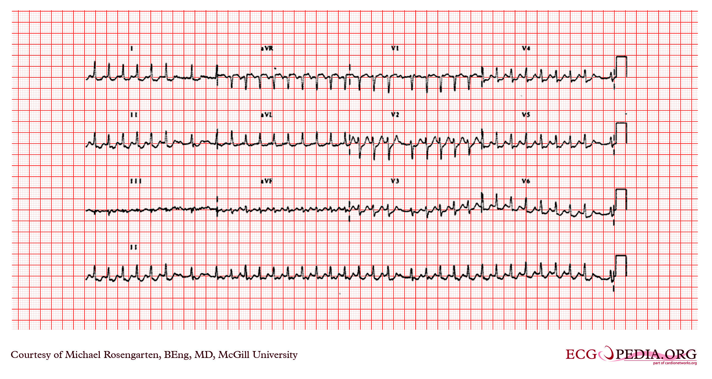
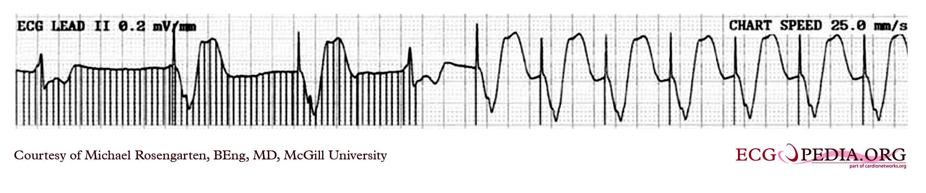
| 16:10, 16 October 2012 | Supraventricular tachycardia1.jpg (file) |  |
4.46 MB | 1 | |
| 15:31, 16 October 2012 | Complete heart block6.jpg (file) | 806 KB | 1 | ||
| 14:52, 16 October 2012 | First Degree AV Block1.jpg (file) |  |
4.44 MB | 1 | |
| 14:48, 16 October 2012 | First Degree AV Block.jpg (file) | 25 KB | Reverted to version as of 21:21, 26 November 2007 | 3 | |
| 20:50, 15 October 2012 | Previous anterior wall myocardial infartion..jpg (file) |  |
4.45 MB | 1 | |
| 20:36, 15 October 2012 | 2 to 1 AV block.jpg (file) |  |
4.46 MB | 1 | |
| 19:37, 15 October 2012 | Pacemaker set to a VVI mode.jpg (file) | 666 KB | 1 | ||
| 16:12, 15 October 2012 | Pacing system non-functional.jpg (file) | 654 KB | 1 | ||
| 15:20, 15 October 2012 | Ventricular Pacemaker 10.jpg (file) | 667 KB | 1 | ||
| 13:44, 15 October 2012 | A fib with LVH.jpg (file) |  |
4.47 MB | 1 | |
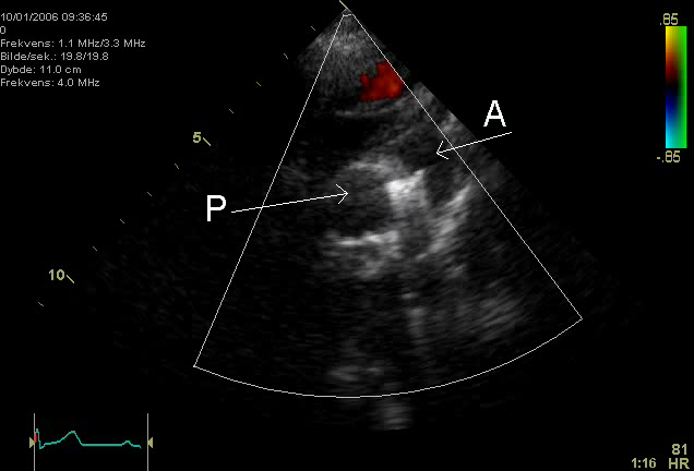
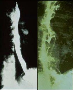
| 03:23, 3 October 2012 | PDA Coil.png.png (file) |  |
106 KB | An echocardiogram of a coiled persisting ductus arteriosus. One can see the aortic arch,the pulmonary artery and the coil between them. http://en.wikipedia.org/wiki/File:PDA_Coil.png | 1 |
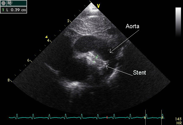
| 03:17, 3 October 2012 | Stent tekst.jpg.jpg (file) |  |
53 KB | An echocardiogram of a stented persisting ductus arteriosus. One can see the aortic arch and the stent leaving. Pulmonary artery not seen. http://en.wikipedia.org/wiki/File:Stent_tekst.jpg | 1 |
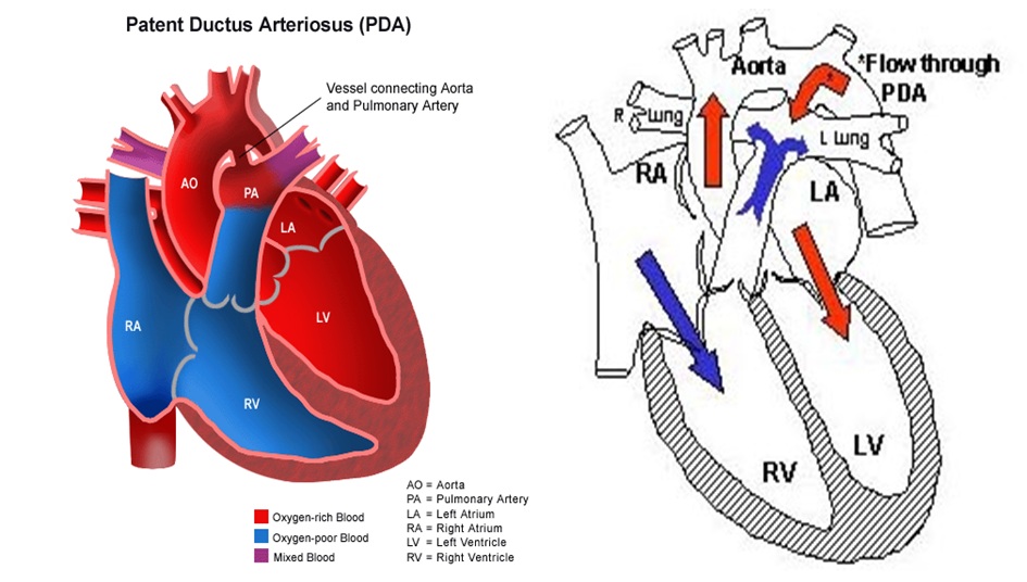
| 21:13, 2 October 2012 | Pathophysiology of patent ductus arteriosus.jpg (file) |  |
123 KB | https://docs.google.com/a/wikidoc.org/viewer?a=v&q=cache:B0mxS1E1mEAJ:thegunnybag.files.wordpress.com/2006/09/patent-ductus-arteriosus-final.ppt+&hl=en&gl=in&pid=bl&srcid=ADGEESgMWyAcACIJyiGzY7Nvocv9YGRHBNGftNHIQv4nuvFwUy8l0Crmad0DO4OLu7H29udJZ-VxyaPkT... | 1 |
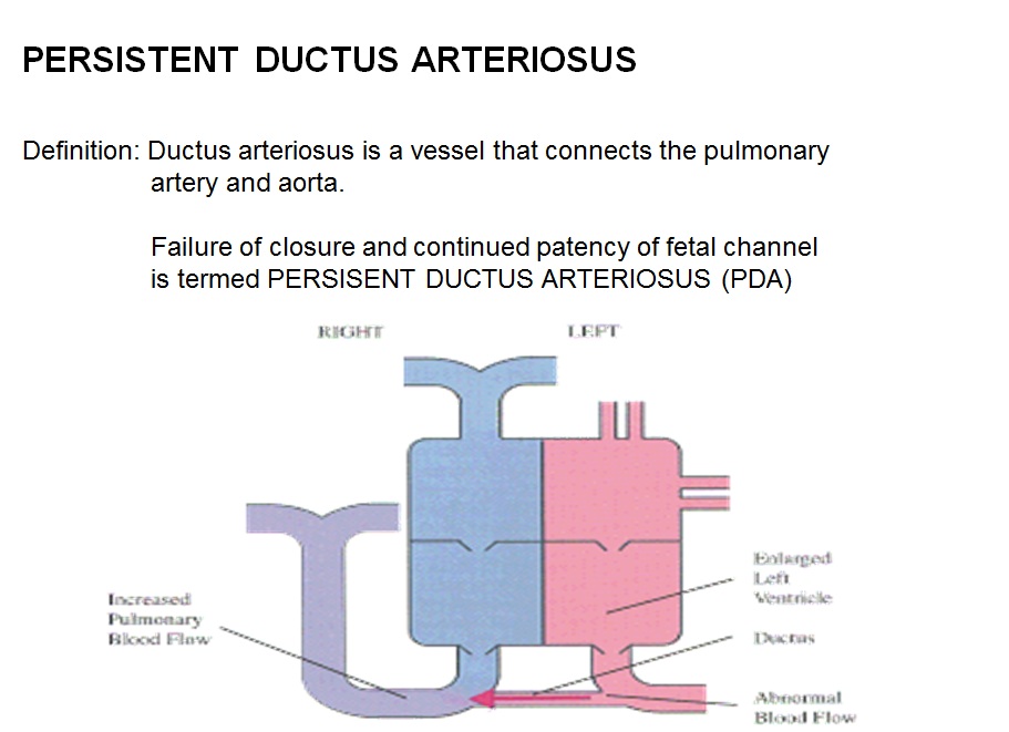
| 21:06, 2 October 2012 | Pathophysiology of patent ductus arteriosus 2.jpg (file) |  |
97 KB | https://docs.google.com/a/wikidoc.org/viewer?a=v&q=cache:B0mxS1E1mEAJ:thegunnybag.files.wordpress.com/2006/09/patent-ductus-arteriosus-final.ppt+&hl=en&gl=in&pid=bl&srcid=ADGEESgMWyAcACIJyiGzY7Nvocv9YGRHBNGftNHIQv4nuvFwUy8l0Crmad0DO4OLu7H29udJZ-VxyaPkT... | 1 |
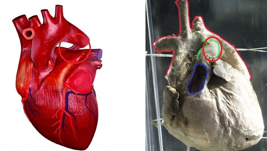
| 20:56, 2 October 2012 | Patent ductus arteriosus (PDA) Gross.jpg (file) | _Gross.jpg) |
115 KB | 3 | |
| 14:14, 7 September 2012 | Normal versus Abnormal Barium study of esophagus.jpg (file) |  |
34 KB | 1 | |
| 13:41, 7 September 2012 | Esophageal varices.jpg (file) |  |
36 KB | 1 |