Uploads by Sujit Routray
Jump to navigation
Jump to search
This special page shows all uploaded files.
| Date | Name | Thumbnail | Size | Description | Versions |
|---|---|---|---|---|---|
| 04:36, 1 April 2016 | Ct image 2 dsrct.jpg (file) |  |
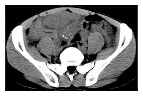
101 KB | Contrast-enhanced CT revealed the heterogeneous mass with obvious enhancement areas and scattered low attenuation. | 1 |
| 04:33, 1 April 2016 | Ct image dsrct.jpg (file) |  |
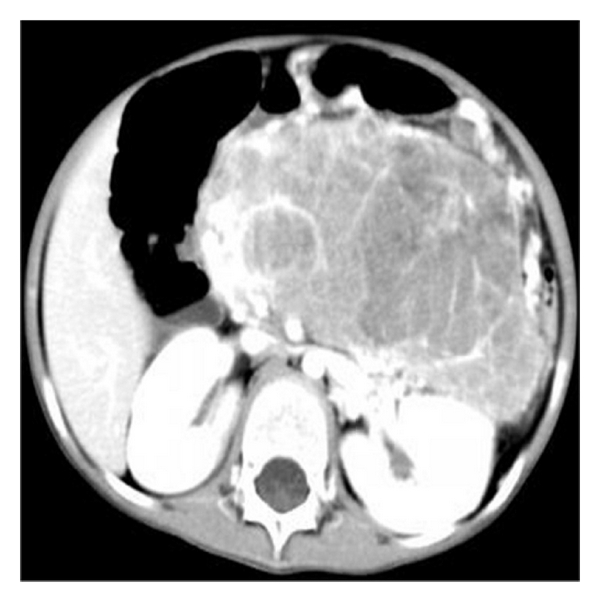
92 KB | Abdominopelvic CT scan revealed diffuse multiple soft-tissue masses in peritoneal and mesenteric surfaces. | 1 |
| 19:18, 29 March 2016 | Prognosis DSRCT.PNG (file) |  |
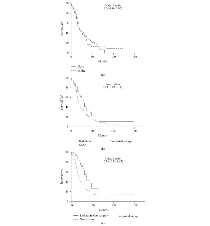
40 KB | (a) Race-based survival of desmoplastic small round cell tumor. There may be a survival disadvantage for African americans compared to Caucasians. Although it did not reach statistical significance, this analysis suggests that African americans are 33%... | 1 |
| 15:22, 29 March 2016 | Gender and race DSRCT.PNG (file) |  |
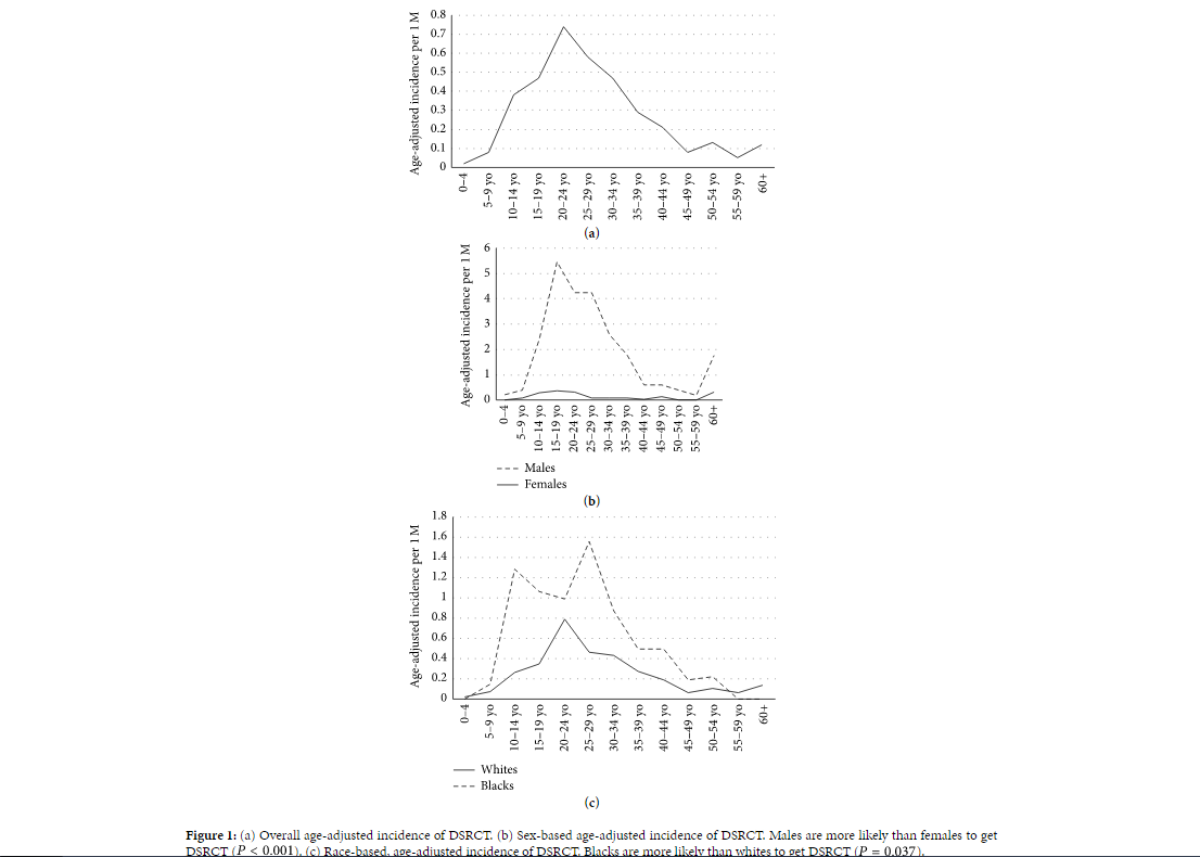
74 KB | (a) Overall age-adjusted incidence of desmoplastic small round cell tumor, (b) Sex-based age-adjusted incidence of desmoplastic small round cell tumor. Males are more likely than females to get desmoplastic small round cell tumor, (c) Race-based, age-a... | 1 |
| 15:01, 29 March 2016 | DSRCT.jpg (file) |  |
1.12 MB | Demographics of desmoplastic small round cell tumor | 1 |
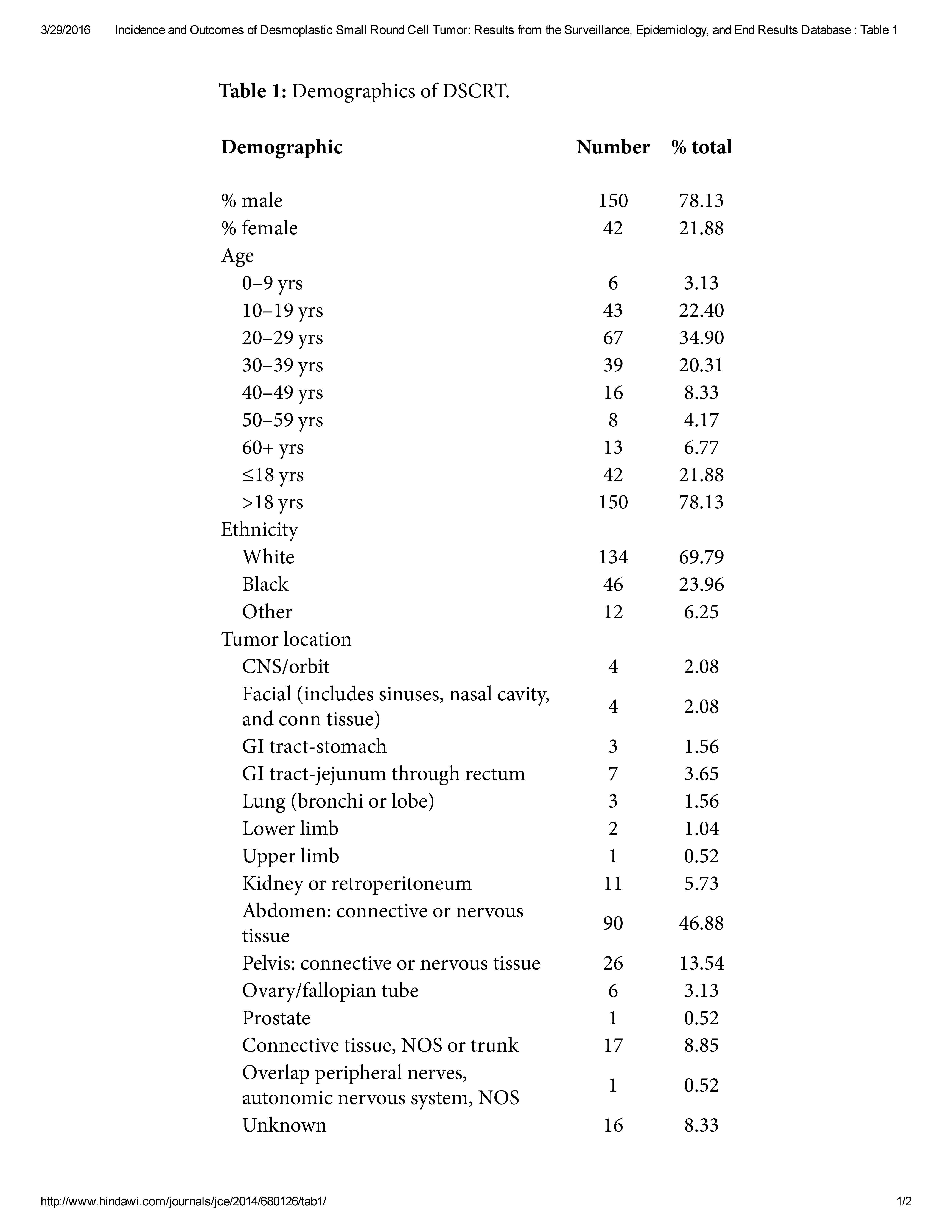
| 14:55, 29 March 2016 | Incidence and Outcomes of Desmoplastic Small Round Cell Tumor Results from the Surveillance, Epidemiology, and End Results Database Table 1.pdf (file) | 64 KB | Demographics of DSCRT. | 1 | |
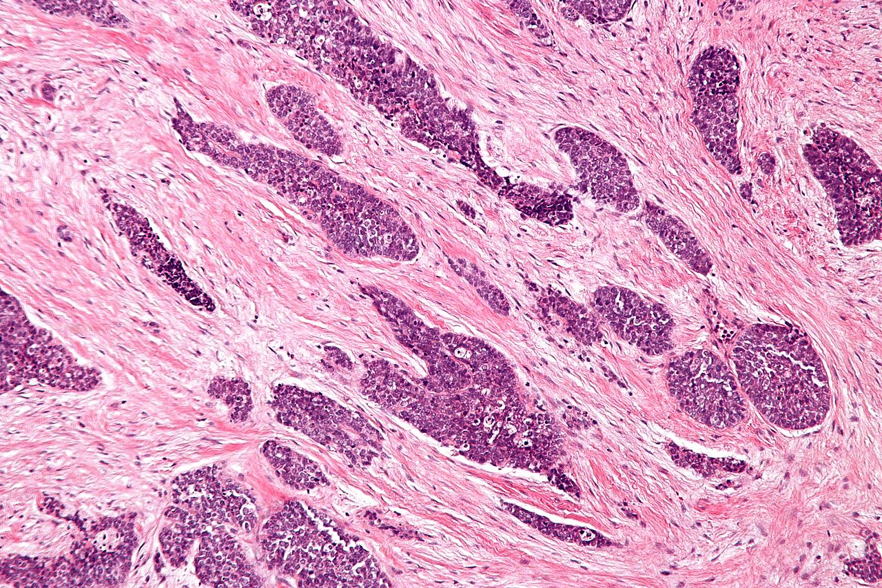
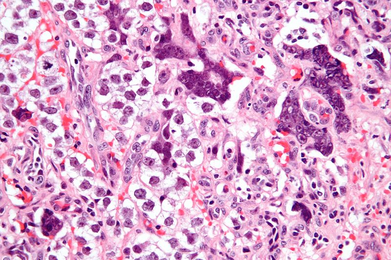
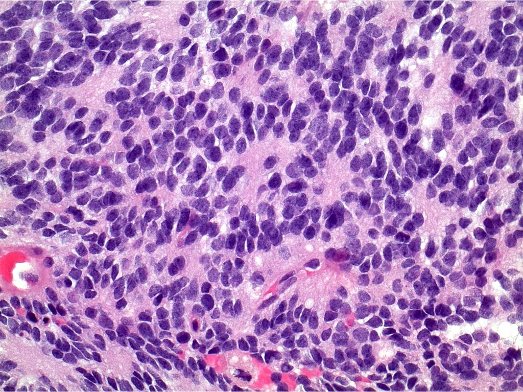
| 14:42, 29 March 2016 | Desmoplastic small round cell tumour - intermed mag.jpg (file) |  |
424 KB | Micrograph of a desmoplastic small round cell tumor, showing the characteristic desmoplastic stroma and angulated nests of small round cells on H&E stain. | 1 |
| 15:22, 14 March 2016 | Ct image of myelofibrosis 1.jpg (file) |  |
48 KB | Image courtesy of Radswiki. Radiopaedia (original file [http://radiopaedia.org/cases/myelofibrosis here]). Creative Commons BY-SA-NC | 1 |
| 15:12, 14 March 2016 | Mri myelofibrosis.jpg (file) |  |
41 KB | Image courtesy of Radswiki. Radiopaedia (original file [http://radiopaedia.org/cases/myelofibrosis-1 here]). Creative Commons BY-SA-NC | 1 |
| 00:58, 7 March 2016 | Capturestage 1 seminoma.PNG (file) |  |
68 KB | Advantages and disadvantages of different management options in the treatment of stage I seminoma | 1 |
| 15:59, 3 March 2016 | 398px-Seminoma of the Testis GROSS PATHOLOGY.jpg (file) |  |
49 KB | Gross specimen of testicle demonstrating a solid, white/tan mass. | 1 |
| 15:35, 3 March 2016 | Testicular seminoma (1) nodal metastasis.jpg (file) | _nodal_metastasis.jpg) |
167 KB | Histopathological image of metastatic seminoma in the inguinal lymph node on hematoxylin & eosin stain. | 1 |
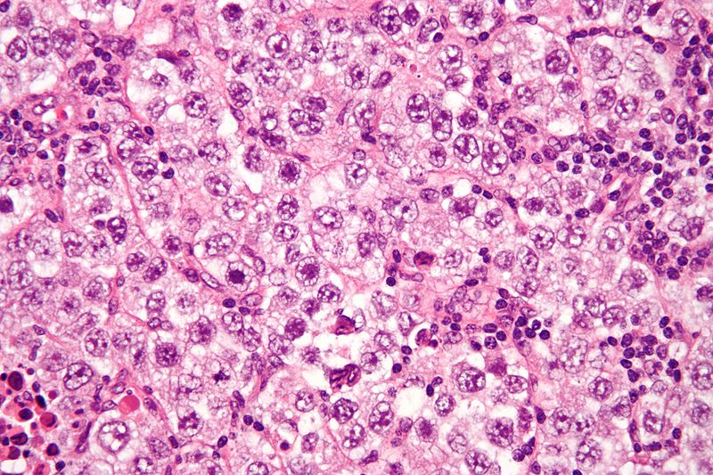
| 15:31, 3 March 2016 | 800px-Seminoma with syncytiotrophoblasts - very high mag.jpg (file) |  |
132 KB | Very high magnification micrograph of a seminoma with syncytiotrophoblasts on H&E stain. Syncytiotrophoblasts are seen in approximately 10-20% of seminomas. They may be associated with an elevated serum beta-hCG. | 1 |
| 15:11, 3 March 2016 | 800px-Seminoma high mag.jpg (file) |  |
152 KB | Microscopic image of seminoma demonstrating fried egg-like cells (clear or eosinophilic cytoplasm, central nucleus) and lymphocytic infiltrate. | 1 |
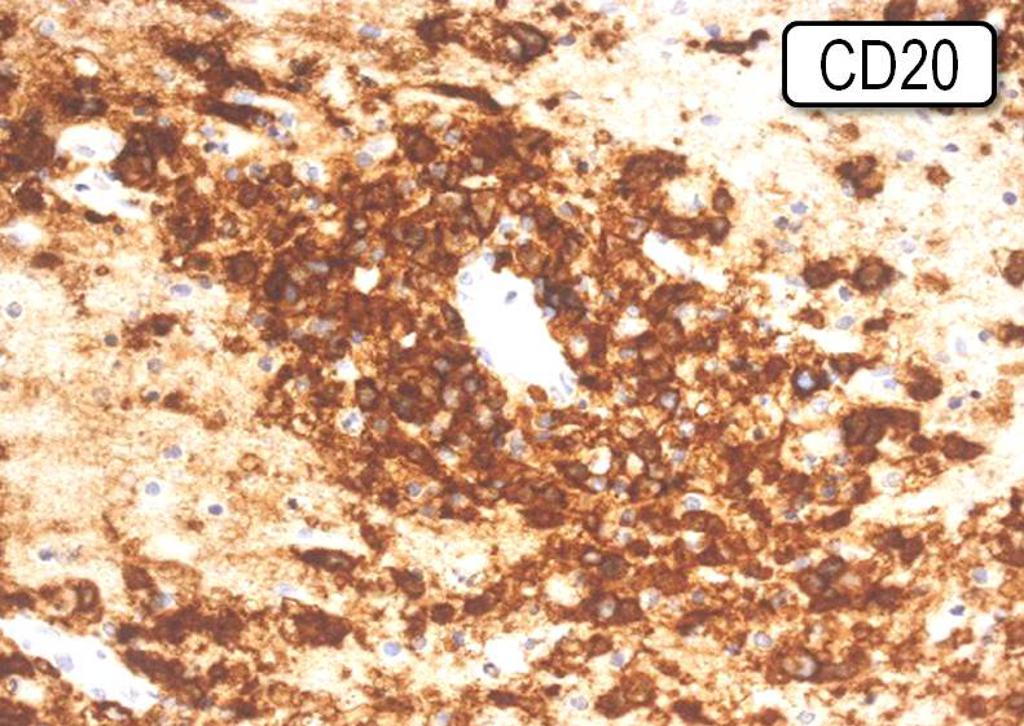
| 15:26, 23 February 2016 | Immunohistochemistry image 1 primary CNS lymphoma.jpg (file) |  |
142 KB | The atypical cells of primary central nervous system lymphoma demonstrating strong surface immunostaining for the B lymphocyte markers, CD20. | 1 |
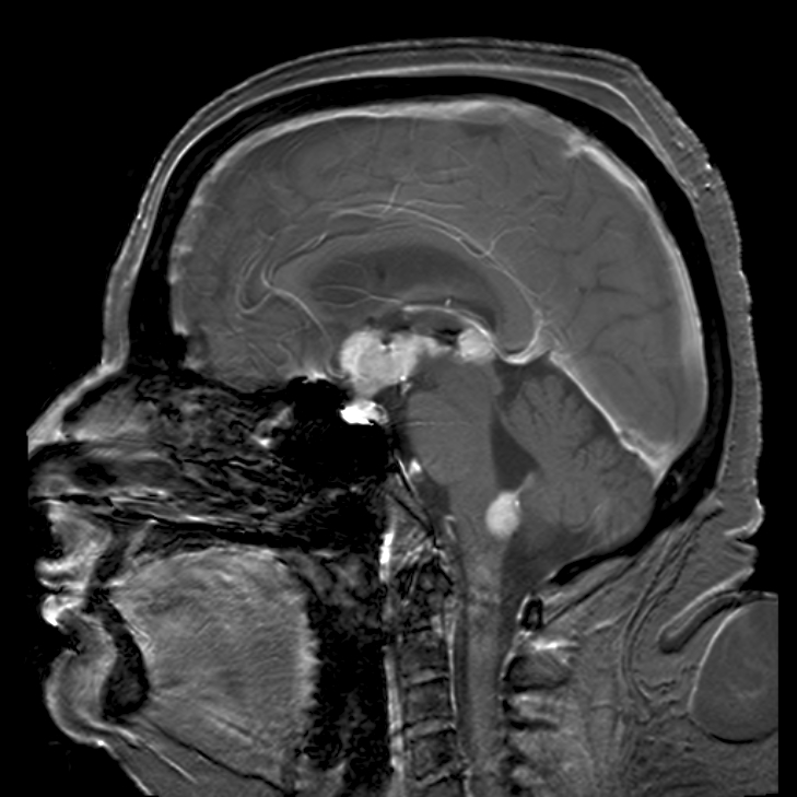
| 14:54, 23 February 2016 | Primary central nervous system B-cell non-Hodgkin lymphoma mri image 1.jpg (file) |  |
193 KB | Brain magnetic resonance imaging demonstrating primary central nervous system B-cell non-Hodgkin lymphoma of the sella turcica and hypothalamus, continuing to the tectum (intensely white areas in the middle). | 1 |
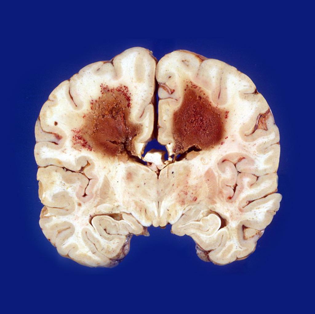
| 20:10, 18 February 2016 | Gross pathological image of primary central nervous system lymphoma image 1.jpg (file) |  |
100 KB | Image courtesy of Dr. A.Prof Frank Gaillard. Radiopaedia (original file [http://radiopaedia.org/cases/primary-cns-lymphoma-in-aids-patient-gross-pathology here]). Creative Commons BY-SA-NC | 2 |
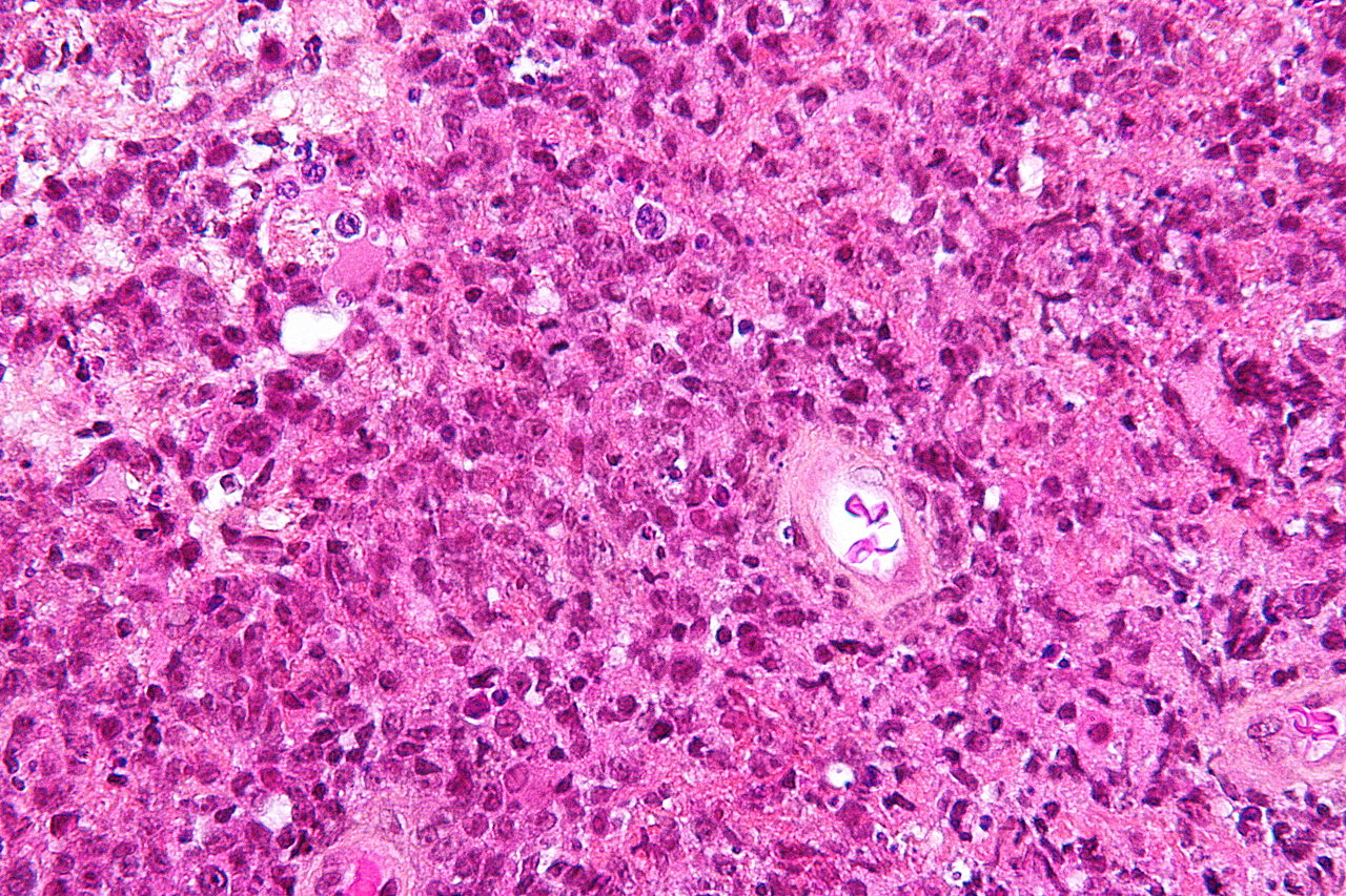
| 19:01, 18 February 2016 | Microscopic pathological image of primary central nervous system lymphoma image 1.jpg (file) |  |
452 KB | Micrograph from a brain biopsy demonstrating a primary CNS lymphoma with the characteristic perivascular distribution composed of large cells with prominent nucleoli, on HPS stain. | 1 |
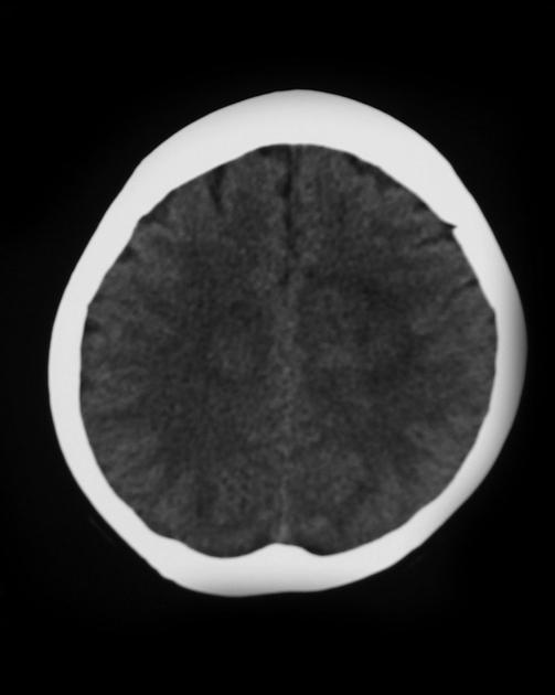
| 15:29, 18 February 2016 | Ct image primary central nervous system lymphoma image 1.jpg (file) |  |
25 KB | Image courtesy of Dr. Frank Gaillard. Radiopaedia (original file [http://radiopaedia.org/cases/cns-lymphoma-primary here]). Creative Commons BY-SA-NC | 1 |
| 18:59, 15 February 2016 | Anatomy of the chest image 1.PNG (file) |  |
34 KB | Anatomy of the chest | 1 |
| 18:39, 15 February 2016 | Lymph nodes image 1.PNG (file) |  |
35 KB | Lymph nodes in the neck and chest | 1 |
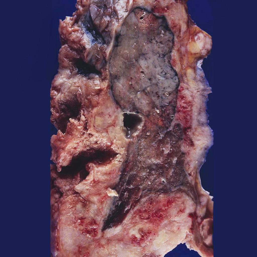
| 17:26, 15 February 2016 | Mesothelioma gross pathology image 1.jpg (file) |  |
146 KB | Image courtesy of Dr. Yale Rosen. Radiopaedia (original file [http://radiopaedia.org/cases/mesothelioma-gross-pathology-1 here]). Creative Commons BY-SA-NC | 1 |
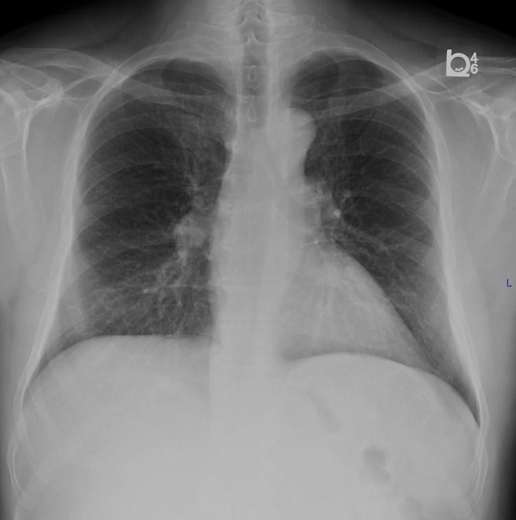
| 21:16, 12 February 2016 | Mesothelioma xray image 1.jpeg (file) |  |
477 KB | Image courtesy of Dr. A.Prof Frank Gaillard. Radiopaedia (original file [http://radiopaedia.org/cases/mesothelioma here]). Creative Commons BY-SA-NC | 1 |
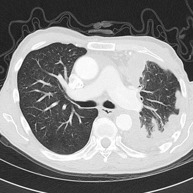
| 20:35, 12 February 2016 | Ct image mesothelioma image 1.jpg (file) |  |
74 KB | Image courtesy of Dr. A.Prof Frank Gaillard. Radiopaedia (original file [http://radiopaedia.org/cases/mesothelioma-1 here]). Creative Commons BY-SA-NC | 1 |
| 19:17, 25 January 2016 | Genetics MALT lymphoma pathophysiology.PNG (file) |  |
72 KB | Genetic alterations in MALT lymphoma. | 1 |
| 20:13, 22 January 2016 | Micropathologyneoplastic meningitis image 2.PNG (file) |  |
408 KB | Microscopic images of the cytospin of the cerebrospinal fluid cells from patient 1: (a) hematoxylin and eosin staining of large, hyperchromatic cells along with erythrocytes, lymphoma monocytoid cells, and eosinophils (asterisks) and (b) an atypical ce... | 1 |
| 20:07, 22 January 2016 | Micropathologyneoplastic meningitis image 1.PNG (file) |  |
458 KB | Light microscopic images of the cytological specimen of cerebrospinal fluid obtained from patient 1: (a) hematoxylin and eosin staining of the hypercellular sample with large, hyperchromatic cells associated with erythrocytes, (b) atypical cells staine... | 1 |
| 15:11, 22 January 2016 | Dx.PNG (file) |  |
92 KB | Proposed algoritm for the diagnosis of neoplastic meningitis. | 1 |
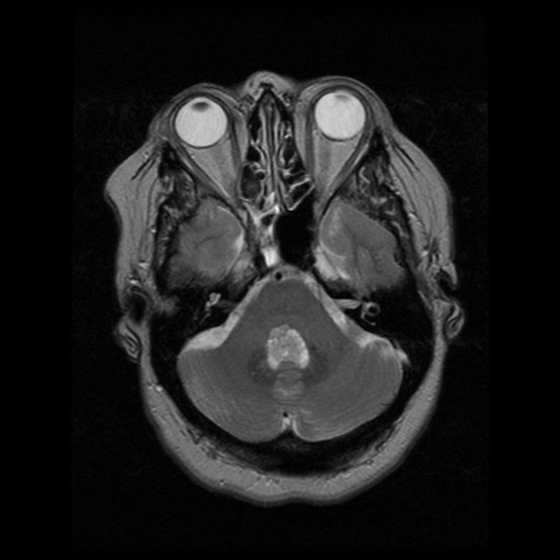
| 18:43, 14 January 2016 | MRI scan picture of choroid plexus papilloma image 2.jpg (file) |  |
51 KB | Image courtesy of Dr. Frank Gaillard. Radiopaedia (original file [http://radiopaedia.org/cases/choroid-plexus-papilloma-6 here]). Creative Commons BY-SA-NC | 1 |
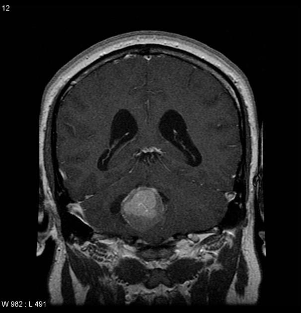
| 18:35, 14 January 2016 | MRI scan picture of choroid plexus papilloma image 1.jpg (file) |  |
38 KB | Image courtesy of Dr. Frank Gaillard. Radiopaedia (original file [http://radiopaedia.org/cases/choroid-plexus-papilloma here]). Creative Commons BY-SA-NC | 1 |
| 14:19, 14 January 2016 | CT scan of choroid plexus papilloma 1.jpg (file) |  |
39 KB | Image courtesy of Dr. Michael Sargent. Radiopaedia (original file [http://radiopaedia.org/cases/choroid-plexus-papilloma-2 here]). Creative Commons BY-SA-NC | 1 |
| 18:16, 21 December 2015 | CT image of atypical teratoid rhabdoid tumor 1.jpg (file) |  |
44 KB | 2 | |
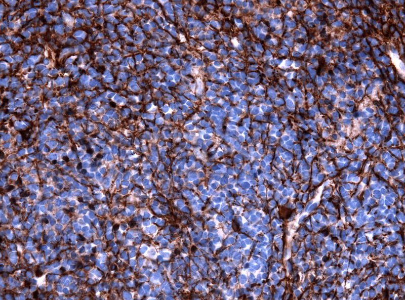
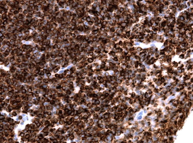
| 19:14, 17 December 2015 | Microscopic features of atrt 1.PNG (file) |  |
858 KB | Histologic features of atypical teratoid rhabdoid tumor. (a) Histologic examination of the tumor reveals diffuse eosinophilic cytoplasmic globules, vesicular chromatin, and scattered large pleomorphic nucleoli (hematoxylin and eosin 40x) and (b) Loss o... | 1 |
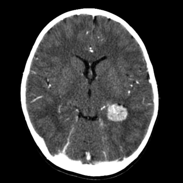
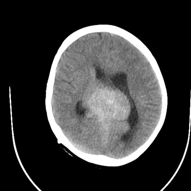
| 20:18, 3 December 2015 | CT image of pineal germinoma 1.jpg (file) |  |
37 KB | Image courtesy of Dr. Hani Al Salam. Radiopaedia (original file [http://radiopaedia.org/cases/germinoma here]). Creative Commons BY-SA-NC | 1 |
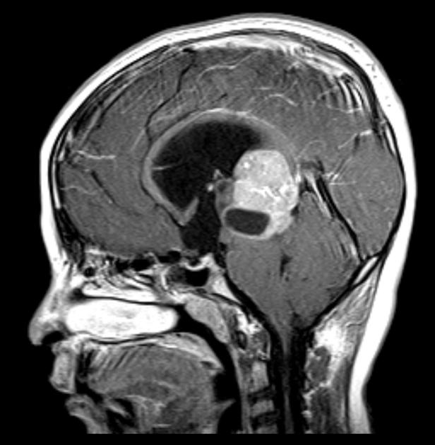
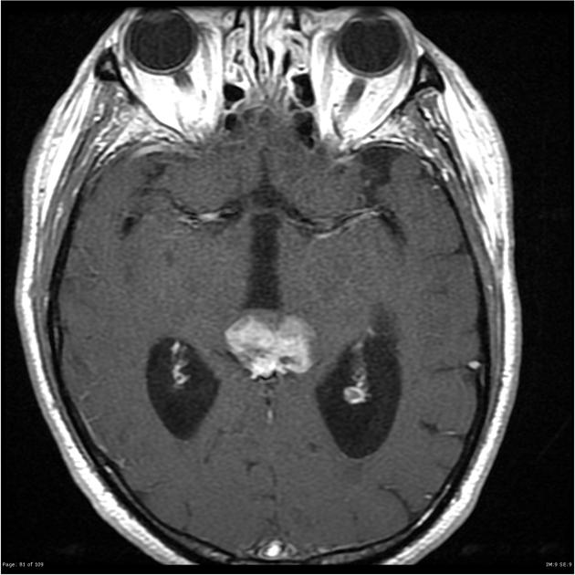
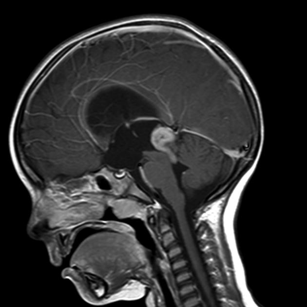
| 20:09, 3 December 2015 | MRI of pineal germinoma 1.jpg (file) |  |
46 KB | Image courtesy of Dr. Frank Gaillard. Radiopaedia (original file [http://radiopaedia.org/cases/pineal-germinoma here]). Creative Commons BY-SA-NC | 1 |
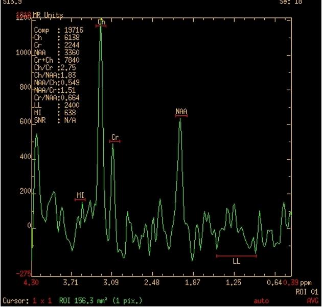
| 21:19, 1 December 2015 | MR spectroscopy.jpg (file) |  |
41 KB | Image courtesy of Dr. Mohammad A. ElBeialy. Radiopaedia (original file [http://radiopaedia.org/cases/pineoblastoma-6 here]). Creative Commons BY-SA-NC | 2 |
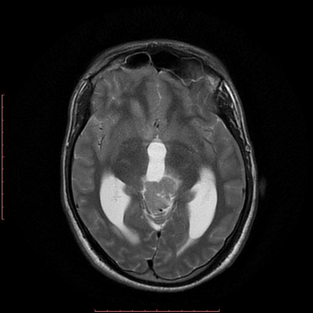
| 20:51, 1 December 2015 | MRI image of pineoblastoma 4.jpg (file) |  |
31 KB | Image courtesy of Dr. Mohammad A. ElBeialy. Radiopaedia (original file [http://radiopaedia.org/cases/pineoblastoma-6 here]). Creative Commons BY-SA-NC | 1 |
| 19:29, 1 December 2015 | MRI image of pineoblastoma 3.jpg (file) |  |
52 KB | Image courtesy of Dr. Frank Gaillard. Radiopaedia (original file [http://radiopaedia.org/cases/pineoblastoma-3 here]). Creative Commons BY-SA-NC | 1 |
| 19:23, 1 December 2015 | MRI image of pineoblastoma 2.jpg (file) |  |
39 KB | Image courtesy of Dr. Michael Sargent. Radiopaedia (original file [http://radiopaedia.org/cases/pineoblastoma-2 here]). Creative Commons BY-SA-NC | 1 |
| 19:19, 1 December 2015 | MRI image of pineoblastoma 1.jpg (file) |  |
23 KB | Image courtesy of Dr. Michael Sargent. Radiopaedia (original file [http://radiopaedia.org/cases/pineoblastoma-2 here]). Creative Commons BY-SA-NC | 1 |
| 19:07, 1 December 2015 | Pineoblastoma ct image 4.jpg (file) |  |
42 KB | Image courtesy of Dr. Bita Abbasi. Radiopaedia (original file [http://radiopaedia.org/cases/pineoblastoma-1 here]). Creative Commons BY-SA-NC | 1 |
| 18:56, 1 December 2015 | Pineoblastoma ct image 3.jpg (file) |  |
44 KB | Image courtesy of Dr. Frank Gaillard. Radiopaedia (original file [http://radiopaedia.org/cases/pineoblastoma-3 here]). Creative Commons BY-SA-NC | 1 |
| 18:47, 1 December 2015 | Microscopic image of pineoblastoma 2.jpg (file) |  |
353 KB | Image courtesy of Dr. Frank Gaillard. Radiopaedia (original file [http://radiopaedia.org/cases/pineoblastoma-3 here]). Creative Commons BY-SA-NC | 1 |
| 18:41, 1 December 2015 | 800px-Pineoblastoma gfap.jpg (file) |  |
164 KB | Immunohistochemical stain of a pineoblastoma demonstrating positivity to GFAP. | 1 |
| 18:38, 1 December 2015 | 800px-Pineoblastoma neurofilament.jpg (file) |  |
148 KB | immunohistochemical stain of a pineoblastoma demonstrating positivity to neurofilament. | 1 |
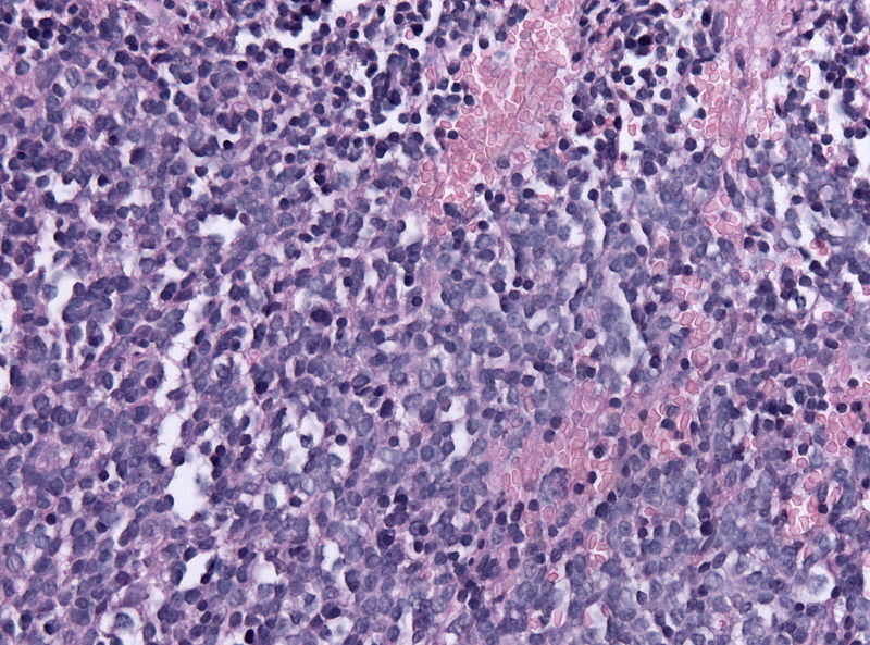
| 18:29, 1 December 2015 | Microscopic image of pineoblastoma 1.jpg (file) |  |
155 KB | Pathology specimen of a pineoblastoma (HE stain, x200 magnification) | 1 |
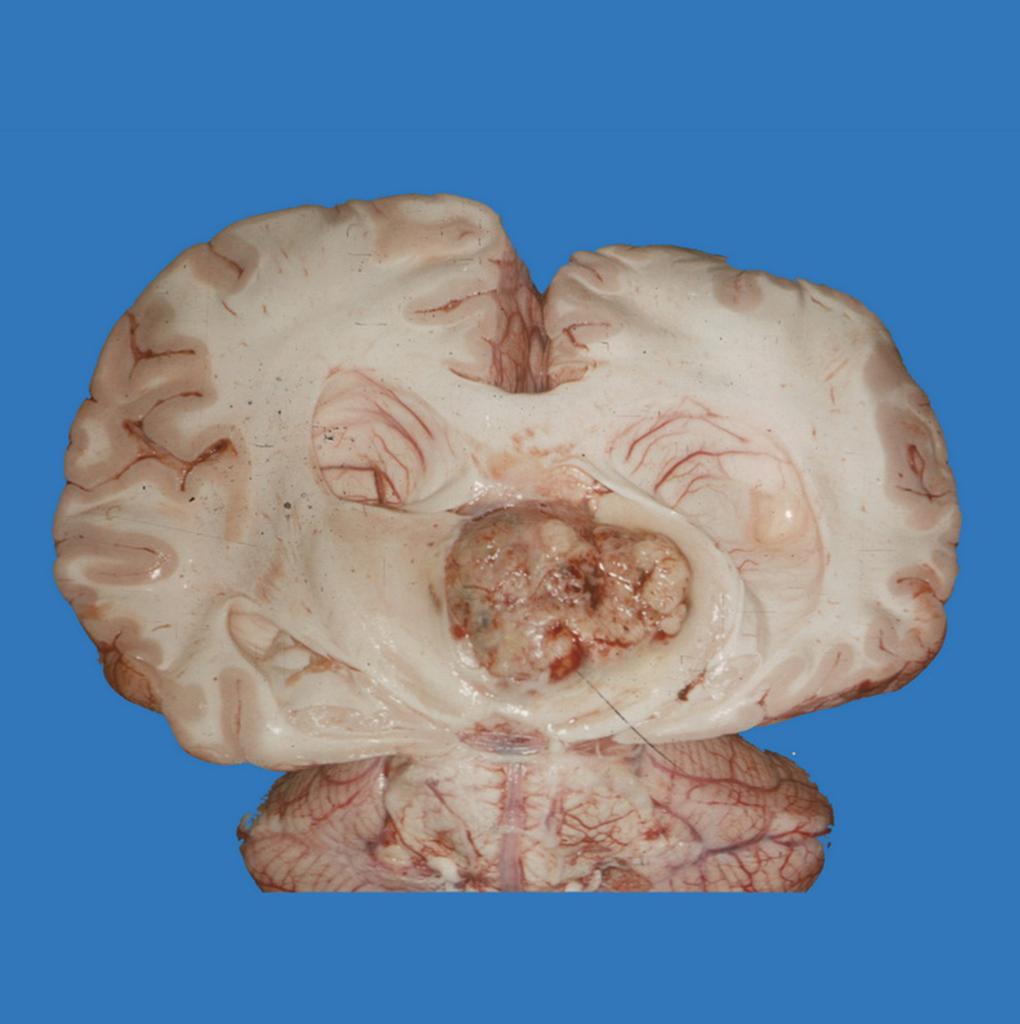
| 18:23, 1 December 2015 | Gross pathology of pineoblastoma.jpg (file) |  |
76 KB | Image courtesy of Dr. Frank Gaillard. Radiopaedia (original file [http://radiopaedia.org/cases/pineoblastoma-gross-pathology here]). Creative Commons BY-SA-NC | 1 |
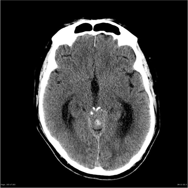
| 18:18, 1 December 2015 | Pineoblastoma ct image 2.jpg (file) |  |
40 KB | Image courtesy of Dr. Michael Sargent. Radiopaedia (original file [http://radiopaedia.org/cases/pineoblastoma-2 here]). Creative Commons BY-SA-NC | 1 |
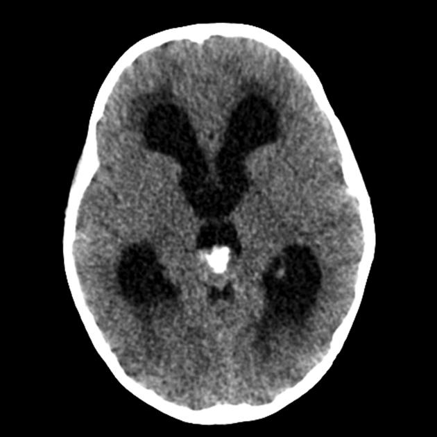
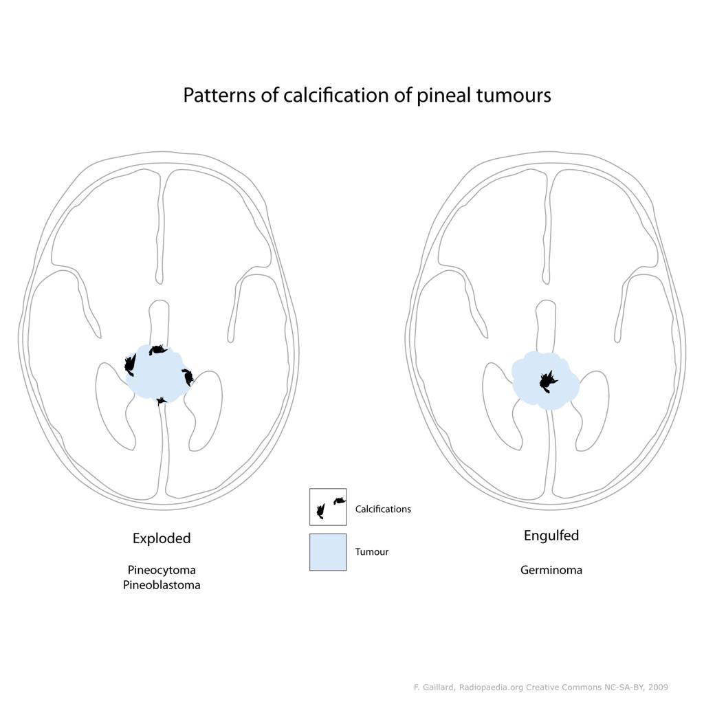
| 18:15, 1 December 2015 | Pineoblastoma ct image 1.jpg (file) |  |
58 KB | Image courtesy of Dr. Frank Gaillard. Radiopaedia (original file [http://radiopaedia.org/cases/pineal-tumour-calcification-illustration here]). Creative Commons BY-SA-NC | 1 |
| 19:31, 24 November 2015 | Ihcptpr2.jpg (file) |  |
181 KB | Photomicrograph of papillary tumor of the pineal region (PTPR) stained positive for epithelial membrane antigen (EMA) at 200x magnification. Image taken using an Olympus Microscope and Analysis-Imaging Software on 12-02-2006 | 1 |