Uploads by Qurrat-ul-ain Abid
Jump to navigation
Jump to search
This special page shows all uploaded files.
| Date | Name | Thumbnail | Size | Description | Versions |
|---|---|---|---|---|---|
| 15:54, 10 April 2019 | Mucinous Cystadenocarcinoma of the Ovary.jpg (file) |  |
238 KB | Multiple cysts in the ovary typical of mucinous cystadenocarcinoma of the ovary | 1 |
| 20:49, 14 January 2019 | Diffuse large B-cell lymphoma of the small intestine (high mag).jpg (file) |  |
175 KB | Diffuse large B-cell lymphoma arising in the small intestine (higher magnification). Notice the malignant lymphoid cells with prominent nucleoli. | 1 |
| 20:42, 14 January 2019 | Diffuse large B-cell lymphoma of the small intestine (intermed mag).jpg (file) |  |
265 KB | Diffuse large B-cell lymphoma of the small intestine. Malignant large lymphoid cells infiltrating the smooth muscle layer of intestinal wall. | 1 |
| 15:12, 14 January 2019 | Gastrointestinal stromal tumor (GIST).jpg (file) |  |
249 KB | Spindle cell type of intestinal gastrointestinal stromal tumor(GIST) showing areas of myxoid changes , hemorrhage and necrosis and surrounding vessels. | 1 |
| 15:05, 14 January 2019 | Periampullary adenocarcinoma.jpg (file) |  |
154 KB | Sample taken from duodenum biopsy reveals Adenocarcinoma of periampullary region, showing dysplastic changes. | 1 |
| 17:25, 28 December 2018 | Jejunal GIST.JPG (file) |  |
39 KB | Small intestinal stromal tumour, endoscopic image. | 1 |
| 17:18, 28 December 2018 | Multiple Carcinoid Tumors of the Small Bowel.jpg (file) |  |
20 KB | Multiple Carcinoid tumor of the small intestine | 1 |
| 17:13, 28 December 2018 | Small intestine neuroendocrine tumour.jpg (file) |  |
119 KB | Small intestine neuroendocrine tumour in low magnification showing neuroednocrine tumor cells(bottom), Paneth cells(red cells at the base of the crypt) and intestinal villi. | 1 |
| 17:01, 28 December 2018 | Duodenal adenocarcinoma.jpg (file) |  |
17 KB | Endoscopic image of adenocarcinoma of duodenum. | 1 |
| 14:16, 28 December 2018 | Macroscopic-appearance-of-small-intestinal-adenocarcinoma-with-carcinomatosis-in-the.jpg (file) |  |
129 KB | Small intestinal adenocarcinoma in the abdominal cavity, as a firm ring-like circumferential thickening on the wall of jejunum (arrow). Numerous tan metastatic nodules (arrowheads) of varying sizes are present on the mesentery and the serosal surfaces... | 1 |
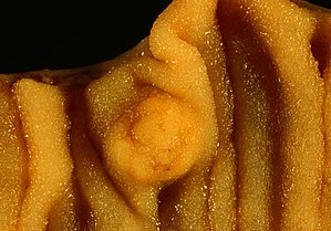
| 14:19, 24 December 2018 | Carcinoid Tumor of the Small Intestine.jpeg (file) |  |
22 KB | Picture of a carcinoid tumour that encroaches into lumen of the small Intestine. Pathology specimen. The prominent folds are plicae circulares, a characteristic of small Intestine. | 1 |