Help
Uploads by Homa Najafi
Jump to navigation
Jump to search
This special page shows all uploaded files.
File list
Items per page:
20
50
100
250
500
Username:
Include old versions of files
Show file list
Date
Name
Thumbnail
Size
Description
Versions
04:01, 5 February 2020
Short axis T2 acute myocarditis.jpeg
(
file
)
37 KB
1
03:59, 5 February 2020
Short axis T1 acute myocarditis.jpeg
(
file
)
46 KB
1
03:58, 5 February 2020
CMR of acute myocarditis.jpeg
(
file
)
54 KB
1
03:55, 5 February 2020
4 chamber CMR of acute myocarditis.jpeg
(
file
)
50 KB
1
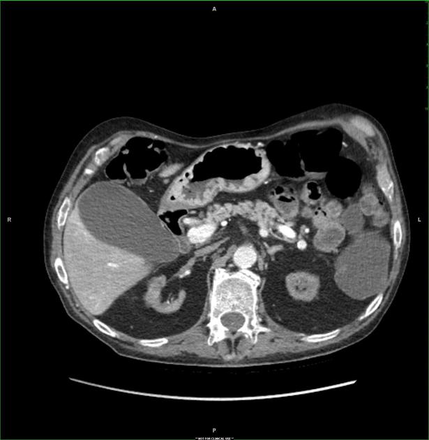
22:22, 3 October 2019
Axial CT scan, venous phase.jpeg
(
file
)
38 KB
1
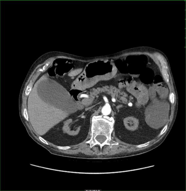
22:21, 3 October 2019
Axial CT scan, arterial phase.jpeg
(
file
)
36 KB
1
22:19, 3 October 2019
Axial Ct scan, non contrast.jpeg
(
file
)
35 KB
1
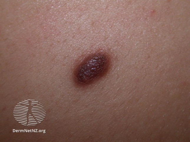
14:42, 27 August 2019
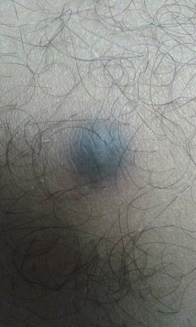
Hyperpigmentation in dermatofibroma.jpg
(
file
)
63 KB
1
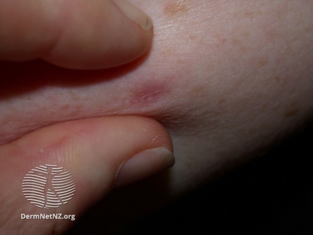
14:39, 27 August 2019
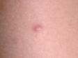
Dimple sign.jpg
(
file
)
40 KB
1
14:05, 27 August 2019
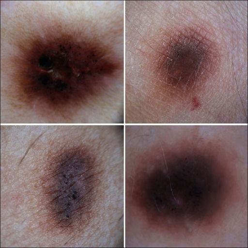
Dermoscopy of dermatofibroma.png
(
file
)
592 KB
1
14:01, 27 August 2019
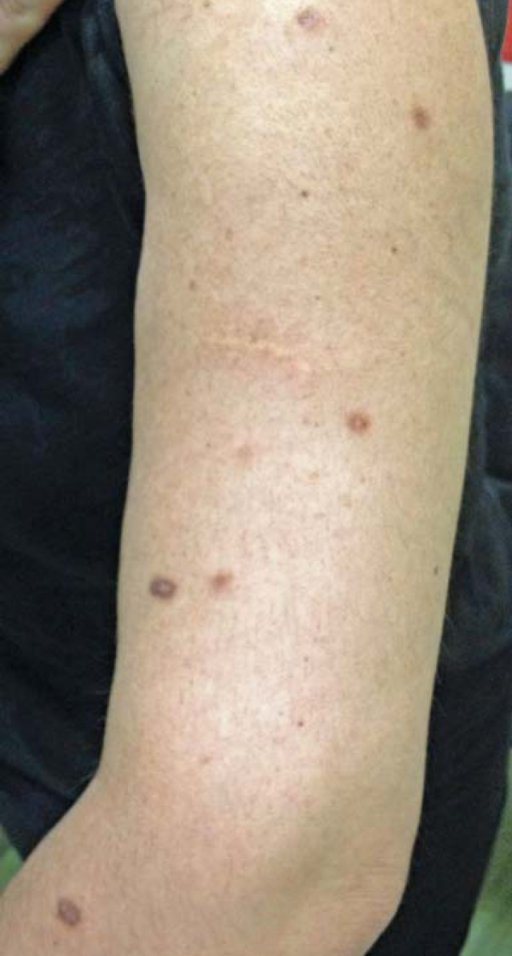
Multiple dermatofibromas.png
(
file
)
739 KB
1
13:58, 27 August 2019
Skin dermatofibroma.jpg
(
file
)
1 KB
1
13:57, 27 August 2019
Dermatofibroma.jpg
(
file
)
656 KB
Reverted to version as of 13:46, 27 August 2019 (UTC)
3
03:47, 27 August 2019
Spindle-shaped fibroblasts, arranged in a storiform pattern.png
(
file
)
504 KB
1
03:44, 27 August 2019
Proliferating histiocytic cells with foamy, granular cytoplasm.png
(
file
)
525 KB
1
18:12, 17 June 2019
InkedMRI renal oncocytoma LI.jpg
(
file
)
41 KB
1
17:59, 17 June 2019
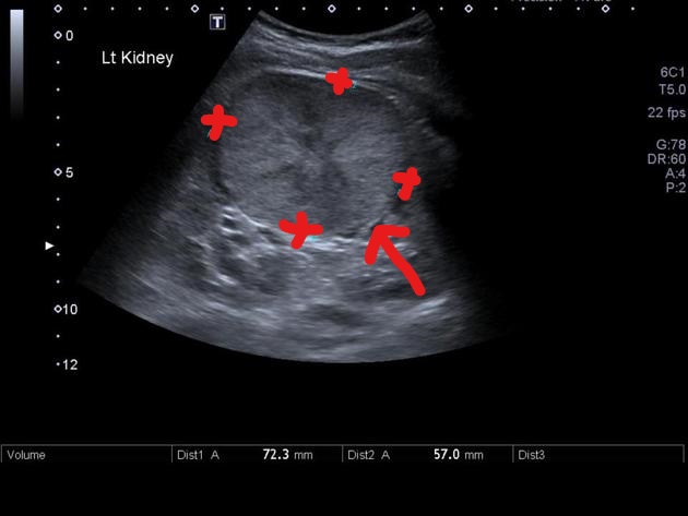
Inkedrenal oncocytoma ultrasound LI.jpg
(
file
)
52 KB
1
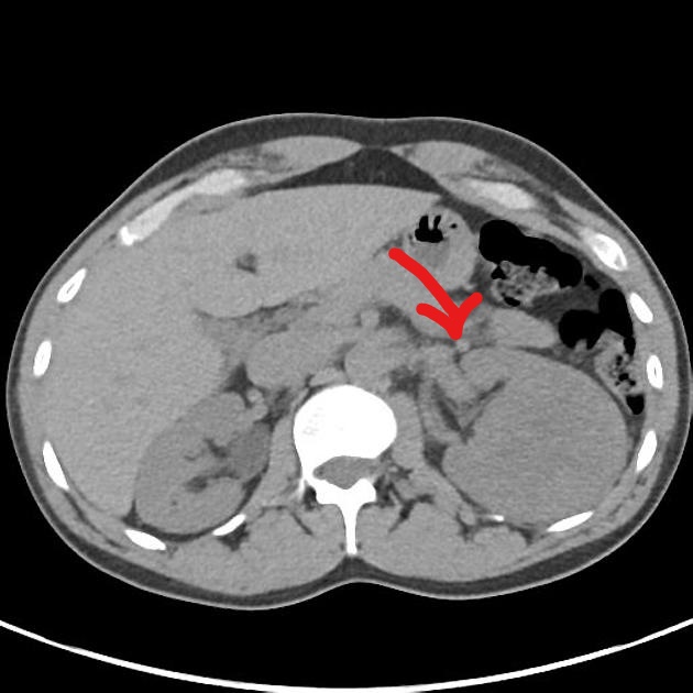
15:56, 17 June 2019
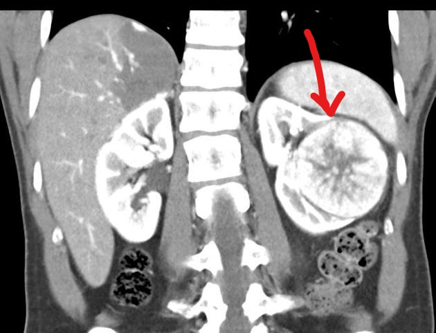
Coronal contrast CT of renal oncocytoma.jpg
(
file
)
79 KB
1
15:54, 17 June 2019
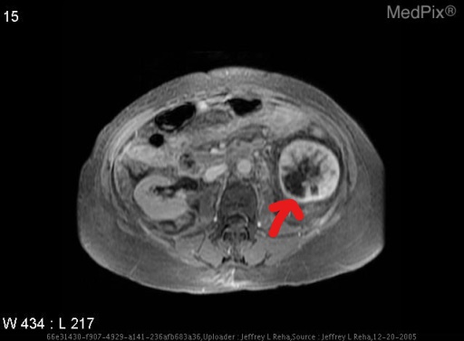
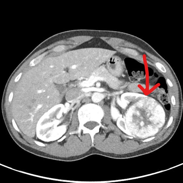
Axial contrast CT of renal oncocytoma.jpg
(
file
)
89 KB
1
15:50, 17 June 2019
InkedAxial non-contrast CT of renal oncocytoma LI.jpg
(
file
)
82 KB
1
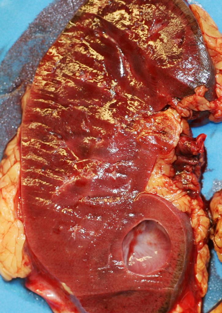
15:38, 17 June 2019
Gross pathology- coronal section.jpg
(
file
)
145 KB
1
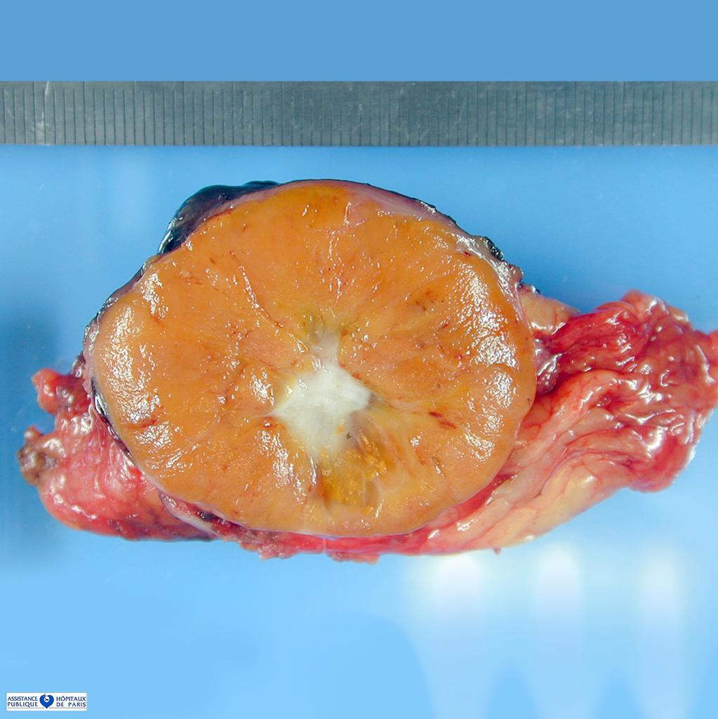
15:32, 17 June 2019
Gross pathology of renal oncocytoma.jpg
(
file
)
111 KB
1
15:09, 17 June 2019
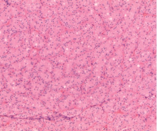
Renal oncocytoma.png
(
file
)
453 KB
Oncocytes are seen
1
14:32, 17 June 2019
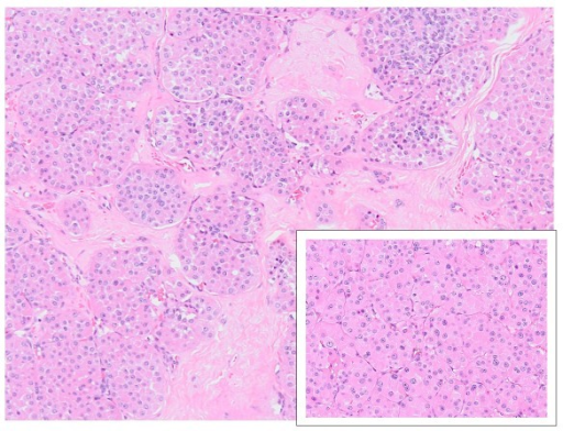
Renal oncocytoma HE.png
(
file
)
470 KB
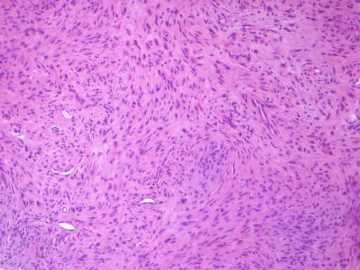
Renal oncocytoma with a nesting growth pattern, eosinophilic cytoplasm, and round nuclei in Hematoxylin and Eosin staining.
1
16:16, 8 May 2019
Nasopharyngeal carcinoma PET LI.jpg
(
file
)
83 KB
1
15:37, 8 May 2019
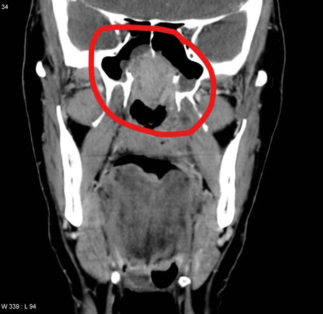
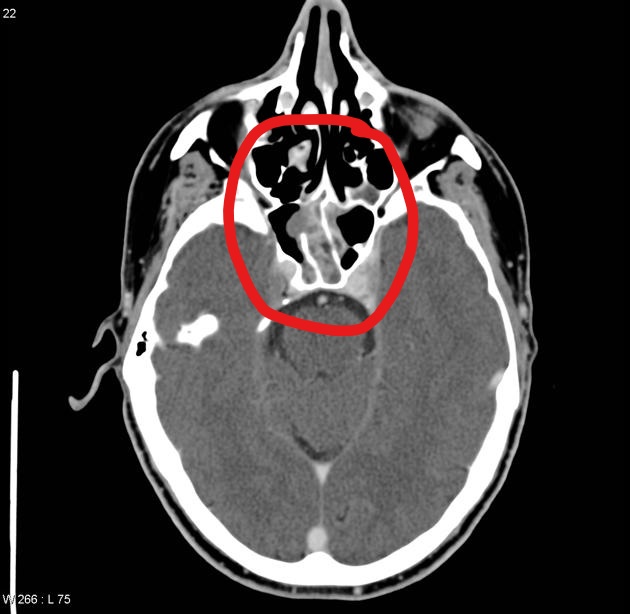
Nasopharyngeal carcinoma CT 3.jpg
(
file
)
76 KB
1
15:36, 8 May 2019
Nasopharyngeal carcinoma CT 2.jpg
(
file
)
78 KB
1
15:34, 8 May 2019
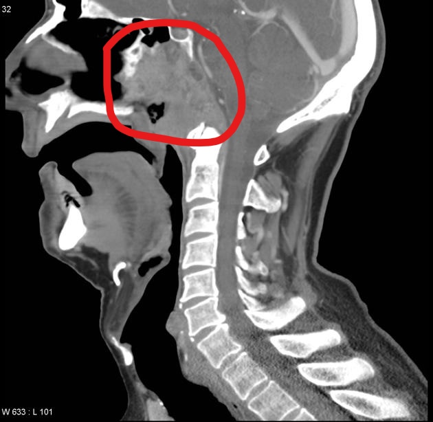
Nasopharyngeal carcinoma CT.jpg
(
file
)
84 KB
1
14:58, 8 May 2019
Nasopharyngeal carcinoma MRI.jpg
(
file
)
78 KB
1
14:53, 8 May 2019
Nasopharyngeal Carcinoma- MRI T1 (2).jpg
(
file
)
72 KB
1
14:46, 8 May 2019
Nasopharyngeal carcinoma-Axial T1 MRI.jpg
(
file
)
72 KB
2
14:17, 8 May 2019
Nasopharyngeal carcinoma MRI-Axial T1C+fat sat.jpg
(
file
)
78 KB
1
13:49, 8 May 2019
PMC4582819 13014 2015 513 Fig1 HTML (1).png
(
file
)
92 KB
1
13:57, 26 April 2019
PMC4195840 13244 2014 349 Fig17 HTML.png
(
file
)
144 KB
1
15:49, 31 December 2018
41689 najafi.jpg
(
file
)
185 KB
1
Cookies help us deliver our services. By using our services, you agree to our use of cookies.
OK
Navigation menu
Personal tools
Log in
Request account
Namespaces
Special page
English
Views
More
Search
Tools
User contributions
Logs
View user groups
Printable version


.jpg)


.png)

