Uploads by Hamid Qazi
Jump to navigation
Jump to search
This special page shows all uploaded files.
| Date | Name | Thumbnail | Size | Description | Versions |
|---|---|---|---|---|---|
| 18:08, 11 April 2018 | Giant cell arteritis -- low mag.jpg (file) |  |
5.51 MB | 1 | |
| 13:53, 29 March 2018 | Lobar-pneumonia-ct-findings.jpg (file) |  |
268 KB | 1 | |
| 04:04, 4 March 2018 | Pneumonia-right-middle-lobe-4.jpg (file) |  |
133 KB | 1 | |
| 16:57, 3 March 2018 | Lung bleb -- extremely low mag.jpg (file) |  |
6.78 MB | 1 | |
| 23:10, 17 February 2018 | Pleurisy and pneumothorax.jpg (file) |  |
139 KB | 1 | |
| 23:07, 17 February 2018 | Pneumothoraxpatho.jpg (file) |  |
94 KB | 1 | |
| 23:00, 17 February 2018 | Bullae and Bleb.jpg (file) |  |
412 KB | 1 | |
| 02:03, 16 February 2018 | Tension-pneumothorax-12.jpg (file) | 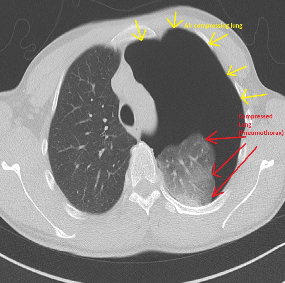 |
269 KB | 1 | |
| 01:52, 16 February 2018 | Tension-pneumothorax-1.jpg (file) | 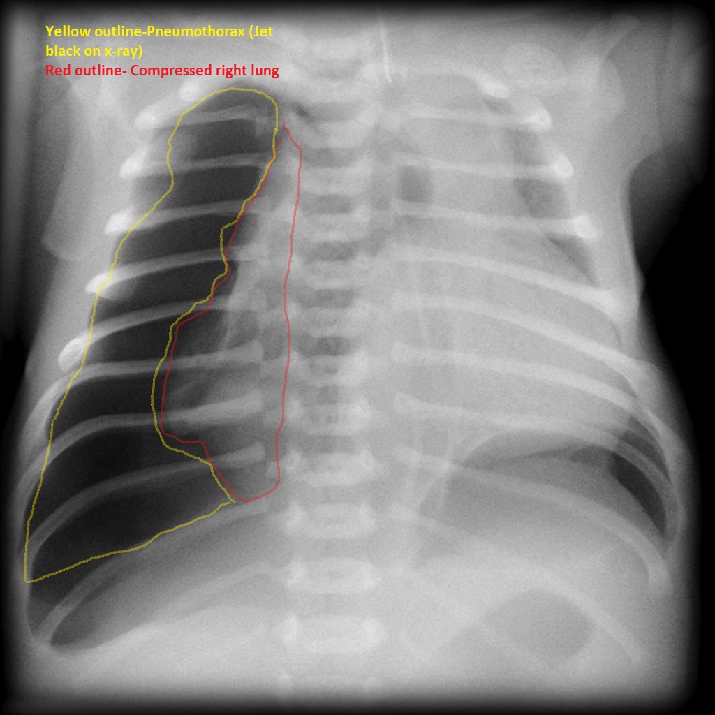 |
185 KB | 1 | |
| 20:46, 9 February 2018 | Cystic-bronchiectasis-causing-pneumothorax.jpg (file) | 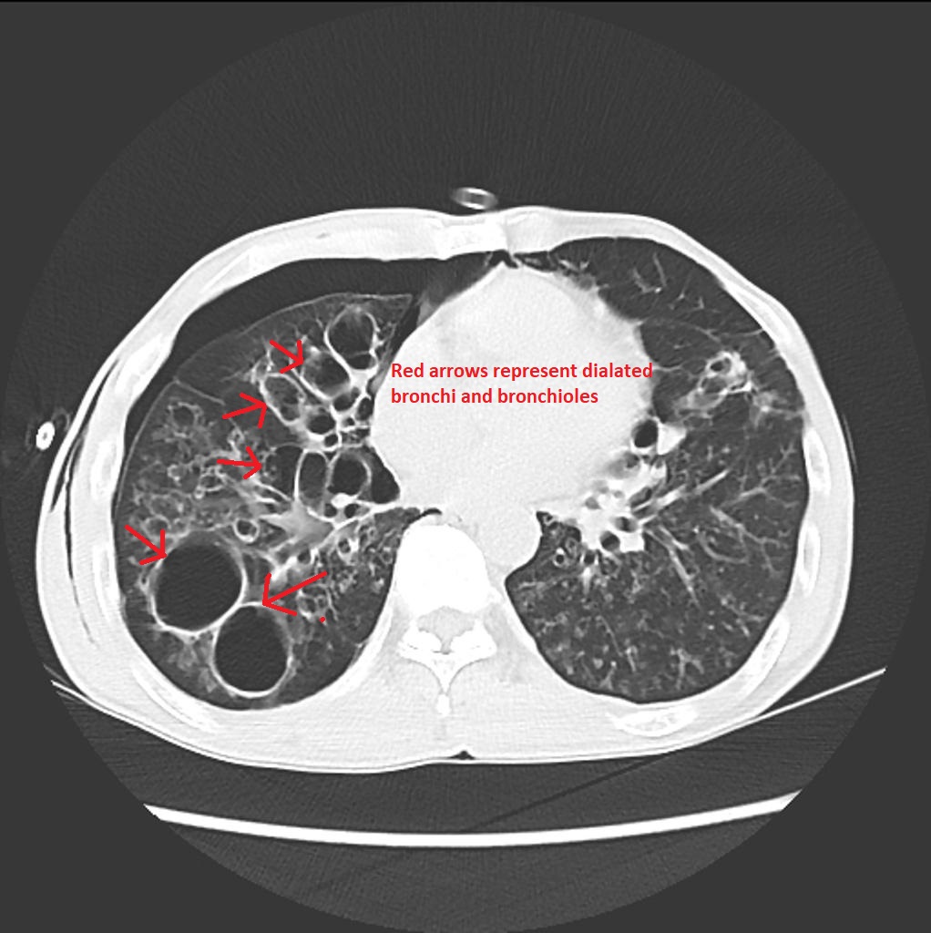 |
179 KB | 1 | |
| 20:36, 9 February 2018 | Bronchiectasis-4.jpg (file) | 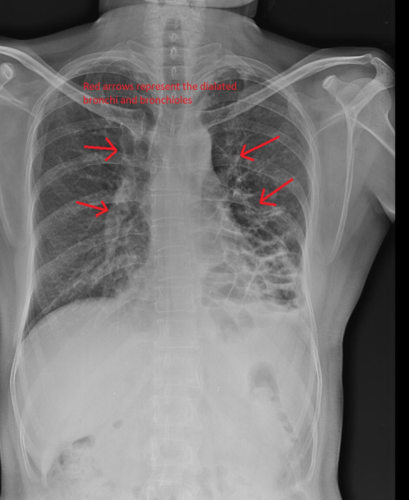 |
116 KB | 1 | |
| 16:30, 9 February 2018 | Bronchiectasis-gross-pathology-1.jpg (file) |  |
253 KB | 4 | |
| 12:51, 8 February 2018 | Hamid1.jpg (file) |  |
1.13 MB | 1 | |
| 15:37, 29 January 2018 | Hernia Common Sites.png (file) |  |
542 KB | 1 | |
| 19:00, 22 January 2018 | IMG 20180122 135707964.jpg (file) |  |
1.76 MB | 2 | |
| 15:50, 22 January 2018 | Umbilical-hernia-1.jpg (file) |  |
80 KB | 1 | |
| 15:45, 22 January 2018 | Umbilical-hernia.jpg (file) |  |
76 KB | 1 | |
| 13:42, 12 January 2018 | Ischemic CT1.jpg (file) |  |
105 KB | 1 | |
| 19:53, 3 January 2018 | Ischaemic-descending-colon.jpg (file) |  |
124 KB | 1 | |
| 18:07, 2 January 2018 | Duodenal-atresia-1.jpg (file) |  |
76 KB | 1 | |
| 16:54, 2 January 2018 | Duodenal-atresia.jpg (file) |  |
60 KB | 1 | |
| 17:23, 27 December 2017 | Double bubble .jpg (file) | 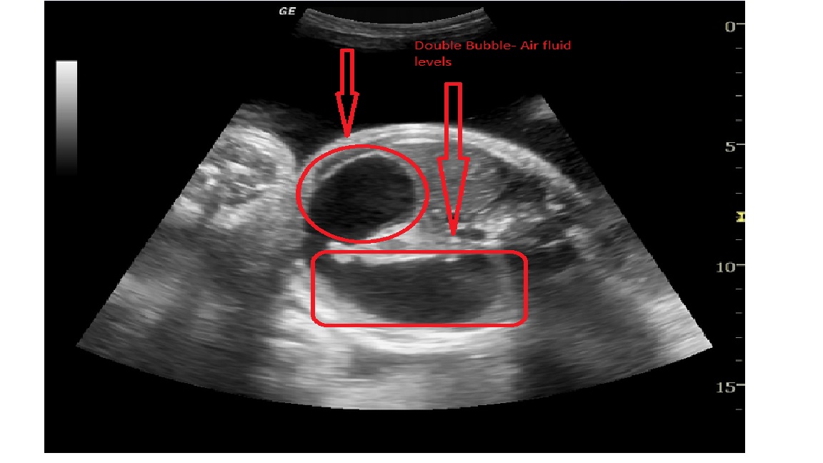 |
118 KB | 1 | |
| 16:26, 27 December 2017 | Duodenal atresia xray.jpg (file) | 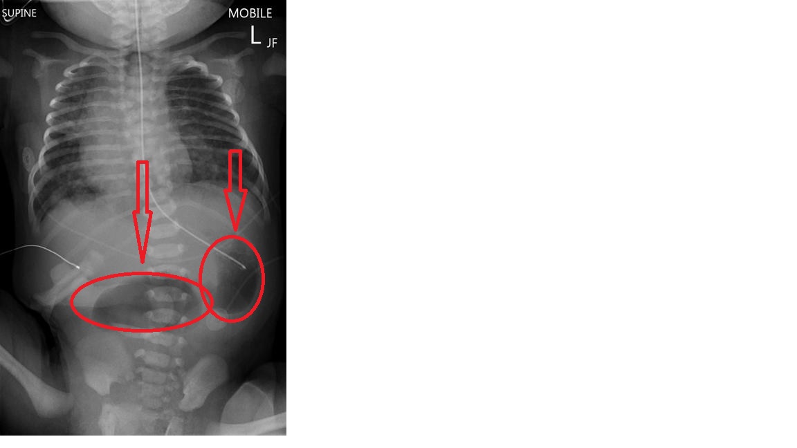 |
59 KB | 1 | |
| 15:33, 26 December 2017 | Duodenum anatomy.jpg (file) | 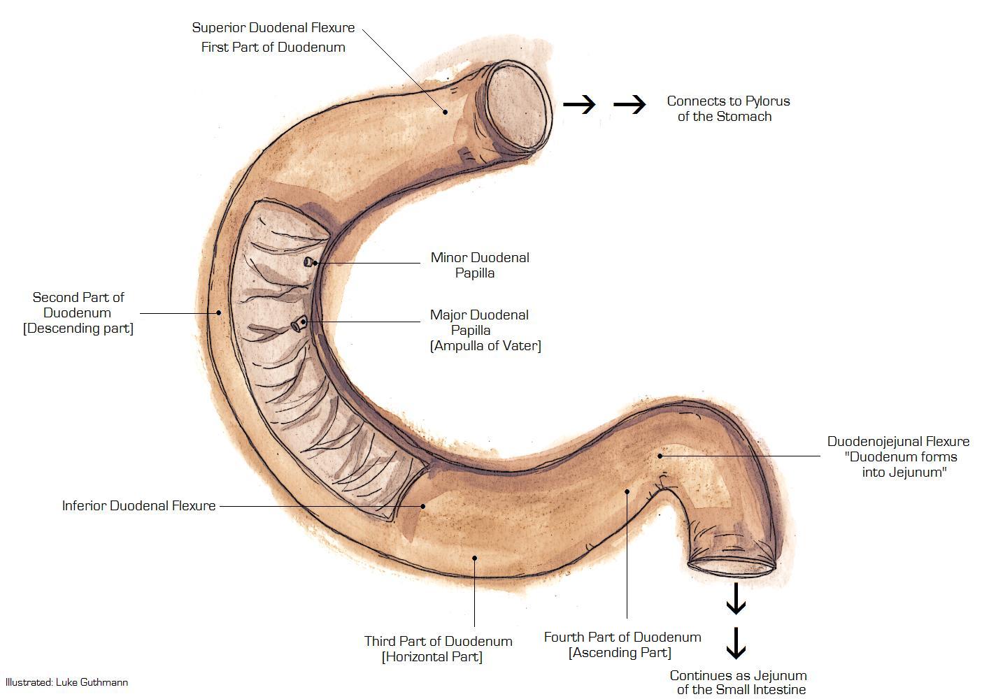 |
130 KB | 1 | |
| 17:04, 21 December 2017 | Polyps in PJS.PNG (file) | 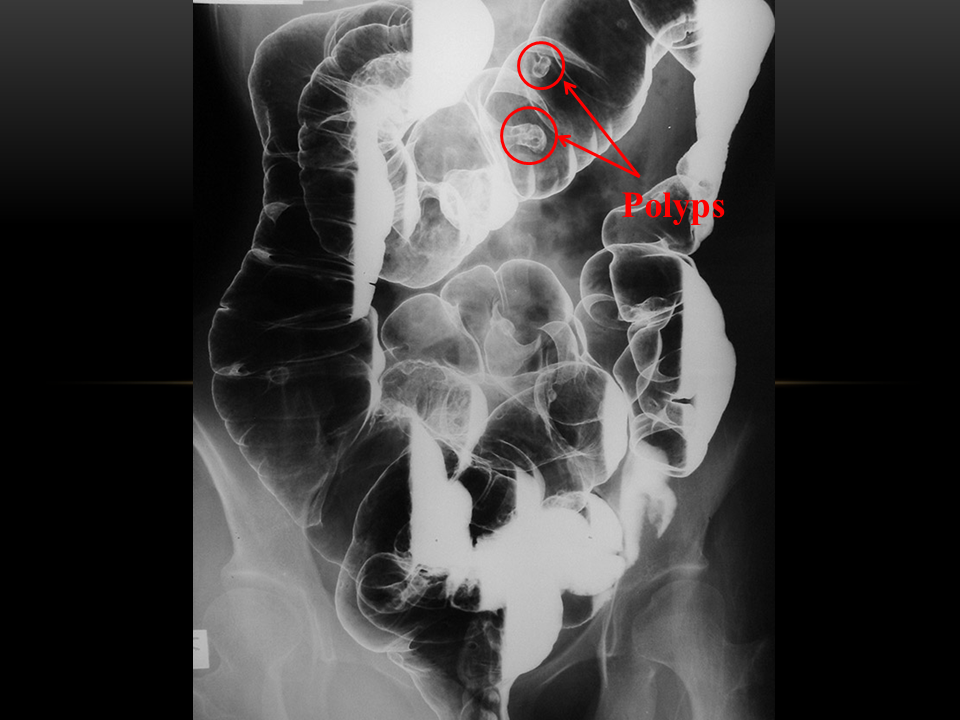 |
433 KB | 1 | |
| 17:03, 21 December 2017 | Multiple polyps and at large mass at the hepatic flexure.jpg (file) | 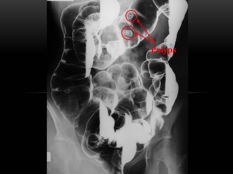 |
51 KB | 5 | |
| 16:39, 19 December 2017 | Peutz-Jeghers Syndrome Hyperpigmentation.jpg (file) |  |
54 KB | 1 | |
| 13:47, 19 December 2017 | Enteroscopy.jpg (file) | 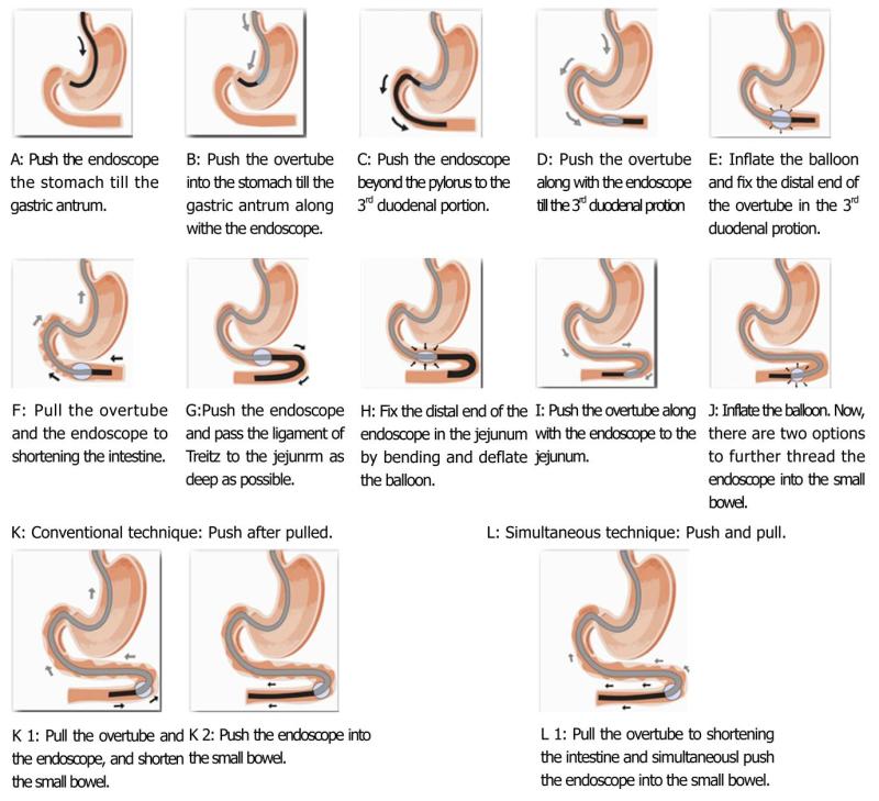 |
90 KB | 1 | |
| 12:48, 19 December 2017 | Peutz-Jeghers syndrome polyp .jpg (file) |  |
2.13 MB | 1 | |
| 19:39, 18 December 2017 | PJS Natural History.jpg (file) | 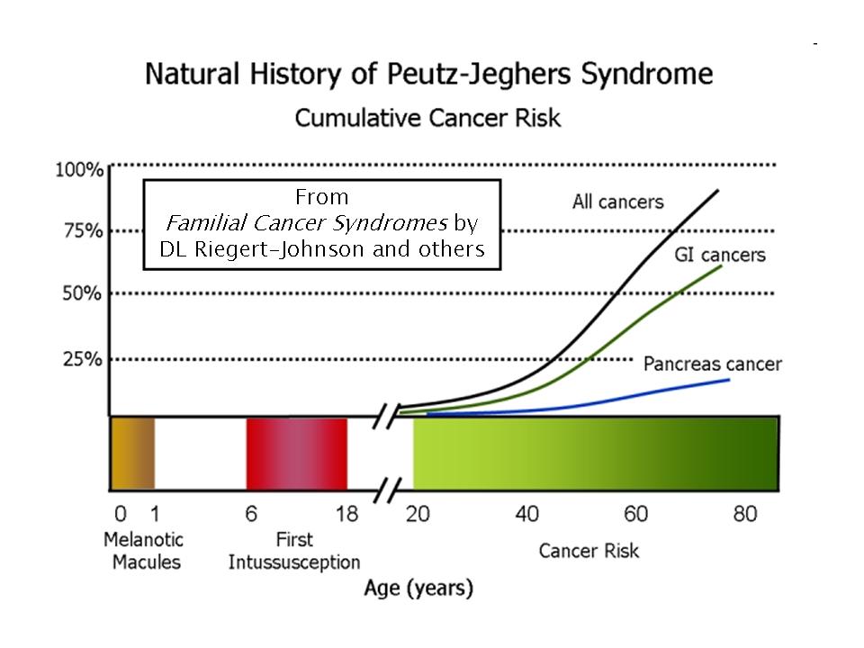 |
64 KB | 1 | |
| 16:48, 18 December 2017 | Colon histology with Peutz-Jeghers polyp.jpg (file) | 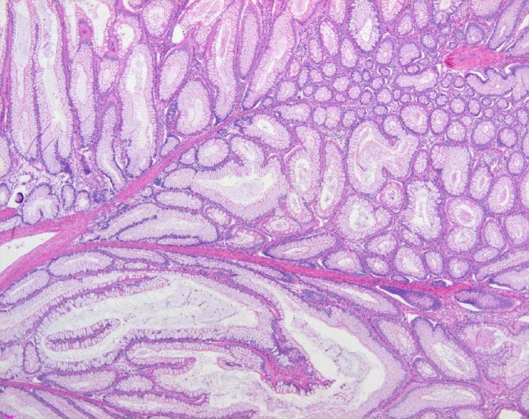 |
113 KB | 1 | |
| 16:34, 18 December 2017 | Peutz-Jeghers syndrome polyp.jpg (file) | 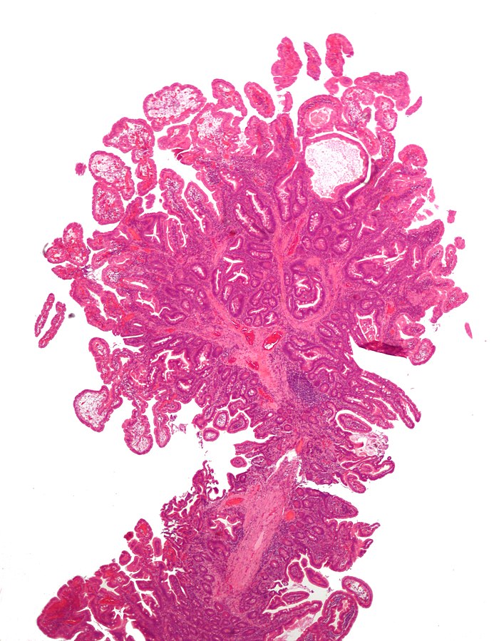 |
170 KB | 1 | |
| 16:50, 14 December 2017 | PJSpolyp.jpg (file) |  |
41 KB | Hamartoma, a typical PJS polyp demonstrating the arborizing pattern of smooth-muscle proliferation. HE staining, magnification 100 ×. (Courtesy of Professor A. Ryska, MD, PhD) | 1 |