Uploads by Dildar Hussain
Jump to navigation
Jump to search
This special page shows all uploaded files.
| Date | Name | Thumbnail | Size | Description | Versions |
|---|---|---|---|---|---|
| 18:38, 1 May 2018 | Pelvic-osteoblastoma.jpg (file) |  |
278 KB | Left acetabular fossa pubic bone expansile predominantly lytic lesion with a rim of reactive sclerosis. Case courtesy of Dr Amr Farouk, <a href="https://radiopaedia.org/">Radiopaedia.org</a>. From the case <a href="https://radiopaedia.org/cases/41171">... | 1 |
| 18:07, 1 May 2018 | Osteoblastoma.jpg (file) |  |
58 KB | Osteoblastoma causing a marked scoliosis in a 7 year old boy. Case courtesy of Dr Angela Byrne, <a href="https://radiopaedia.org/">Radiopaedia.org</a>. From the case <a href="https://radiopaedia.org/cases/7593">rID: 7593</a> | 1 |
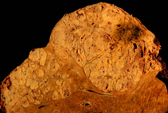
| 12:50, 21 February 2018 | Hepatocellular carcinoma 1.jpg (file) |  |
38 KB | <a href="https://commons.wikimedia.org/wiki/File%3AHepatocellular_carcinoma_1.jpg">via Wikimedia Commons</a> | 2 |
| 00:06, 17 February 2018 | Squamous cell Ca.jpg (file) |  |
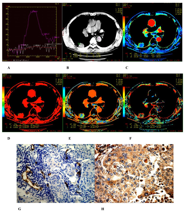
197 KB | (A-H) Hamartoma found in apicoposterior segment of superior of the left lung of a 54-year-old male. (A) Time density curve. (B-F) (original image, BF, BV, MTT, PS) type I parametric maps, PS value was moderate (12.029). (G) CD34 staining shows a few of... | 1 |
| 23:33, 16 February 2018 | Peripheral pulmonary nodules1.jpg (file) |  |
203 KB | (A-H) Poorly differentiated adenocarcinoma found in the apicoposterior segment of superior lobe of the left lung of a 56 year-old male. (A) Time density curve. (B-F) (original image, BF, BV, MTT, PS) typeI parametric maps, PS value is higher (30.883).... | 1 |
| 18:13, 16 February 2018 | IJRI-25-109-g016.jpg (file) |  |
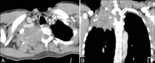
26 KB | Superior sulcus tumor. Axial (A) and coronal (B) CT scans show a large mass in the apex of the right lung causing destruction of the first and second ribs (arrows) with erosion of the right half of the vertebral body (arrowheads) suggestive of a superi... | 1 |
| 16:48, 16 February 2018 | IJRI-25-109-g015.jpg (file) |  |
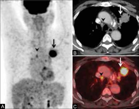
57 KB | Incremental value of FDG PET/CT in baseline staging. MIP image of FDG PET scan shows intense tracer concentration in the right hemithorax (arrow, A) corresponding to a right lung mass (arrow, B). Also seen are two FDG-avid foci in the abdomen (arrowhea... | 1 |
| 16:46, 16 February 2018 | IJRI-25-109-g014.jpg (file) |  |
66 KB | Pleural effusion and role of FDG PET/CT. Enhancing lung masses seen on CT scans in two different patients (arrows in A and C) with minimal pleural effusions (arrowheads in A and C). Corresponding PET/CT scans show intense FDG-avid metastatic pleural de... | 1 |
| 16:26, 16 February 2018 | IJRI-25-109-g009.jpg (file) |  |
54 KB | FDG PET in nodal disease false-positive study. Maximum intensity projection (MIP) image shows an FDG-avid primary lung tumor on the right side (arrow, A) and multiple foci of FDG uptake in the mediastinum (arrowhead, A). CT scan shows enhancing, primar... | 1 |
| 16:18, 16 February 2018 | IJRI-25-109-g008.jpg (file) |  |
47 KB | FDG PET in nodal disease. Maximum intensity projection (MIP) image shows an FDG-avid primary lung tumor on the left side (arrow, A) and a focus of FDG uptake in the mediastinum (arrowhead, A). CT scan shows enhancing, spiculated primary tumor (arrow, B... | 1 |
| 16:07, 16 February 2018 | IJRI-25-109-g006.jpg (file) |  |
39 KB | Role of FDG PET/CT in primary tumor delineation. Irregular soft tissue opacity seen on coronal CT scan (arrow, A) with no obvious demarcation between the tumor and surrounding consolidation. PET/CT shows the FDG-avid tumor (arrow, B) separate from the... | 1 |
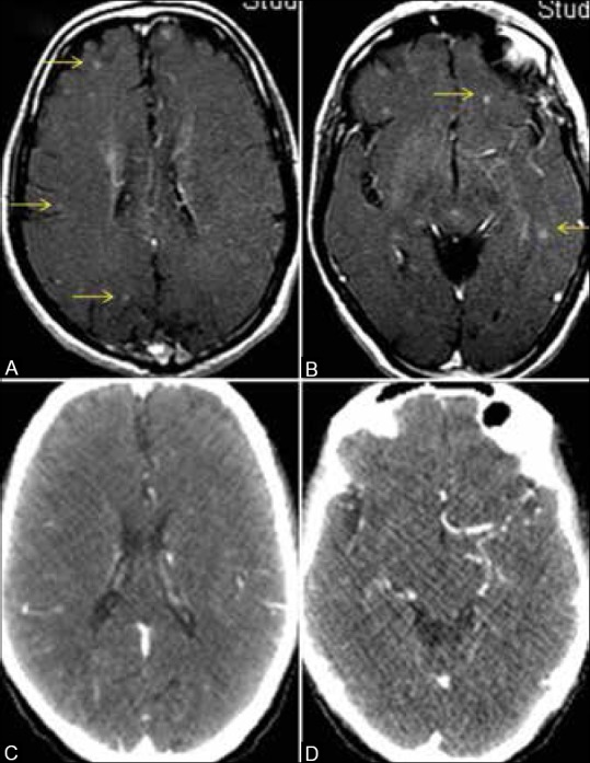
| 18:19, 15 February 2018 | IJRI-25-109-g012.jpg (file) |  |
92 KB | Brain metastases in asymptomatic patient, CT scan versus MRI. MRI brain in a patient of lung cancer shows multiple tiny enhancing foci scattered in the parenchyma bilaterally (arrows in A and B) suggestive of metastatic lesions. Corresponding contrast... | 1 |
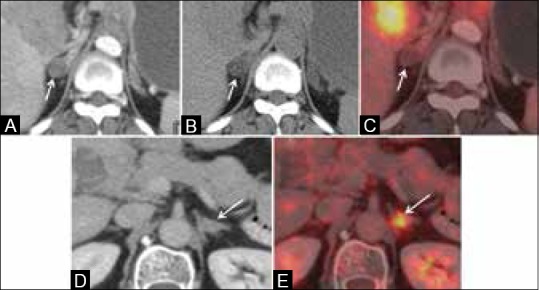
| 18:13, 15 February 2018 | IJRI-25-109-g011.jpg (file) |  |
37 KB | Adrenal adenoma versus metastasis. Enhancing solid adrenal nodule on CT scan in a case of lung cancer (arrow, A) suggestive of metastatic deposit. Unenhanced CT scan shows fatty attenuation within the nodule with an HU value of 0 suggesting the possibi... | 1 |
| 18:12, 15 February 2018 | IJRI-25-109-g010.jpg (file) |  |
43 KB | Metastatic disease. Bilateral pleural effusions-M1a (arrow, A), lung metastases-M1a (arrows, B), adrenal metastasis-M1b (arrow, C), vertebral metastasis-M1b (arrow, D), brain metastasis-M1b (arrow, E), liver metastases-M1b (arrows, F) | 1 |
| 16:52, 15 February 2018 | IJRI-25-109-g005.jpg (file) |  |
67 KB | Stage T4 tumors. T4 tumor due to invasion of pulmonary artery (arrow, A), descending aorta (arrow, B), vertebral body (arrow, C), superior vena cava with thrombus (arrow, D) | 1 |
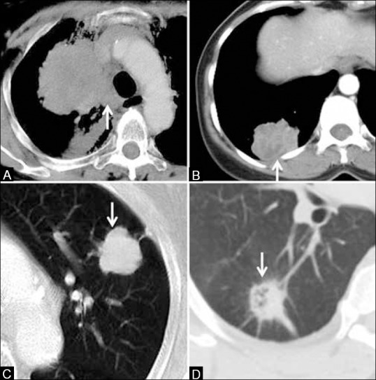
| 16:45, 15 February 2018 | IJRI-25-109-g004.jpg (file) |  |
75 KB | Stage T3 tumors. T3 tumor due to size >7 cm in size (arrow, A), eroding the ribs (arrow, B), infiltrating the mediastinal pleura but not the vessels (arrow, C), and causing atelectasis of the entire lung (arrowhead, D) | 1 |
| 16:41, 15 February 2018 | IJRI-25-109-g003.jpg (file) |  |
61 KB | Stage T1 and T2 tumors. Stage T1 tumor due to size <3 cm (arrow, A). Stage T2 endobronchial tumor (arrowhead) causing pneumonitis restricted to the upper lobe (arrow) in B. T2a tumor >3 cm but <5 cm (arrow, C). T2b tumor >5 cm but <7 cm (arrow in D) | 1 |
| 16:26, 15 February 2018 | IJRI-25-109-g002.jpg (file) |  |
62 KB | Lung cancers with atypical radiological pattern. Squamous cell cancer presenting as a cavitating mass (arrow, A). Adenocarcinoma presenting as dense consolidation (arrow, B). Bronchoalveolar carcinoma (adenocarcinoma in situ) presenting as ground-glass... | 1 |
| 16:04, 15 February 2018 | IJRI-25-109-g001.jpg (file) |  |
65 KB | 1 | |
| 15:17, 9 January 2018 | Massive HCC.gif (file) |  |
1.4 MB | 1 | |
| 15:07, 9 January 2018 | Diffuse HCC.gif (file) |  |
1.4 MB | 1 | |
| 14:50, 9 January 2018 | Hcc16.gif (file) |  |
1.28 MB | 1 | |
| 21:06, 16 December 2017 | 512px-Jaundice08.jpg (file) |  |
53 KB | 1 | |
| 17:19, 14 December 2017 | Gallstones png.png (file) |  |
71 KB | 1 | |
| 00:53, 10 December 2017 | Dildar Hussain.jpg (file) |  |
124 KB | 1 | |
| 00:39, 10 December 2017 | Syed Muhammad Dildar Hussain.jpg (file) |  |
101 KB | 1 |