Sandbox elastofibroma
Jump to navigation
Jump to search
| Sandbox elastofibroma | |
 | |
|---|---|
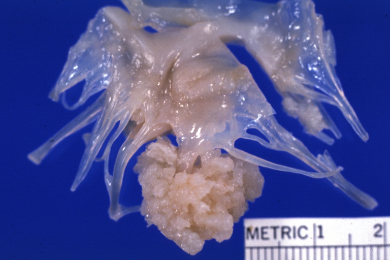
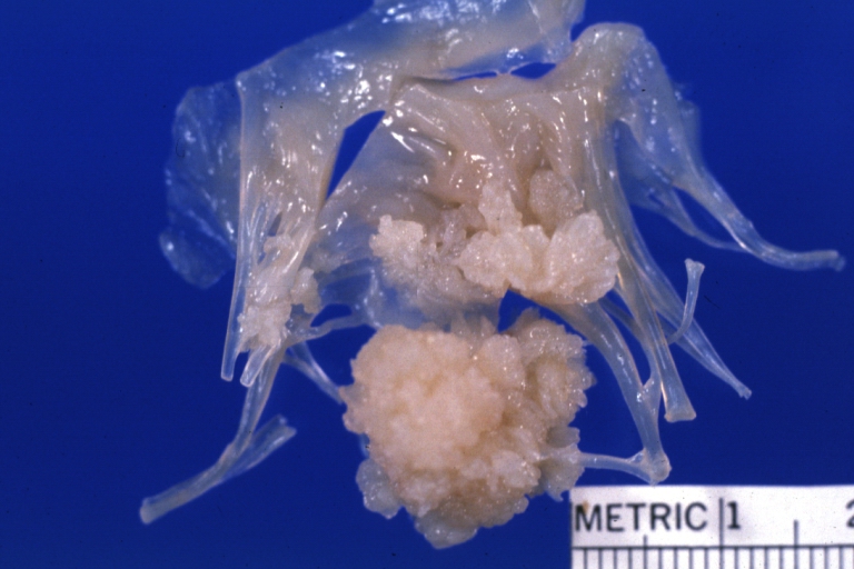
| Elastofibroma: Gross, an excellent mitral valve photo of this benign lesion. Image courtesy of Professor Peter Anderson DVM PhD and published with permission © PEIR, University of Alabama at Birmingham, Department of Pathology |
Patholophysiology
Microscopy
In general, the tumor is an ill defined, nonencapsulated, rubbery, and firm, white lesion with interspersed fat. The tumors can be quite large (up to 20 cm), although most are around 5 cm. [1]
By microscopic view, there is an admixture of heavy dense bands of collagenous tissue dissected by fat and abnormal elastic fibers. The elastic fibers are often quite large and are easily identified. The elastic fibers are coarse, thick, and darkly eosinophilic, often fragmented into globules, creating a "string of pearls" or "pipe cleaner" appearance. Because of degeneration, the elastic fibers will appear as globules with a serrated or "prickled" edge. [1]
Histochemistry
The elastic fibers will be highlighted by a Weigert or von Gieson elastic stains. [2]
-
 Elastofibroma
 -
 Elastofibroma
 -
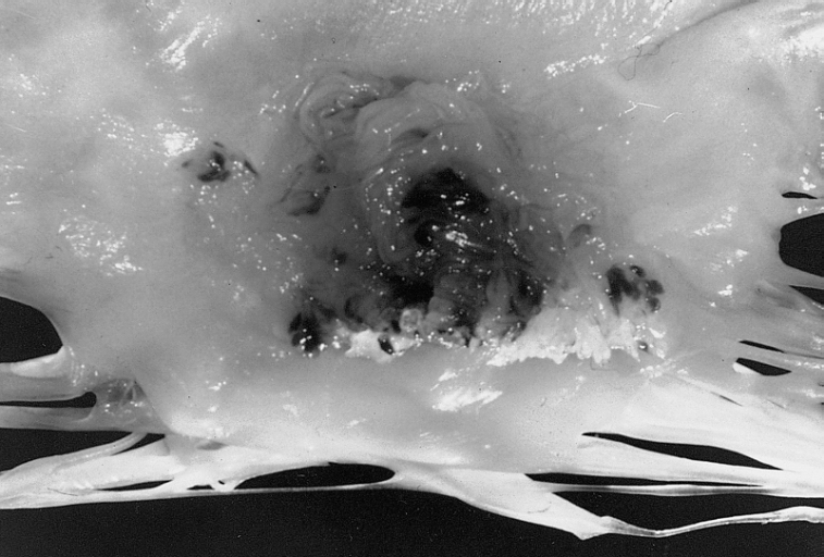 Papillary Fibroelastoma: When located on the mitral valve, these tumors are usually on the anterior leaflet of the atrial surface.


- ↑ 1.0 1.1 PMID 17462431 (PMID 17462431)
Citation will be completed automatically in a few minutes. Jump the queue or expand by hand - ↑ PMID 1636353 (PMID 1636353)
Citation will be completed automatically in a few minutes. Jump the queue or expand by hand