Pulmonary contusion CT
|
Pulmonary contusion Microchapters |
|
Diagnosis |
|---|
|
Treatment |
|
Case Studies |
|
Pulmonary contusion CT On the Web |
|
American Roentgen Ray Society Images of Pulmonary contusion CT |
|
Risk calculators and risk factors for Pulmonary contusion CT |
Editor-In-Chief: C. Michael Gibson, M.S., M.D. [1]
CT
Computed tomography (CT scanning) is a more sensitive test for pulmonary contusion, and it can identify abdominal, chest, or other injuries that accompany the contusion. In one study, chest X-ray detected pulmonary contusions in 16.3% of people with serious blunt trauma, while CT detected them in 31.2% of the same people.[1] Unlike X-ray, CT scanning can detect the contusion almost immediately after the injury. However, in both X-ray and CT a contusion may become more visible over the first 24–48 hours after trauma as bleeding and edema into lung tissues progress.[2] CT scanning also helps determine the size of a contusion, which is useful in determining whether a patient needs mechanical ventilation; a larger volume of contused lung on CT scan is associated with an increased likelihood that ventilation will be needed. CT scans also help differentiate between contusion and pulmonary hematoma, which may be difficult to tell apart otherwise.[3] However, pulmonary contusions that are visible on CT but not chest X-ray are usually not severe enough to affect outcome or treatment.
-
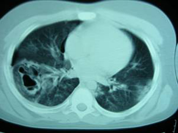 A chest CT scan revealing pulmonary contusions, pneumothorax, and pseudocysts
 -
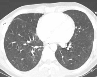 This CT scan, taken 22 days after pulmonary contusion with major chest trauma, shows that the contusion has completely resolved.


(Images shown below courtesy of RadsWiki)
-
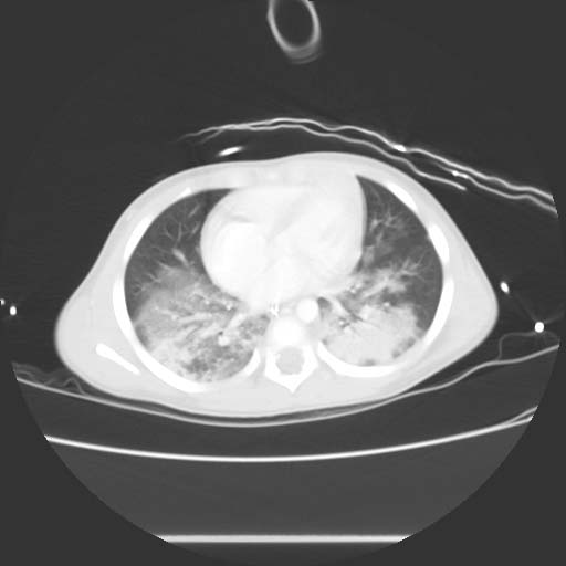 CT image demonstrates bilateral pulmonary contusions
 -
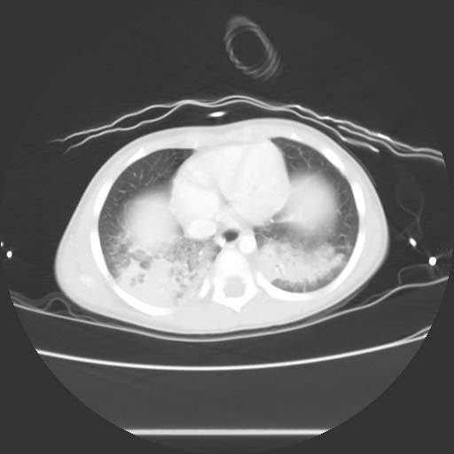 CT image demonstrates bilateral pulmonary contusions


References
- ↑
Keel M, Meier C (2007). "Chest injuries - what is new?". Current Opinion in Critical Care. 13 (6): 674–679. doi:10.1097/MCC.0b013e3282f1fe71. PMID 17975389. Unknown parameter
|month=ignored (help) - ↑
Miller LA (2006). "Chest wall, lung, and pleural space trauma". Radiologic Clinics of North America. 44 (2): 213–224, viii. doi:10.1016/j.rcl.2005.10.006. PMID 16500204. Unknown parameter
|month=ignored (help) - ↑ Grueber GM, Prabhakar G, Shields TW (2005). "Blunt and penetrating injuries of the chest wall, pleura, and lungs". In Shields TW. General Thoracic Surgery. Philadelphia, PA: Lippincott Williams & Wilkins. p. 959. ISBN 0-7817-3889-X.