Pinworm
| style="background:#Template:Taxobox colour;"|Pinworm | ||||||||||||||
|---|---|---|---|---|---|---|---|---|---|---|---|---|---|---|
 Pinworms(U.S.)/Threadworms(U.K.) (Enterobius vermicularis).
| ||||||||||||||
| style="background:#Template:Taxobox colour;" | Scientific classification | ||||||||||||||
| ||||||||||||||
| Species | ||||||||||||||
|
Enterobiasis Microchapters |
|
Diagnosis |
|---|
|
Treatment |
|
Case Studies |
|
Pinworm On the Web |
|
American Roentgen Ray Society Images of Pinworm |
Editor-In-Chief: C. Michael Gibson, M.S., M.D. [1]
Overview
The pinworm (genus Enterobius), also known as threadworm (in the United Kingdom and Australia) or seatworm, is a parasitic worm. It is a nematode (roundworm) and a common intestinal parasite or helminth, especially in humans.[5] The medical condition associated with pinworm infestation is known as enterobiasis[6] (a type of helminthiasis) or less precisely as oxyuriasis in reference to the family Oxyuridae.[7]
Throughout this article, the word "pinworm" refers to Enterobius. In British usage, however, pinworm refers to Strongyloides, while Enterobius is called threadworm.[8]
Classification
The pinworm (genus Enterobius) is a type of roundworm (nematode), and three species of pinworm have been identified with certainty.[9] Humans are hosts only to Enterobius vermicularis (formerly Oxyurias vermicularis).[10] Chimpanzees are host to Enterobius anthropopitheci, which is morphologically distinguishable from the human pinworm.[3] Hugot (1983) claims there is another species affecting humans, Enterobius gregorii, which is supposedly a sister species of E. vermicularis, and has a slightly smaller spicule (i.e., sexual organ).[11] Its existence is controversial however; Totkova et al. (2003) consider there to be insufficient evidence,[4] and Hasegawa et al. (2006) contend that E. gregorii is a younger stage of E. vermicularis.[2][3] Regardless of its status as a distinct species, E. gregorii is considered clinically identical to E. vermicularis.[10]
Morphology

The adult female has a sharply pointed posterior end, is 8 to 13 mm long, and 0.5 mm thick.[12] The adult male is considerably smaller, measuring 2 to 5 mm long and 0.2 mm thick, and has a curved posterior end.[12] The eggs are translucent[12] and have a surface that adheres to objects.[13] The eggs measure 50 to 60 μm by 20 to 30 μm, and have a thick shell flattened on one side.[12] The small size and colourlessness of the eggs make them invisible to the naked eye, except in barely visible clumps of thousands of eggs. Eggs may contain a developing embryo or a fully developed pinworm larva.[12] The larvae grow to 140–150 μm in length.[13]
Distribution
The pinworm has a worldwide distribution,[14] and is the most common helminth (i.e., parasitic worm) infection in the United States, western Europe, and Oceania.[15][16] In the United States, a study by the Center of Disease Control reported an overall incidence rate of 11.4% among people of all ages.[16] Pinworms are particularly common in children, with prevalence rates in this age group having been reported as high as 61% in India, 50% in England, 39% in Thailand, 37% in Sweden, and 29% in Denmark.[16] Finger sucking has been shown to increase both incidence and relapse rates,[16] and nail biting has been similarly associated.[17] Because it spreads from host to host through contamination, pinworms are common among people living in close contact, and tends to occur in all people within a household.[14] The prevalence of pinworms is not associated with gender,[14] nor with any particular social class, race, or culture.[16] Pinworms are an exception to the tenet that intestinal parasites are uncommon in affluent communities.[16] The earliest known instance of the pinworms associated with humans is evidenced by pinworm eggs found in coprolite, carbon dated to 7837 BC at western Utah;[13] however 240 million years ago parasitic pinworm nematodes already infested pre-mammalian cynodonts: a fossilized egg was detected in fossil dung.[18]
Lifecycle
The entire lifecycle, from egg to adult, takes place in the human gastrointestinal tract of a single human host,[12][13] from about 2–4 weeks[19] or about 4–8 weeks.[16]
The lifecycle begins with eggs being ingested.[13] The eggs hatch in the duodenum (i.e., first part of the small intestine).[20] The emerging pinworm larvae grow rapidly to a size of 140 to 150 μm,[19] and migrate through the small intestine towards the colon.[13] During this migration, they moult twice and become adults.[13][16] Females survive for 5 to 13 weeks, and males about 7 weeks.[13] The male and female pinworms mate in the ileum (i.e., last part of the small intestine),[13] whereafter the male pinworms usually die,[20] and are passed out with stool.[21] The gravid female pinworms settle in the ileum, caecum (i.e., beginning of the large intestine), appendix and ascending colon,[13] where they attach themselves to the mucosa[16] and ingest colonic contents.[14]
Almost the entire body of a gravid female becomes filled with eggs.[20] The estimations of the number of eggs in a gravid female pinworm range from about 11,000[13] to 16,000.[16] The egg-laying process begins about five weeks after initial ingestion of pinworm eggs by the human host.[13] The gravid female pinworms migrate through the colon towards the rectum at a rate of 12 to 14 cm per hour.[13] They emerge from the anus, and while moving on the skin near the anus, the female pinworms deposit eggs either through (1) contracting and expelling the eggs, (2) dying and then disintegrating, or (3) bodily rupture due to the host scratching the worm.[20] After depositing the eggs, the female becomes opaque and dies.[21] The reason the female emerges from the anus is to obtain the oxygen necessary for the maturation of the eggs.[21]
Infection
E. vermicularis causes the medical condition enterobiasis, whose primary symptom is itching in the anal area.[22] Albendazole or mebendazole is the first-line treatment of pinworm infection. Pyrantel pamoate is alternative.
Transmission
Pinworms spread through human-to-human transmission, by ingesting (i.e., swallowing) infectious pinworm eggs and/or by anal insertion.[16][20] The eggs are hardy and can remain viable (i.e., infectious) in a moist environment up to three weeks.[16][21] They do not tolerate heat well, but can survive in low temperatures: two-thirds of the eggs are still viable after 18 hours at −8 °C (18 °F).[21]
After the eggs have been initially deposited near the anus, they are readily transmitted to other surfaces through contamination.[20] The surface of the eggs is sticky when laid,[13][21] and the eggs are readily transmitted from their initial deposit near the anus to fingernails, hands, night-clothing and bed linen.[19] From here, eggs are further transmitted to food, water, furniture, toys, bathroom fixtures and other objects.[13][16][20] Household pets often carry the eggs in their fur, while not actually being infected.[23] Dust containing eggs can become airborne and widely dispersed when dislodged from surfaces, for instance when shaking out bed clothes and linen.[16][21][23] Consequently, the eggs can enter the mouth and nose through inhalation, and be swallowed later.[16][19][20][21] Although pinworms do not strictly multiply inside the body of their human host,[19] some of the pinworm larvae may hatch on the anal mucosa, and migrate up the bowel and back into the gastrointestinal tract of the original host[16][19] in a process called retroinfection.[16][21] When this retroinfection occurs, it can lead to a heavy parasitic load and ensures the pinworm infestation continues[16] or can be not clinically significant.[21] Despite the limited, 13-week lifespan of individual pinworms,[13] autoinfection (i.e., infection from the original host to itself), either through the anus-to-mouth route or through retroinfection, usually necessitates repeated treatment, at 2-week intervals, in order to remove the infection completely.[24]
Gallery
-
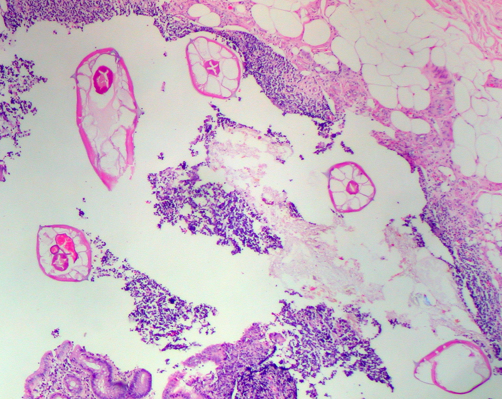 Pinworms are sometimes diagnosed incidentally by pathology. Micrograph of pinworms in the appendix, H&E stain
 -
-
 Egg under a light microscope
 -
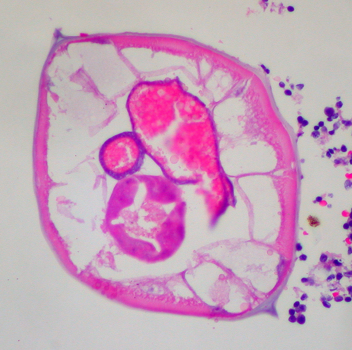
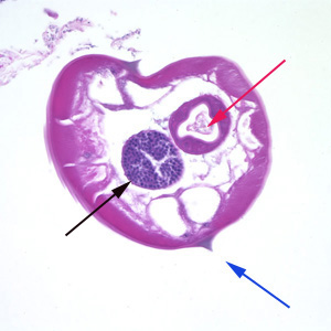 Pinworms are sometimes diagnosed incidentally by pathology: Micrograph of male pinworm in cross section, alae (blue arrow), intestine (red arrow) and testis (black arrow), H&E stain
 -
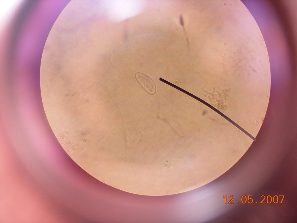
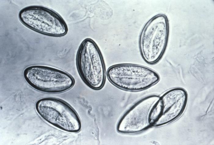 Pinworm eggs are easily seen under a microscope.
 -
This micrograph reveals the cephalic alae in the head region of E. vermicularis.
-
![This image reveals some of the cytoarchitectural features seen in a lymph node specimen that had been extracted from a patient suspected of a Hantavirus illness. From Public Health Image Library (PHIL). [25]](/images/5/59/Pinworm04.jpeg) This image reveals some of the cytoarchitectural features seen in a lymph node specimen that had been extracted from a patient suspected of a Hantavirus illness. From Public Health Image Library (PHIL). [25]
This image reveals some of the cytoarchitectural features seen in a lymph node specimen that had been extracted from a patient suspected of a Hantavirus illness. From Public Health Image Library (PHIL). [25] -
![Photomicrograph reveals some of the ultrastructural details of an Enterobius vermicularis egg, otherwise known as the human pinworm. From Public Health Image Library (PHIL). [25]](/images/d/d6/Pinworm03.jpeg) Photomicrograph reveals some of the ultrastructural details of an Enterobius vermicularis egg, otherwise known as the human pinworm. From Public Health Image Library (PHIL). [25]
Photomicrograph reveals some of the ultrastructural details of an Enterobius vermicularis egg, otherwise known as the human pinworm. From Public Health Image Library (PHIL). [25]
.jpg)
.jpg)


![This image reveals some of the cytoarchitectural features seen in a lymph node specimen that had been extracted from a patient suspected of a Hantavirus illness. From Public Health Image Library (PHIL). [25]](/index.php/File:Pinworm04.jpeg)
![Photomicrograph reveals some of the ultrastructural details of an Enterobius vermicularis egg, otherwise known as the human pinworm. From Public Health Image Library (PHIL). [25]](/index.php/File:Pinworm03.jpeg)
See also
References
- ↑ 1.0 1.1 Hasegawa et al. 2005.
- ↑ 2.0 2.1 Hasegawa et al. 1998
- ↑ 3.0 3.1 3.2 Hasegawa et al. 2006
- ↑ 4.0 4.1 Totkova et al. 2003
- ↑ Encyclopædia Britannica.
- ↑ Merriam-Webster: Enterobiasis
- ↑ Merriam-Webster: Oxyuriasis
- ↑ Vanderkooi 2000, p. B-152 & B-225
- ↑ NCBI taxonomy database 2009
- ↑ 10.0 10.1 dpdx 2009
- ↑ Hugot 1983
- ↑ 12.0 12.1 12.2 12.3 12.4 12.5 Gutiérrez 2005, p. 354.
- ↑ 13.00 13.01 13.02 13.03 13.04 13.05 13.06 13.07 13.08 13.09 13.10 13.11 13.12 13.13 13.14 13.15 Cook 1994, p. 1159
- ↑ 14.0 14.1 14.2 14.3 Gutiérrez 2005, p. 355.
- ↑ http://www.betterhealth.vic.gov.au/bhcv2/bhcarticles.nsf/pages/Worms_pinworms
- ↑ 16.00 16.01 16.02 16.03 16.04 16.05 16.06 16.07 16.08 16.09 16.10 16.11 16.12 16.13 16.14 16.15 16.16 16.17 Burkhart & burkhart 2005, p. 837
- ↑ Cook 1994, p. 1160
- ↑ "Scientists find 240 million-year-old parasite that infected mammals’ ancestor : accessed 8 December 2014.
- ↑ 19.0 19.1 19.2 19.3 19.4 19.5 Cook et al. 2009, p. 1516
- ↑ 20.0 20.1 20.2 20.3 20.4 20.5 20.6 20.7 Garcia 1999, p. 246
- ↑ 21.00 21.01 21.02 21.03 21.04 21.05 21.06 21.07 21.08 21.09 Caldwell 1982, p. 307.
- ↑ "Enterobiasis leads to itching". Retrieved 20 August 2011.
- ↑ 23.0 23.1 Caldwell 1982, p. 308.
- ↑ http://www.webmd.com/children/tc/pinworms-topic-overview?page=2
- ↑ 25.0 25.1 "Public Health Image Library (PHIL)".
- Hasegawa H, Ikeda Y, Fujisaki A; et al. (December 2005). "Morphology of chimpanzee pinworms, Enterobius (Enterobius) anthropopitheci (Gedoelst, 1916) (Nematoda: Oxyuridae), collected from chimpanzees, Pan troglodytes, on Rubondo Island, Tanzania". The Journal of Parasitology. 91 (6): 1314–7. doi:10.1645/GE-569R.1. PMID 16539010.
- "Pinworm". Encyclopædia Britannica. Retrieved 8 April 2009.
- "Enterobiasis". Merriam-Webster's Medical Dictionary. Merriam-Webster. Retrieved 8 April 2009.
- "Oxyuriasis". Merriam-Webster's Medical Dictionary. Merriam-Webster. Retrieved 8 April 2009.
- Totkova A, Klobusicky M, Holkova R, Valent M (2003). "Enterobius gregorii—reality or fiction?" (PDF). Bratislavské Lekárske Listy. 104 (3): 130–3. PMID 12940699.
- "Enterobius". NCBI taxonomy database. National Center for Biotechnology Information, U.S. National Library of Medicine. 2009. Retrieved 8 April 2009.
- "Enterobiasis". DPDx. Division of Parasitic Diseases, Centers for Disease Control and Prevention. Retrieved 8 April 2009.
- Nakano T, Okamoto M, Ikeda Y, Hasegawa H (December 2006). "Mitochondrial cytochrome c oxidase subunit 1 gene and nuclear rDNA regions of Enterobius vermicularis parasitic in captive chimpanzees with special reference to its relationship with pinworms in humans". Parasitology Research. 100 (1): 51–7. doi:10.1007/s00436-006-0238-4. PMID 16788831.
- Hugot JP (1983). "[Enterobius gregorii (Oxyuridae, Nematoda), a new human parasite]". Annales de Parasitologie Humaine et Comparée (in French). 58 (4): 403–4. PMID 6416131.
- Hasegawa H, Takao Y, Nakao M, Fukuma T, Tsuruta O, Ide K (February 1998). "Is Enterobius gregorii Hugot, 1983 (Nematoda: Oxyuridae) a distinct species?". The Journal of Parasitology. 84 (1): 131–4. doi:10.2307/3284542. PMID 9488350.
- Gutiérrez, Yezid (2000). Diagnostic pathology of parasitic infections with clinical correlations (PDF) (Second ed.). Oxford University Press. pp. 354–366. ISBN 0-19-512143-0. Retrieved 21 August 2009.
- Cook, Gordon C; Zumla, Alimuddin I (2009). Manson's tropical diseases (22nd ed.). Saunders Elsevier. pp. 1515–1519. ISBN 978-1-4160-4470-3. Retrieved 18 November 2009.
- "B80: Enterobiasis". International Statistical Classification of Diseases and Related Health Problems (ICD) 10th Revision. World Health Organization. 2007. Retrieved 5 December 2009.
- Cook GC (September 1994). "Enterobius vermicularis infection". Gut. 35 (9): 1159–62. doi:10.1136/gut.35.9.1159. PMC 1375686. PMID 7959218.
- Garcia, Lynne Shore (2009). Practical guide to diagnostic parasitology. American Society for Microbiology. pp. 246–247. ISBN 1-55581-154-X. Retrieved 5 December 2009.
- Burkhart CN, Burkhart CG (October 2005). "Assessment of frequency, transmission, and genitourinary complications of enterobiasis (pinworms)". International Journal of Dermatology. 44 (10): 837–40. doi:10.1111/j.1365-4632.2004.02332.x. PMID 16207185.
- Caldwell JP (February 1982). "Pinworms (Enterobius Vermicularis)". Canadian Family Physician. 28: 306–9. PMC 2306321. PMID 21286054.
- Vanderkooi M (2000). Village Medical Manual (5th ed.).