Patella
|
WikiDoc Resources for Patella |
|
Articles |
|---|
|
Most recent articles on Patella |
|
Media |
|
Evidence Based Medicine |
|
Clinical Trials |
|
Ongoing Trials on Patella at Clinical Trials.gov Clinical Trials on Patella at Google
|
|
Guidelines / Policies / Govt |
|
US National Guidelines Clearinghouse on Patella
|
|
Books |
|
News |
|
Commentary |
|
Definitions |
|
Patient Resources / Community |
|
Directions to Hospitals Treating Patella Risk calculators and risk factors for Patella
|
|
Healthcare Provider Resources |
|
Causes & Risk Factors for Patella |
|
Continuing Medical Education (CME) |
|
International |
|
|
|
Business |
|
Experimental / Informatics |
Editor-In-Chief: C. Michael Gibson, M.S., M.D. [1]
Overview
The patella or kneecap is a thick, triangular bone which articulates with the femur and covers and protects the front of the knee joint. It is the largest sesamoid bone in the human body. It is attached to the tendon of the quadriceps femoris muscle, which contracts to straighten the leg. The vastus intermedialis muscle is attached to the base of patella. The vastus lateralis and vastus medialis are attached to lateral and medial borders of patella respectively.
The patella is stabilised by the insertion of vastus medialis and the prominence of the anterior femoral condyles, which prevent lateral dislocation during flexion. The retinacular fibres of the patella also stabilise it during exercise.
The primary functional role of the patella is knee extension. The patella increases the leverage that the tendon can exert on the femur by increasing the angle at which it acts.
The patella ossifies between the ages 2-6 years. In some people it may be absent congenitally or hypoplastic. In 2% of the population there is a bipartite patella, which is usually asymptomatic.
Regarding non-human animals, the patella is fully developed only in placental mammals; marsupials have only rudimentary, non-ossified patellae.[1]
See also
Additional images
-
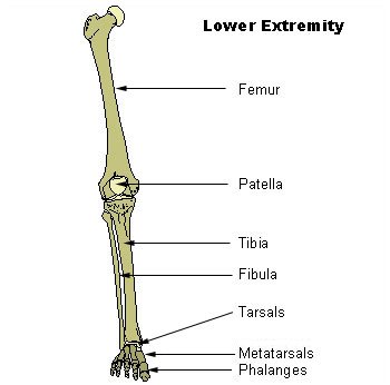 Lower extremity
 -
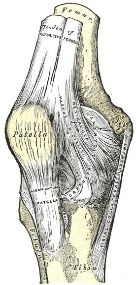 Right knee-joint. Anterior view.
 -
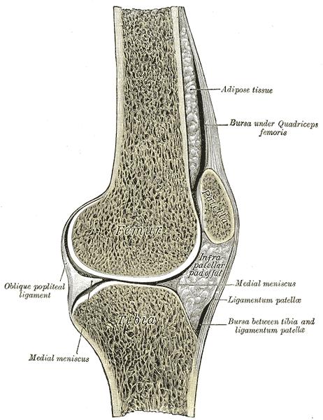 Sagittal section of right knee-joint.
 -
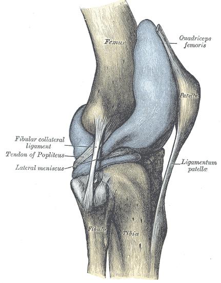 Capsule of right knee-joint (distended). Lateral aspect.
 -
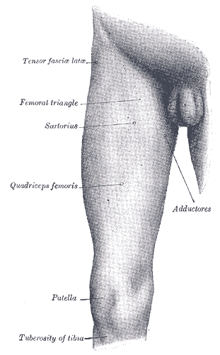 Front and medial aspect of right thigh.
 -
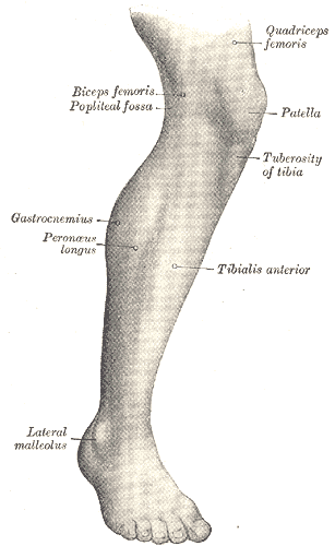 Lateral aspect of right leg.


References
- ↑ Herzmark MH (1938). "The Evolution of the Knee Joint" (PDF). J Bone Joint Surg Am. 20: 77–84. Retrieved 2007-11-17.