Osteomyelitis imaging findings
Jump to navigation
Jump to search
|
Osteomyelitis Microchapters |
|
Diagnosis |
|---|
|
Treatment |
|
Case Studies |
|
Osteomyelitis imaging findings On the Web |
|
American Roentgen Ray Society Images of Osteomyelitis imaging findings |
|
Risk calculators and risk factors for Osteomyelitis imaging findings |
Editor-In-Chief: C. Michael Gibson, M.S., M.D. [1]
Imaging Findings
Diagnosis of osteomyelitis is often based on radiologic results showing a lytic center with a ring of sclerosis, though bone cultures are normally required to identify the specific pathogen.
- Conventional radiographic evaluation of acute osteomyelitis is insufficient because bone changes are not evident for 14–21 days after the onset of infection.
- Although MR imaging is the accepted modality of choice for the early detection and surgical localization of osteomyelitis, in the emergency department, CT is usually more readily available for establishing the diagnosis. [1]
- At CT, features of bacterial osteomyelitis include overlying soft-tissue swelling, periosteal reaction, medullary low-attenuation areas or trabecular coarsening, and focal cortical erosions.
MRI
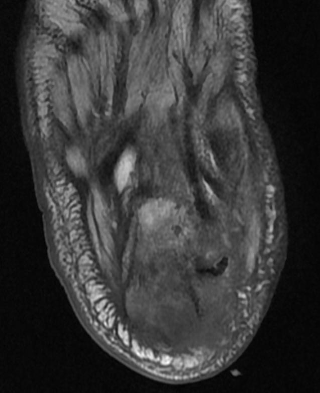
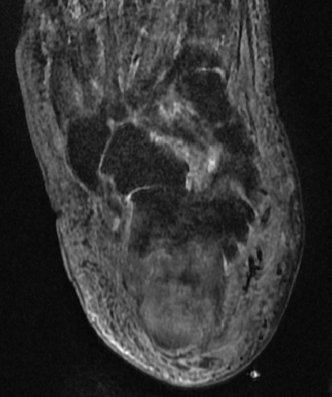
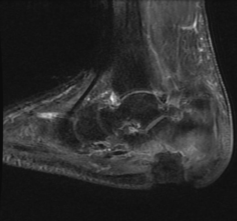
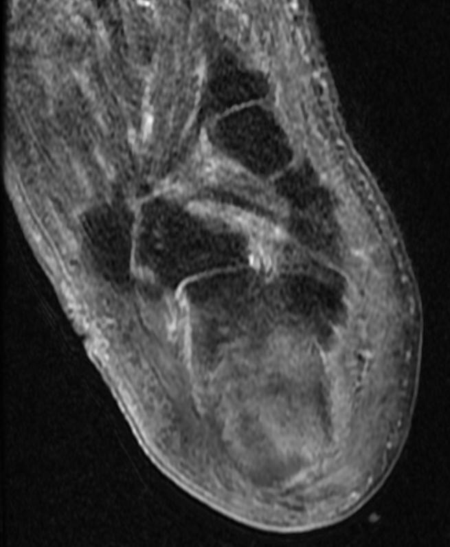
Patient #1 Extensive calcaneal osteomyelitis. Note soft tissue ulceration and cellulitis
-
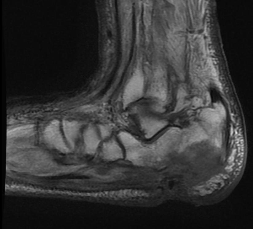 T1
T1 -
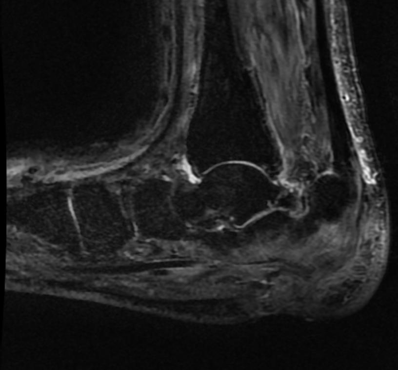 STIR
STIR -
 T1
T1 -
 STIR
STIR -
 T1 fat sat contrast
 -
 T1 fat sat contrast


Bone Scan
Patient #2
-
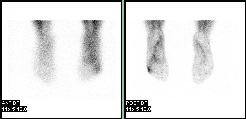 Blood pool
 -
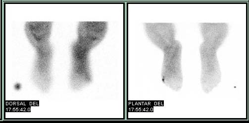 Delayed


{{#ev:youtube|X2ShDUfeso0}}
References
- ↑ Laura M. Fayad, John A. Carrino, and Elliot K. Fishman. Musculoskeletal Infection: Role of CT in the Emergency Department. RadioGraphics 2007 27: 1723-1736.