Osteochondroma ultrasound
Jump to navigation
Jump to search
|
Osteochondroma Microchapters |
|
Diagnosis |
|---|
|
Treatment |
|
Case Studies |
|
Osteochondroma ultrasound On the Web |
|
American Roentgen Ray Society Images of Osteochondroma ultrasound |
|
Risk calculators and risk factors for Osteochondroma ultrasound |
Editor-In-Chief: C. Michael Gibson, M.S., M.D. [1]Associate Editor(s)-in-Chief: Maria Fernanda Villarreal, M.D. [2]
Overview
On ultrasound, the osteochondroma cartilage cap is visualized accurately as a hypoechoic region bounded by bone.[1]
Ultrasound
- Ultrasound findings associated with osteochondroma, include:[1][2]
Gallery
-
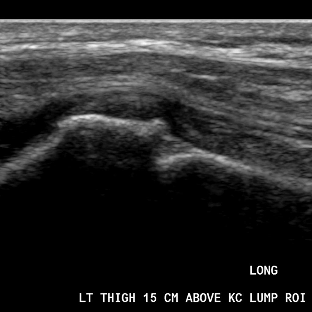 Image shows a hypoechoic layer that corresponds with the osteochondroma cartilage cap.

References
- ↑ 1.0 1.1 Osteochondroma. Dr Yuranga Weerakkody. Radiopedia. http://radiopaedia.org/articles/osteochondroma Accessed on January 28, 2016
- ↑ Malghem J, Vande Berg B, Noël H, Maldague B (1992). "Benign osteochondromas and exostotic chondrosarcomas: evaluation of cartilage cap thickness by ultrasound". Skeletal Radiology. 21 (1): 33–7. PMID 1546334.