Japanese encephalitis pathophysiology
|
Japanese encephalitis Microchapters |
|
Diagnosis |
|---|
|
Treatment |
|
Case Studies |
|
Japanese encephalitis pathophysiology On the Web |
|
American Roentgen Ray Society Images of Japanese encephalitis pathophysiology |
|
Risk calculators and risk factors for Japanese encephalitis pathophysiology |
Editor-In-Chief: C. Michael Gibson, M.S., M.D. [1]; Associate Editor(s)-in-Chief: Anthony Gallo, B.S. [2]
Overview
Japanese encephalitis virus is usually transmitted via mosquitos to the human host. Japanese encephalitis virus contains positive-sense viral RNA; this RNA has its genome directly utilized as if it were mRNA, producing a single protein which is modified by host and viral proteins to form the various proteins needed for replication. Transmission to humans requires mosquito species capable of creating a "bridge" between infected animals and uninfected humans; this occurs when humans become part of the enzootic cycle. The incubation period is 5-15 days. Humans are dead-end hosts for the virus, meaning there is an insufficient amount of Japanese encephalitis virus in the blood stream to infect a mosquito; there is also no evidence of person to person spread.
Pathophysiology
Japanese encephalitis virus is usually transmitted via mosquitos to the human host. Japanese encephalitis virus contains positive-sense viral RNA; this RNA has its genome directly utilized as if it were mRNA, producing a single protein which is modified by host and viral proteins to form the various proteins needed for replication. The following table is a summary of the Japanese encephalitis virus:[1][2]
| Characteristic | Data |
|---|---|
| Nucleic acid | RNA |
| Sense | ssRNA(+) |
| Virion | Enveloped |
| Capsid | Spherical |
| Symmetry | Yes; T=3-like organization; icosahedral-like |
| Capsid monomers | Unknown |
| Envelope length (diameter) | 50 nm |
| Additional envelope information | Mature virons contain 2 virus-encoded membrane proteins (M and E); immature virons contain a protein precursor |
| Genome shape | Linear |
| Genome length | 10-11 kb |
| Nucleotide cap | Yes |
| Polyadenylated tail | No; a loop structure is formed instead |
| Incubation period | 5-15 days |
Japanese encephalitis is contracted by the bite of an infected mosquito, primarily Culex tritaeniorhynchus. Japanese encephalitis virus circulates between a mosquito vector and either pigs or water birds (such as herons) in Southeast Asia.[3] Transmission to humans requires mosquito species capable of creating a "bridge" between infected animals and uninfected humans; this occurs when humans become part of the enzootic cycle. The incubation period is 5-15 days.[4] Humans are dead-end hosts for the virus, meaning there is an insufficient amount of Japanese encephalitis virus in the blood stream to infect a mosquito; there is also no evidence of person to person spread.[5]
Japanese encephalitis virus is transmitted in the following pattern:[1]
- Attachment of the viral envelope protein E to host receptors mediates internalization into the host cell by clathrin-mediated endocytosis, or by apoptotic mimicry.
- Fusion of virus membrane with host endosomal membrane. RNA genome is released into the cytoplasm.
- The positive-sense ssRNA virus is translated into a polyprotein, which is cleaved into all structural and non-structural proteins necessary for RNA synthesis (replication and transcription).
- Replication takes place at the surface of endoplasmic reticulum in cytoplasmic viral factories. A dsRNA genome is synthesized from the genomic ssRNA(+).
- The dsRNA genome is transcribed/replicated, thereby providing viral mRNAs/new ssRNA(+) genomes.
- Virus assembly occurs at the endoplasmic reticulum. The virion buds via the host endosomal sorting complexes required for transport (ESCRT), and is sent to the Golgi apparatus.
- The prM protein is cleaved in the Golgi, thereby maturing the virion which is fusion competent.
- New virions are released by exocytosis.
On microscopic histopathological analysis, the enveloped, spherical, and icosahedral-like virion shape are characteristic findings of Japanese encephalitis.
Gallery
-
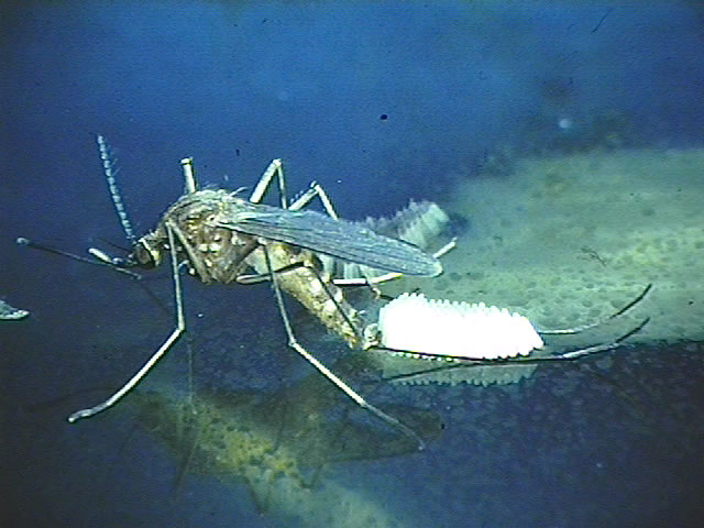 Culex mosquito laying eggs. (Photograph by Richard G. Weber)
 -
![Diagram illustrates the methods by which the arbovirus, Japanese encephalitis virus (JEV) reproduces and amplifies itself in the avian populations, and is subsequently transmitted to human beings as the dead end host. From Public Health Image Library (PHIL). [6]](/images/6/6a/Flavivirus02.jpeg) Diagram illustrates the methods by which the arbovirus, Japanese encephalitis virus (JEV) reproduces and amplifies itself in the avian populations, and is subsequently transmitted to human beings as the dead end host. From Public Health Image Library (PHIL). [6]
Diagram illustrates the methods by which the arbovirus, Japanese encephalitis virus (JEV) reproduces and amplifies itself in the avian populations, and is subsequently transmitted to human beings as the dead end host. From Public Health Image Library (PHIL). [6]

![Diagram illustrates the methods by which the arbovirus, Japanese encephalitis virus (JEV) reproduces and amplifies itself in the avian populations, and is subsequently transmitted to human beings as the dead end host. From Public Health Image Library (PHIL). [6]](/index.php/File:Flavivirus02.jpeg)
References
- ↑ 1.0 1.1 Flavivirus. SIB Swiss Institute of Bioinformatics. (2015) http://viralzone.expasy.org/viralzone/all_by_species/24.html Accessed on April 12, 2016
- ↑ Japanese encephalitis - Frequently Asked Questions. CDC Centers for Disease Control and Prevention. (2015) http://www.cdc.gov/japaneseencephalitis/qa/index.html Accessed on April 12, 2016
- ↑ Japanese encephalitis. National Health Service United Kingdom (2016). http://www.nhs.uk/Conditions/Japanese-encephalitis/Pages/Introduction.aspx Accessed on April 12, 2016.
- ↑ Japanese Encephalitis Symptoms and Treatment. Centers for Disease Control and Prevention (CDC), National Center for Emerging and Zoonotic Infectious Diseases, Division of Vector-Borne Diseases. (2015) http://www.cdc.gov/japaneseencephalitis/symptoms/ Accessed on April 12, 2016.
- ↑ Japanese encephalitis - Fact sheet No 386. World Health Organization (WHO) (2015) http://www.who.int/mediacentre/factsheets/fs386/en/ Accessed on April 12, 2016
- ↑ "Public Health Image Library (PHIL)".