File:Thin Basement Membrane Disease.jpg
Jump to navigation
Jump to search
No higher resolution available.
Thin_Basement_Membrane_Disease.jpg (685 × 523 pixels, file size: 121 KB, MIME type: image/jpeg)
Summary
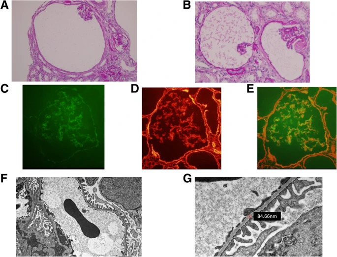
1(A) and 1(B) showing light microscopy of Thin basement membrane disease with cystic dilation. 1(C), 1(D), 1(E) showing GBM using different Immunofluoroscence 1(F), 1(G) showing thin GBM without electron deposition using Electron microscopy Courtesy: https://bmcnephrol.biomedcentral.com/articles/10.1186/s12882-019-1451-6/figures/2
File history
Click on a date/time to view the file as it appeared at that time.
| Date/Time | Thumbnail | Dimensions | User | Comment | |
|---|---|---|---|---|---|
| current | 06:56, 20 October 2020 | | 685 × 523 (121 KB) | Marufa Marium (talk | contribs) | 1(A) and 1(B) showing light microscopy of Thin basement membrane disease with cystic dilation. 1(C), 1(D), 1(E) showing GBM using different Immunofluoroscence 1(F), 1(G) showing thin GBM without electron deposition using Electron microscope Courtesy: https://bmcnephrol.biomedcentral.com/articles/10.1186/s12882-019-1451-6/figures/2 |
You cannot overwrite this file.
File usage
The following 3 pages use this file: