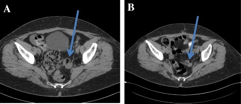
File:CT image of ovarian granulosa cell tumor.jpg
CT_image_of_ovarian_granulosa_cell_tumor.jpg (473 × 205 pixels, file size: 29 KB, MIME type: image/jpeg)
Summary
Patient 1 Computer tomography images of recurrent ovarian granulosa cell tumour. a The progression of tumour (local tumour recurrence (blue arrow), diffuse peritoneal carcinosis with ascites and inguinal pathological lymph node), and b the regression of local recurrence tumour (blue arrow),Bufa A, Farkas N, Preisz Z, et al. Diagnostic relevance of urinary steroid profiles on ovarian granulosa cell tumors: two case reports. J Med Case Rep. 2017;11(1):166. Published 2017 Jun 22. doi:10.1186/s13256-017-1324-1,https://www.ncbi.nlm.nih.gov/pmc/articles/PMC5480180/
File history
Click on a date/time to view the file as it appeared at that time.
| Date/Time | Thumbnail | Dimensions | User | Comment | |
|---|---|---|---|---|---|
| current | 19:39, 6 May 2019 |  | 473 × 205 (29 KB) | Maneesha Nandimandalam (talk | contribs) | Patient 1 Computer tomography images of recurrent ovarian granulosa cell tumour. a The progression of tumour (local tumour recurrence (blue arrow), diffuse peritoneal carcinosis with ascites and inguinal pathological lymph node), and b the regression o... |
You cannot overwrite this file.
File usage
The following page uses this file: