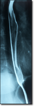
Esophagogram
Overview
|
WikiDoc Resources for Esophagogram |
|
Articles |
|---|
|
Most recent articles on Esophagogram Most cited articles on Esophagogram |
|
Media |
|
Powerpoint slides on Esophagogram |
|
Evidence Based Medicine |
|
Clinical Trials |
|
Ongoing Trials on Esophagogram at Clinical Trials.gov Clinical Trials on Esophagogram at Google
|
|
Guidelines / Policies / Govt |
|
US National Guidelines Clearinghouse on Esophagogram
|
|
Books |
|
News |
|
Commentary |
|
Definitions |
|
Patient Resources / Community |
|
Patient resources on Esophagogram Discussion groups on Esophagogram Patient Handouts on Esophagogram Directions to Hospitals Treating Esophagogram Risk calculators and risk factors for Esophagogram
|
|
Healthcare Provider Resources |
|
Causes & Risk Factors for Esophagogram |
|
Continuing Medical Education (CME) |
|
International |
|
|
|
Business |
|
Experimental / Informatics |

A barium swallow is a medical imaging procedure used to examine the upper GI (gastrointestinal) tract, which includes the oesophagus and, to a lesser extent, the stomach.
Principle
Barium sulphate is a type of Contrast medium that is visible to x-rays. As the patient swallows the Barium suspension, it coats the oesphagus with a thin layer of the barium. This enables the hollow structure to be imaged.
Examination
The patient is asked to drink a suspension of barium sulfate. Fluoroscopy images are taken as the barium is swallowed. This is typically at a rate of 2 or 3 frames pers second. The patient is asked to swallow the Barium a number of times, whilst standing in different positions, i.e. AP, oblique and lateral, to assess the 3D structure as best possible.
Pathology
Pathologies detected on a Barium Swallow include:
- Achalasia
- Oesophageal pouch
- Cancer of oesophagus
- Tracheoesophageal fistula
- Schatzki ring
- Reflux
- Zenker's diverticulum
- Hiatus hernia