Ebstein's anomaly of the tricuspid valve associated abnormalities
|
Ebsteins anomaly of the tricuspid valve Microchapters | |
|
Diagnosis | |
|---|---|
|
Treatment | |
|
Case Studies | |
|
Ebstein's anomaly of the tricuspid valve associated abnormalities On the Web | |
|
American Roentgen Ray Society Images of Ebstein's anomaly of the tricuspid valve associated abnormalities | |
|
FDA on Ebstein's anomaly of the tricuspid valve associated abnormalities | |
|
CDC on Ebstein's anomaly of the tricuspid valve associated abnormalities | |
|
Ebstein's anomaly of the tricuspid valve associated abnormalities in the news | |
|
Blogs on Ebstein's anomaly of the tricuspid valve associated abnormalities | |
Editor-In-Chief: C. Michael Gibson, M.S., M.D. [1] and Claudia P. Hochberg, M.D. [2]
Associate Editor-In-Chief: Cafer Zorkun, M.D., Ph.D. [3]
Associated Abnormalities
While Ebstein's anomaly is defined as the congenital displacement of the tricuspid valve towards the apex of the right ventricle, it is often associated with other abnormalities.
Anatomy
Typically, there are anatomic abnormalities of the tricuspid valve, with enlargement of the anterosuperior leaflet of the valve which is often adherent to the right ventricular free wall.
About 50% of individuals with Ebstein's anomaly have an associated shunt between the right and left atriums, either an atrial septal defect ASD or a patent foramen ovale PFO.
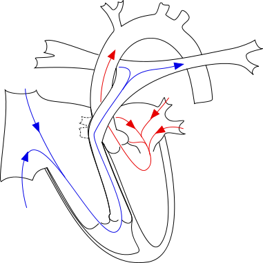
- The tricuspid valve leaflets are displaced toward the RV apex.
- The RV cavity is dilated and the free wall is thin.
- May be associated with LV dysfunction and mitral valve prolapse (MVP).
- It has also been associated with pulmonic stenosis (PS), VSD, pulmonary hypertension, bicuspid aortic valve and right sided aortic arch.
-It is commonly associated with congenitally corrected transposition of the great vessels and occasionally with tetralogy of Fallot.
- Is often associated with one or more accessory conduction pathways in 25%, and WPW in 13%. Usually this is a right sided accessory pathway.
- The presence of an ASD permits right-to-left shunting and cyanosis. The degree of cyanosis is related to the degree of tricuspid regurgitation (TR), tricuspid stenosis (TS) and the compliance of the RV.
-
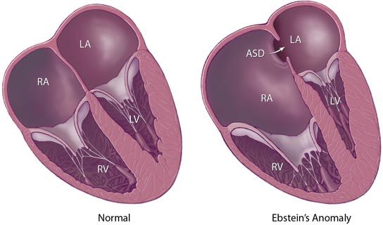 Graphical represntation of Ebstein's Anomaly from the Mayo Clinic website (note there is also an ASD on this diagram)
 -
 Ebstein Anomaly


References