Deep vein thrombosis MRI
| Resident Survival Guide |
Editor(s)-In-Chief: The APEX Trial Investigators, C. Michael Gibson, M.S., M.D. [1]; Associate Editor(s)-In-Chief: Cafer Zorkun, M.D., Ph.D. [2] ; Kashish Goel, M.D.; Assistant Editor(s)-In-Chief: Justine Cadet
|
Deep Vein Thrombosis Microchapters |
|
Diagnosis |
|---|
|
Treatment |
|
Special Scenario |
|
Trials |
|
Case Studies |
|
Deep vein thrombosis MRI On the Web |
|
Risk calculators and risk factors for Deep vein thrombosis MRI |
Overview
MRI can be used for the diagnosis of deep vein thrombosis (DVT); however, it is not a routine test in DVT because of cost and inaccessibility. MRI can be used in MR venography or in direct thrombus imaging.[1] MRI may be performed to diagnose DVT if compression ultrasound is not feasible because of the presence of a cast or excessive swelling, or when the the ultrasound results are non-diagnostic.
MRI
MRI can be used in multiple ways:[1]
- MR venography without contrast
- MR venography with IV gadolinium contrast
- Direct thrombus imaging (visualizing thrombus against a suppressed background)
In a double blinded study involving 85 patients suspected of having DVT, magnetic resonance venography (MRV) had a 100% sensitivity and 96% specificity, as compared with contrast venography.[2] In this study, DVT was documented by contrast venography in 27 (27%) venous systems. Results of MRV and contrast venography were identical in 98 (97%) of 101 venous systems, whereas results of duplex scanning and contrast venography were identical in 40 (98%) of 41 venous systems. All DVTs identified by contrast venography were detected by MRV and duplex scanning. Thus it was concluded that MRV is an accurate noninvasive venographic technique for the detection of DVT.
Uses
- MRI may be performed to diagnose DVT if compression ultrasound is not feasible because of the presence of a cast or excessive swelling, or when the the ultrasound results are non-diagnostic.[1]
- MRI can be used when contrast venography cannot be performed due to a patient being allergic to iodinated contrast or if the patient is in acute renal insufficiency.
Examples
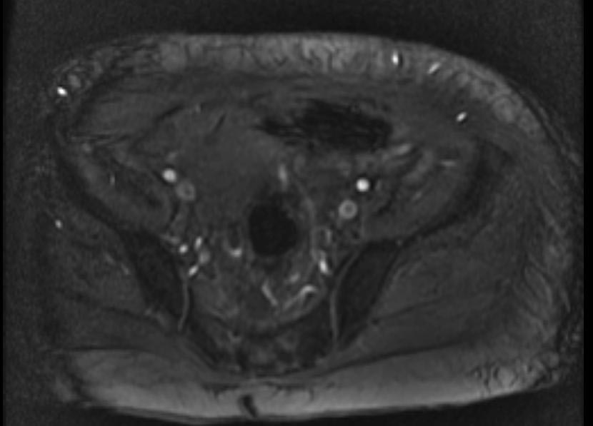
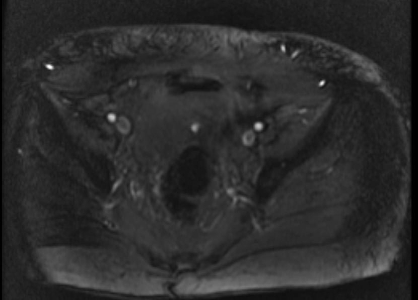
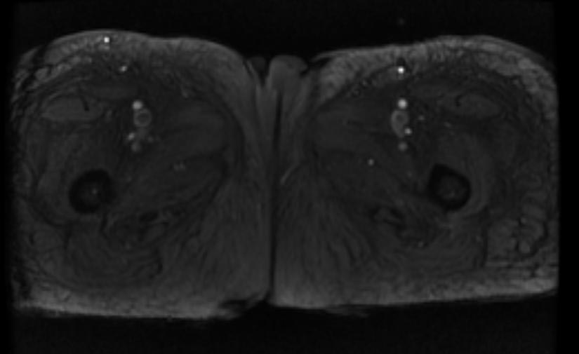
- 2D TOF GRE MRV images: Bilateral deep vein thromboses
References
- ↑ 1.0 1.1 1.2 Kearon C, Akl EA, Comerota AJ, Prandoni P, Bounameaux H, Goldhaber SZ; et al. (2012). "Antithrombotic therapy for VTE disease: Antithrombotic Therapy and Prevention of Thrombosis, 9th ed: American College of Chest Physicians Evidence-Based Clinical Practice Guidelines". Chest. 141 (2 Suppl): e419S–94S. doi:10.1378/chest.11-2301. PMC 3278049. PMID 22315268.
- ↑ Carpenter JP, Holland GA, Baum RA, Owen RS, Carpenter JT, Cope C (1993). "Magnetic resonance venography for the detection of deep venous thrombosis: comparison with contrast venography and duplex Doppler ultrasonography". J Vasc Surg. 18 (5): 734–41. PMID 8230557.


