Corrective
|
Transposition of the great vessels Microchapters |
|
Classification |
|---|
|
Differentiating Transposition of the great vessels from other Diseases |
|
Diagnosis |
|
Treatment |
|
Surgery |
|
Case Studies |
|
Corrective On the Web |
|
American Roentgen Ray Society Images of Corrective |
Editor-In-Chief: C. Michael Gibson, M.S., M.D. [1]
Associate Editors-In-Chief: Keri Shafer, M.D. [2]; Atif Mohammad, M.D., Priyamvada Singh, MBBS
Corrective
Arterial switch

The Jatene procedure surgery is the preferred, and most frequently used, method of correcting d-TGA; ideally, it is performed on an infant between 8-14 days old.
The heart and vessels are accessed via median sternotomy, and a cardiopulmonary bypass machine is used; as this machine needs its "circulation" to be filled with blood, a child will require a blood transfusion for this surgery. The procedure involves transecting both the aorta and pulmonary artery; the coronary arteries are then detached from the aorta and reattached to the neo-aorta, before "swapping" the upper portion of the aorta and pulmonary artery to the opposite arterial root. Including the anaesthesia and immediate post operative recovery, this surgery takes an average of approximately six to eight hours to complete.
Some arterial switch recipients may present with post-operative pulmonary stenosis, which would then be repaired with angioplasty, pulmonary stenting via heart cath or median sternotomy, and/or xenograft.

Atrial switch
In some cases, it is not possible to perform an arterial switch, either because of late diagnosis, sepsis, or a contraindicative coronary artery pattern. In the case of sepsis or late diagnosis, a delayed Arterial Switch can sometimes be made possible by PAB, which may also require a concomitant construction of an aortic-to-pulmonary artery shunt.
When an arterial switch is impossible, an atrial switch will be attempted using either the Senning or Mustard procedure. Both methods involve creating a baffle to redirect red and blue blood flow to the appropriate artery. Since the late 1970s the Mustard procedure has been preferred.
Post-operative
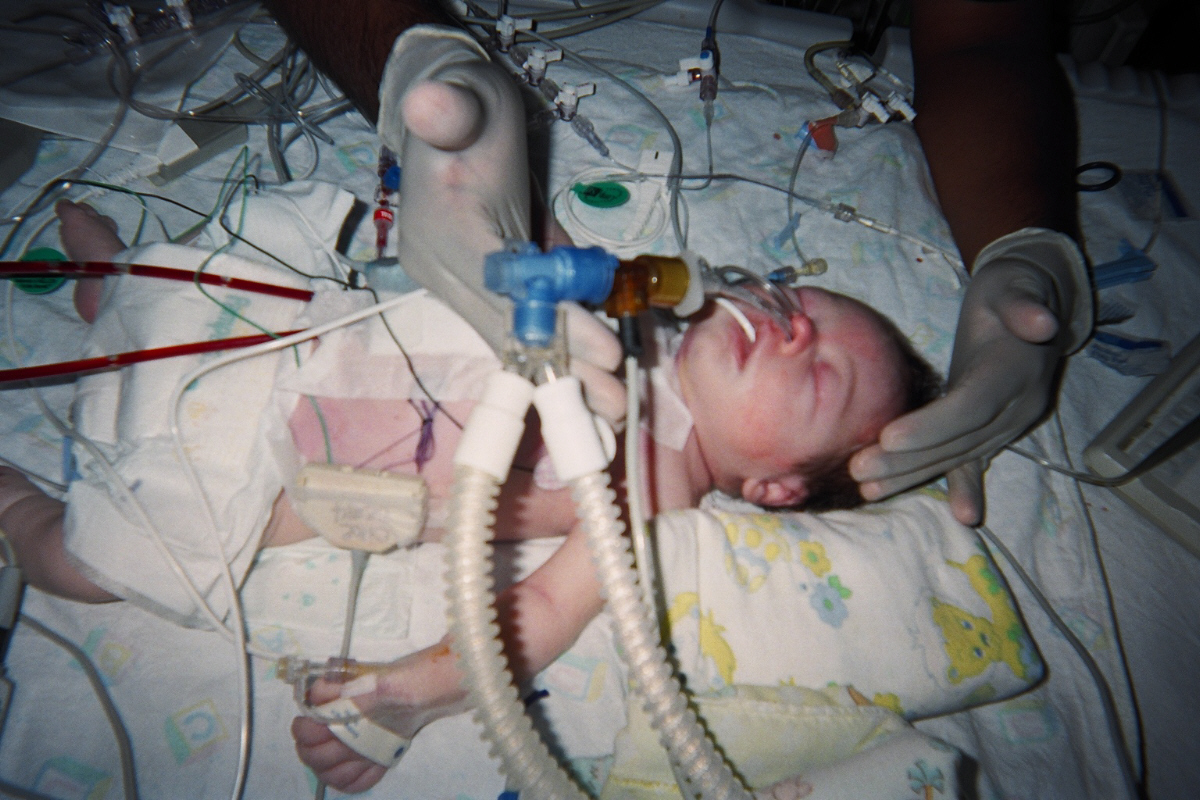
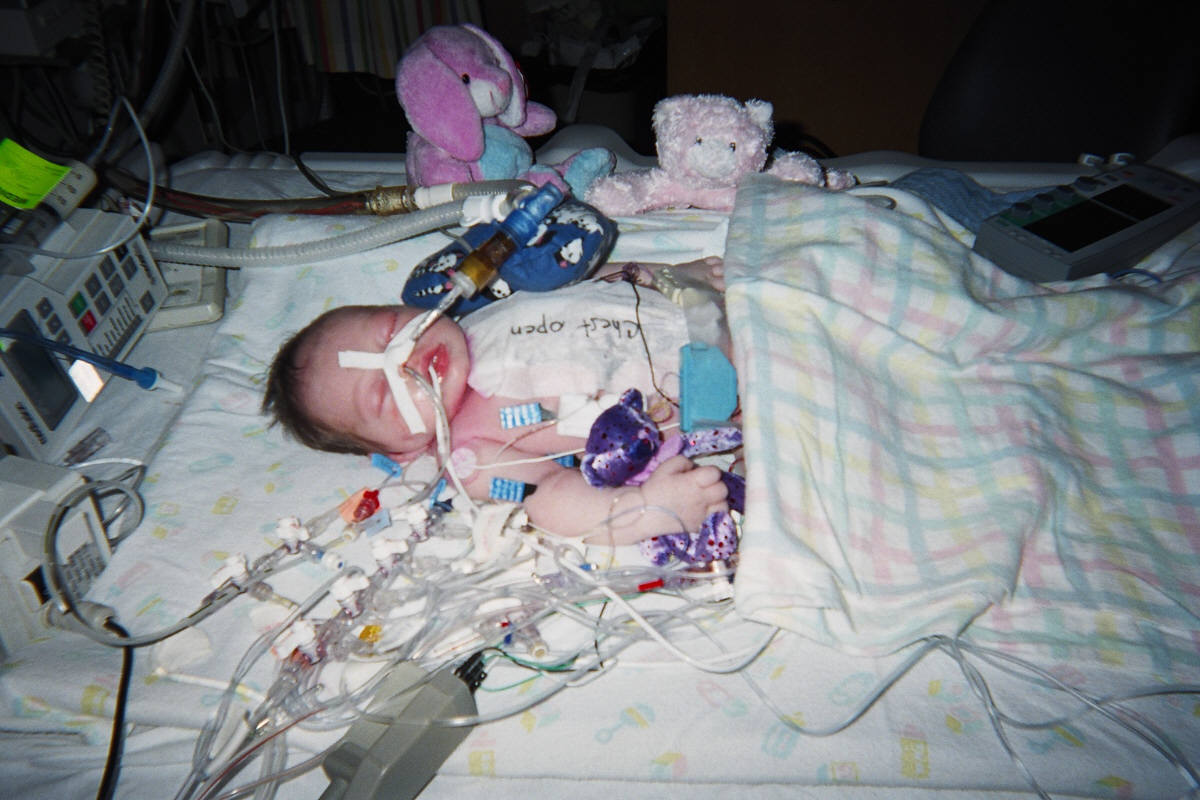
Following corrective surgery but prior to cessation of anaesthesia, two small incisions are made immediately below the sternotomy incision which provide exit points for chest tubes used to drain fluid from the thoracic cavity, with one tube placed at the front and another at the rear of the heart.
The patient returns to the ICU post-operatively for recovery, maintenance, and close observation; recovery time may vary, but tends to average approximately two weeks, after which the patient may be transferred to a Transitional Care Unit (TCU), and eventually to a cardiac ward.
Post-operative care is very similar to the palliative care received, with the exception that the patient no longer requires PGE or the surgical palliation procedures. Additionally, the patient is kept on a cooling blanket for a period of time to prevent fever, which could cause brain damage. The sternum is not closed immediately which allows extra space in the thoracic cavity, preventing excess pressure on the heart, which swells considerably following the surgery; the sternum and incision are closed after a few days, when swelling is sufficiently reduced.