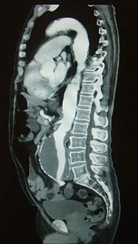
Computed tomography angiography

Editor-In-Chief: C. Michael Gibson, M.S., M.D. [1]
Overview
Computed tomography angiography (CTA), is an examination that uses x-rays to visualize blood flow in arterial and venous vessels throughout the body. This ranges from arteries serving the brain to those bringing blood to the lungs, kidneys, arms and legs.
Technique
CTA combines the use of x-rays with computerized analysis of the images. Beams of x-rays are passed from a rotating device through the area of interest in the patient's body from several different angles to create cross-sectional images, which then are assembled by computer into a three-dimensional picture of the area being studied. The scan is performed simultaneously with a high-speed contrast media injection using a technique called bolus tracking. Compared to catheter angiography, which involves placing a sizable catheter and injecting contrast material into a large artery or vein, CTA is a much less invasive and more patient-friendly procedure. The contrast material is injected into a small peripheral vein by using a small needle or cannula. This type of exam has been used to screen large numbers of individuals for arterial disease.
Most patients undergo CT angiography without being admitted to a hospital.
4D CT
4D CT digital subtracted angiogram has the ability to see whole brain dynamic flow of contrast from arterial to venous.
Clinical Applications
CTA is commonly used to:
- Examine the pulmonary arteries in the lungs to rule out pulmonary embolism, a serious but treatable condition. This is called a CTPA.
- Visualize blood flow in the renal arteries in patients with hypertension and those suspected of having kidney disorders. Stenosis of a renal artery is a correctable cause of hypertension in some patients. A special computerized method of viewing the images makes renal CT angiography a very accurate examination. Also done in prospective kidney donors.
- Identify aneurysms in the aorta or in other major blood vessels. Aneurysms are diseased areas of a weakened blood vessel wall that bulges out — like a bulge in a tire. Aneurysms are life-threatening because they can rupture.
- Identify dissection in the aorta or its major branches. Dissection means that the layers of the artery wall peel away from each other — like the layers of an onion. Dissection can cause pain and can be life-threatening.
- Identify a small aneurysm or arteriovenous malformation inside the brain that can be life-threatening.
- Detect atherosclerotic disease that has narrowed the arteries to the legs.
- Detect thrombosis in veins, for example large veins in the pelvis and legs. Such clots can travel to the lungs and result in pulmonary embolism.
CTA is also used to detect narrowing or obstruction of arteries in the pelvis and in the carotid arteries,which bring blood from the heart to the brain. When a stent has been placed to restore blood flow in a diseased artery, CTA will show whether it is serving its purpose. Examining arteries in the brain may help reach a correct diagnosis in patients who complain of headaches, dizziness, ringing in the ears or fainting. Injured patients may benefit from CTA if there is a possibility that one or more arteries have been damaged. In patients with a tumor, it may be helpful for the surgeon to know the details of arteries feeding the growth.
Benefits and Risks
Benefits
- CTA can be used to examine blood vessels in many key areas of the body, including the brain, kidneys, pelvis, and the lungs. The procedure is able to detect narrowing of blood vessels in time for corrective therapy to be done.
- This method displays the anatomical detail of blood vessels more precisely than magnetic resonance imaging (MRI) or ultrasound. Today, many patients can undergo CTA in place of a conventional catheter angiogram.
- CTA is a useful way of screening for arterial disease because it is safer and much less time-consuming than catheter angiography and is a cost-effective procedure. There is also less discomfort because contrast material is injected into an arm vein rather than into a large artery in the groin.
Risks
- There is a risk of an allergic reaction—which may be serious—whenever contrast material containing iodine is injected. If you have a history of allergy to x-ray dye, your radiologist may advise that you take special medication for 24 hours before CTA to lessen the risk of allergic reaction. Another option is to undergo a different exam that does not call for contrast material injection.
- CTA should be avoided in patients with kidney disease or severe diabetes, because x-ray contrast material can further harm kidney function.
- If a large amount of x-ray contrast material leaks out under the skin where the IV is placed, skin damage can result. If you feel any pain in this area during contrast material injection, you should immediately inform the technologist.
- If you are breastfeeding at the time of the exam, you should ask your radiologist how to proceed. It may help to pump breast milk ahead of time and keep it on hand for use after CTA contrast material has cleared from your body, although it is not normally a problem.
- Women should always inform their doctor or x-ray technologist if there is any possibility that they are pregnant.