Granulocytic sarcoma

(Redirected from Chloroma)
Jump to navigation Jump to search


Editor-In-Chief: C. Michael Gibson, M.S., M.D. [1] Associate Editor(s)-in-Chief: Parnian Jabbari

WikiDoc Resources for Granulocytic sarcoma

Articles

Most recent articles on Granulocytic sarcoma

Most cited articles on Granulocytic sarcoma

Review articles on Granulocytic sarcoma

Articles on Granulocytic sarcoma in N Eng J Med, Lancet, BMJ

Media

Powerpoint slides on Granulocytic sarcoma

Images of Granulocytic sarcoma

Photos of Granulocytic sarcoma

Podcasts & MP3s on Granulocytic sarcoma

Videos on Granulocytic sarcoma

Evidence Based Medicine

Cochrane Collaboration on Granulocytic sarcoma

Bandolier on Granulocytic sarcoma

TRIP on Granulocytic sarcoma

Clinical Trials

Ongoing Trials on Granulocytic sarcoma at Clinical Trials.gov

Trial results on Granulocytic sarcoma

Clinical Trials on Granulocytic sarcoma at Google

Guidelines / Policies / Govt

US National Guidelines Clearinghouse on Granulocytic sarcoma

NICE Guidance on Granulocytic sarcoma

NHS PRODIGY Guidance

FDA on Granulocytic sarcoma

CDC on Granulocytic sarcoma

Books

Books on Granulocytic sarcoma

News

Granulocytic sarcoma in the news

Be alerted to news on Granulocytic sarcoma

News trends on Granulocytic sarcoma

Commentary

Blogs on Granulocytic sarcoma

Definitions

Definitions of Granulocytic sarcoma

Patient Resources / Community

Patient resources on Granulocytic sarcoma

Discussion groups on Granulocytic sarcoma

Patient Handouts on Granulocytic sarcoma

Directions to Hospitals Treating Granulocytic sarcoma

Risk calculators and risk factors for Granulocytic sarcoma

Healthcare Provider Resources

Symptoms of Granulocytic sarcoma

Causes & Risk Factors for Granulocytic sarcoma

Diagnostic studies for Granulocytic sarcoma

Treatment of Granulocytic sarcoma

Continuing Medical Education (CME)

CME Programs on Granulocytic sarcoma

International

Granulocytic sarcoma en Espanol

Granulocytic sarcoma en Francais

Business

Granulocytic sarcoma in the Marketplace

Patents on Granulocytic sarcoma

Experimental / Informatics

List of terms related to Granulocytic sarcoma

Overview

Granulocytic sarcoma (GS, also known as chloroma) was first discovered by Allen Burns, a British physician, in 1811. The term chloroma was first used by King to address the greenish appearance of the tumor due to myeloperoxidase. The association of the GS with acute myeloid leukemia (AML) was first recognized by Dock in 1902. GS can be classified into two categories based on its co-occurence with other malignancies. Infiltration of the tumor with myeloblasts is the main characteristic of the tumor on H&E stain. GS rises from primitive precursors of granulocytes. The disease is an extramedullary manifestation of myeloid diseases, however, it can occur as a primary disease. Aggregation of myeloblasts, promyelocytes and myelocytes outside of the bone marrow presents itself as these solid tumors. Tumors can occur at any site and can appear as green, gray, white or brown masses. GS must be differentiated from other diseases that can present as extramedullary solid tumors. All patients with GS must be evaluated for concurrent or future malignancies as GS can occur in the course of or prior to other malignancies. The prevalence of GS is approximately 2 per 1,000,000 individuals worldwide. The most important risk factor for development of GS is genetic mutations and susceptibility. Symptoms of GS may include the following: Symptoms due to mass effect such as deafness, ptosis, altered vision, intestinal obstruction, headache, neck pain, abdominal pain, and constitutional symptoms. Chemotherapy is the main stain of treatment in patients with GS. Even patients with isolated GS must receive systemic treatment to better the prognosis.

Historical Perspective

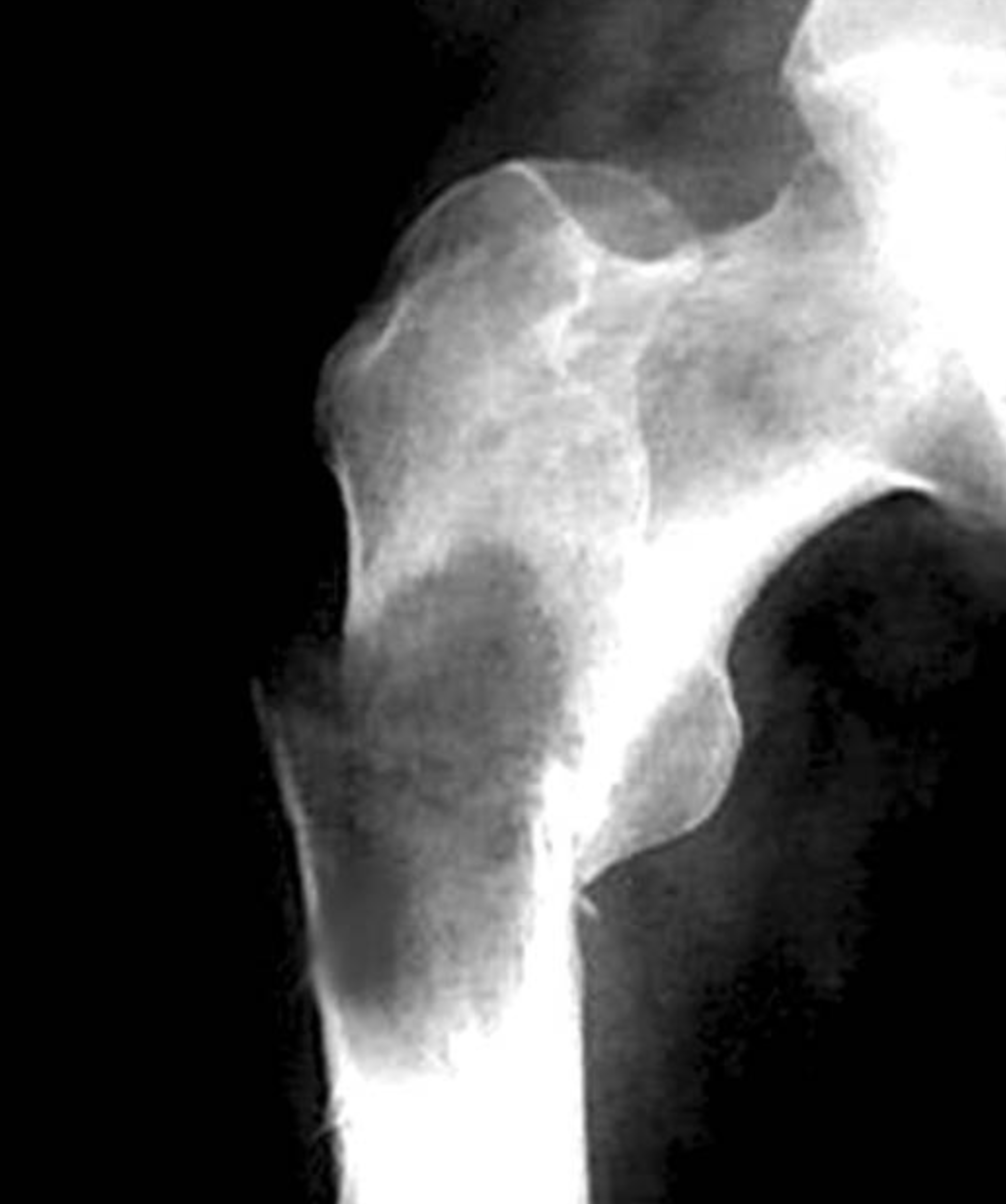
  • Plain radiograph of femur in a patient with GS. Courtesy of image: pathology outlines (http://www.pathologyoutlines.com/topic/bonemyeloidsarcoma.html)
    Granulocytic sarcoma (GS, also known as chloroma) was first discovered by Allen Burns, a British physician, in 1811 [1].
  • The term chloroma was first used by King to address the greenish appearance of the tumor due to myeloperoxidase.
  • The association of the GS with acute myeloid leukemia (AML) was first recognized by Dock in 1902 [2].
  • The term "granulocytic sarcoma" was suggested by Rappaport in 1967 to grant generalisability to it [3].

Classification

Pathophysiology

  • Infiltration of the tumor with myeloblasts is the main characteristic of the tumor on H&E stain.
  • GS rises from primitive precursors of granulocytes.
  • The disease is an extramedullary manifestation of myeloid diseases, however, it can occur as a primary disease.
  • Aggregation of myeloblasts, promyelocytes and myelocytes outside of the bone marrow presents itself as these solid tumors.
  • Tumors can occur at any site and can appear as green, gray, white or brown masses.

Differentiating GS from other Diseases

GS must be differentiated from other diseases that can present as extramedullary solid tumors, such as:

All patients with GS must be evaluated for concurrent or future malignancies as GS can occur in the course of or prior to other malignancies.

Epidemiology and Demographics

  • The prevalence of GS is approximately 2 per 1,000,000 individuals worldwide.
  • Most of the cases of GS are case reports and the disease is extremely rare.

Age

  • Patients of all age groups may develop GS.
  • GS associated with AML occurs more commonly in children.

Gender

  • GS affects both men and women.
  • Due to the rarity of the disease it is not clear whether there is a gender predilection for it.

Race

  • There is no racial predilection for GS.

Risk Factors

  • Risk factors for GS are usually chromosomal aberrations and include[4]:

Natural History, Complications and Prognosis

  • GS evolve over time, that depends on the co-occurence of the disease with other malignancies.
  • GS may present before evidences of other malignancies manifest or after these malignancies are evident.
  • Symptoms of the isolated GS depends on the location and the site of the tumor.
  • Majority of cases are associated with AML or other myeloproliferative/myelodysplastic syndromes.
  • Majority of GS tumors are found in the soft tissues such as the peritoneum, lymph nodes, CNS and skin. They are also found in bone and periosteum.
  • Early clinical features include weight loss, fatigue. Other manifestations of the tumor depend on its size and location.
  • Prognosis of GS depends on its association with other malignacies. In cases of isolated GS the prognosis is good. However, GS associated with myeloproliferative disorders has poor prognosis.
  • Prognosis of isolated GS with chromosome 8 abnormalities is worse than other cases of isolated GS.

Diagnosis

Diagnostic Criteria

  • There are no predefined criteria for diagnosis of granulocytic sarcoma.
  • Granulocytic sarcoma must be suspected in patients with AML or myelodysplastic syndromes. Diagnosis must be confirmed with histopathologic study of the specimen.

Symptoms

Symptoms of GS may include the following:

  • Symptoms due to mass effect such as deafness, ptosis, altered vision, intestinal obstruction, etc.
  • Headache, neck pain, abdominal pain,etc. based on the site of the tumor
  • Constitutional symptoms such as fever, fatigue, etc.

Physical Examination

Patients with GS can present with varying presentations.

Physical examination may be remarkable for:

Laboratory Findings

Imaging Findings

  • In cases of CNS involvement, magnetic resonance imaging (MRI) or CT scan of the CNS can reveal extra-axial masses.
  • In cases with soft tissue involvement, sonogram of the tissue can reveal the mass.

GS appears as[5]:

  • Hyperdense/isodence to brain/muscle in CT scan without enhancement
  • Isointense/hyperintense on T2-weighted MRI
  • Isointemse/hypointense on T1-weighted MRI
Histopathologic micrograph of GS. Infiltration with myeloblasts which can be seen in the course of AML. Courtesy of image: Wikipedia (By Nephron - Own work, CC BY-SA 3.0, https://commons.wikimedia.org/w/index.php?curid=15893726)

Abdominal plain radiogram can reveal obstruction, intussusception, etc.

Echocardiogram may reveal mobile masses in any heart chamber in cases of heart involvement.

Chest radiographs can show lymph node enlargement and consolidation.

Other Diagnostic Studies

Treatment

Medical Therapy

  • Chemotherapy is the main stain of treatment in patients with GS. Even patients with isolated GS must receive systemic treatment to better the prognosis.
  • Patients receiving cytarabine have better prognosis[4].
  • Patients with chemotherpeutic regimens accepted for AML had longer period of progression to AML.

Treatment includes different regimens[6]:

All of these therapeutic agents act through DNA damage and interferes with DNA synthesis.

Bone marrow transplantation

Radiation

  • Radiation can be considered as an adjunctive therapy[7].
  • Combination of chemotherapy and radiotherapy can be considered in patients with CNS involvement or when rapid regression of symptoms is required.

Surgery

  • Surgery alone is not a good treatment strategy for GS.
  • Surgery can be considered prior to chemotherapy in patients where debulking can better the prognosis and help with symptom relief.[8]
  • Surgery can also have a diagnostic role in cases where excision of the mass provides specimen for histopatjologic diagnosis.

Prevention

  • There are no primary preventive measures available for GS.

References

  1. Burns, Allen. "Observations of surgical anatomy, in Head andNeck". London, England, Royce, 1811: 364–366.
  2. Dock G, Warthin AS. "A new case of chloroma withleukemia". Trans Assoc Am Phys, 1904. 19:64: 115.
  3. Rappaport H (1967). Tumors of the hematopoietic system, inAtlas of Tumor Pathology, Section III. Washington: Fascicle 8. ArmedForces Institute of Pathology. pp. 241–247.
  4. 4.0 4.1 Daniela Dörfel; et al. (2016). "Cardiac Myeloid Sarcoma: Multimodality Radiologic Imaging Features and Pathologic Correlation". The American Journal of Medicine. 129: e117–e120.
  5. Guermazi; et al. (2002). "Granulocytic sarcoma (chloroma): imaging findings in adults and children". American Journal of Roentgenology. 178: 319–25.
  6. Yilmaz, A. F., Saydam, G., Sahin, F., & Baran, Y. (2013). "Granulocytic sarcoma: a systematic review". American Journal of Blood Research.
  7. 7.0 7.1 Richard L. Bakst, Martin S. Tallman, Dan Douer and Joachim Yahalom (2011). "How I treat extramedullary acute myeloid leukemia". Blood. 118: 3785–3793.
  8. Antic D; et al. (2013). "Is there a "gold" standard treatment for patients with isolated myeloid sarcoma?". Biomed Pharmacother. 67: 72–77.

Template:WS Template:WH