Basilar membrane
Template:Infobox Anatomy The basilar membrane within the cochlea of the inner ear is a stiff structural element that separates two liquid-filled tubes that run along the coil of the cochlea, the scala media and the scala tympani (see figure).
Function
Endolymph/perilymph separation
The fluids in these two tubes, the endolymph and the perilymph are very different chemically, biochemically, and electrically. Therefore they have to be kept strictly separated. This separation is the main function of Reissner's membrane (between scala vestibuli and scala media), and is one of the functions of the basilar membrane in the hearing organ of all land vertebrates.
A base for the sensory cells
The basilar membrane is also the base for the sensory cells of hearing, the hair cells (see figure). This function gave the basilar membrane its name, and it is again present in all land vertebrates. Due to its location, the basilar membrane places the hair cells in a position where they are adjacent to both the endolymph and the perilymph, which is a precondition of hair cell function.
Frequency dispersion
A third, evolutionarily younger, function of the basilar membrane is strongly developed in the cochlea of most mammalian species and weakly developed in some bird species. It is the function of frequency dispersion of incoming sound waves. In brief, the membrane is tapered and it is stiffer at one end than at the other. The dispersion of fluid waves causes sound input of a certain frequency to vibrate some locations of the membrane more than other locations. As shown in experiments by Nobel Prize laureate Georg von Békésy, high frequencies lead to maximum vibrations at the basal end of the cochlear coil (narrow, stiff membrane), and low frequencies lead to maximum vibrations at the apical end of the cochlear coil (wide, more compliant membrane). This "place-frequency map" can be described quantitatively by the Greenwood Function and its variants.
Anatomy
The basilar membrane is a pseudo-resonant structure[1] that, like strings on an instrument, varies in width and stiffness. The "string" of the basilar membrane is not a set of parallel strings, as in a guitar, but a long structure that has different properties (width, stiffness, mass, damping, and the dimensions of the ducts that it couples to) at different points along its length. The motion of the basilar membrane is generally described as a traveling wave.[2] The parameters of the membrane at a given point along its length determine its characteristic frequency (CF), the frequency at which it is most sensitive to sound vibrations. The Basilar membrane is widest (0.42–0.65 mm) and least taut at the apex of the cochlea, and narrowest (0.08–0.16 mm) and most taut at the base.[3] High-frequency sounds localize near the base of the cochlea (near the round and oval windows), while low-frequency sounds localize near the apex.
Additional images
-
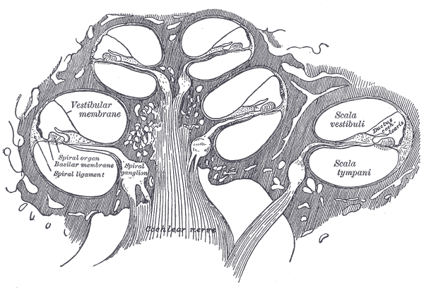 Diagrammatic longitudinal section of the cochlea.
 -
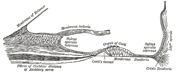 Floor of cochlear duct.
 -
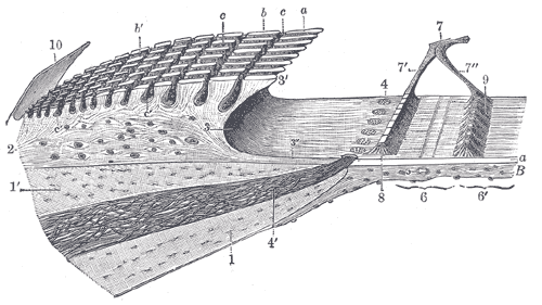 Spiral limbus and basilar membrane.
 -
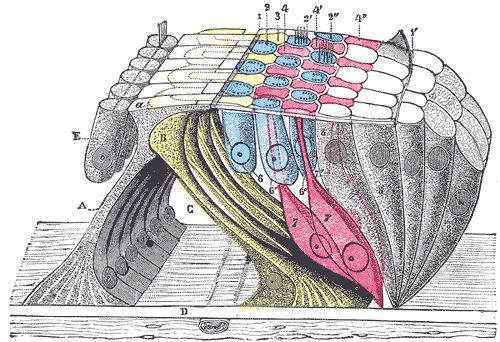
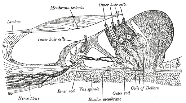 Section through the spiral organ of Corti (magnified)
 -
 The reticular lamina and subjacent structures.


References
- ↑ M. Holmes and J. D. Cole, "Pseudoresonance in the cochlea, ' in: Mechanics of Hearing, E. de Boer and M. A. Viergever (editors), Proceedings of the IUTAM/ICA Symposium, Delft (1983), pp. 45-52.
- ↑ Richard R. Fay, Arthur N. Popper, and Sid P. Bacon (2004). Compression: From Cochlea to Cochlear Implants. Springer. ISBN 0387004963.
- ↑ Oghalai JS. The cochlear amplifier: augmentation of the traveling wave within the inner ear. Current Opinion in Otolaryngology & Head & Neck Surgery. 12(5):431-8, 2004
- Fritzsch B: The water-to-land transition: Evolution of the tetrapod basilar papilla; middle ear, and auditory nuclei. In: Douglas B. Webster, Richard R. Fay, Arthur N. Popper, editors (1992). The Evolutionary biology of hearing. Berlin: Springer-Verlag. pp. 351–375. ISBN 0-387-97588-8.
- Nageris B, Adams JC, Merchant SN (1996). "A human temporal bone study of changes in the basilar membrane of the apical turn in endolymphatic hydrops". The American journal of otology. 17 (2): 245–52. PMID 8723956.
- Kössl M, Vater M (2000). "Consequences of outer hair cell damage for otoacoustic emissions and audio-vocal feedback in the mustached bat". J. Assoc. Res. Otolaryngol. 1 (4): 300–14. PMID 11547810.
- Nilsen KE, Russell IJ (1999). "Timing of cochlear feedback: spatial and temporal representation of a tone across the basilar membrane". Nat. Neurosci. 2 (7): 642–8. doi:10.1038/10197. PMID 10404197.
- Nilsen KE, Russell IJ (2000). "The spatial and temporal representation of a tone on the guinea pig basilar membrane". Proc. Natl. Acad. Sci. U.S.A. 97 (22): 11751–8. doi:10.1073/pnas.97.22.11751. PMID 11050205.