Central retinal artery
Editor-In-Chief: C. Michael Gibson, M.S., M.D. [1]
The central retinal artery (retinal artery) branches off the ophthalmic artery, running inferior to the optic nerve within its dural sheath to the eyeball.
Anatomy
It pierces the optic nerve close to the eyeball, sending branches over the internal surface of the retina, and these terminal branches are the only blood supply to the larger part of it.
The central part of the retina where the light rays are focussed after passing through the pupil and the lens is a circular area called the macula. The center of this circular area is the fovea. The fovea and a small area surrounding it are not supplied by the central retinal artery or its branches.
The central retinal artery still supplies all the nerve fibers that form the optic nerve that carries the visual information to the occipital lobe cerebral cortex.
Pathology
Thus if the central retinal artery gets occluded, there is complete loss of vision in that eye even though the fovea is not affected. The entire retina (with the exception of the fovea) becomes pale and swollen and opaque while the central fovea still appears reddish (this is because the choroid color shows through). This is the basis of the famous "Cherry red spot" seen on examination of the retina on funduscopy of a central retinal artery occlusion (CRAO).
In some cases - approximately 20% of the population - there is a branch of the ciliary circulation called the cilio-retinal artery which supplies the retina between the macula and the optic nerve, including the nerve fibers from the foveal photoreceptors. If this artery is present, the central vision will be preserved even in case of CRAO.
-
![Central Retinal Artery Occlusion (embolus; painless, unilateral vision loss)[1]](/images/5/5a/Central_Retinal_Artery_Occlusion.jpg) Central Retinal Artery Occlusion (embolus; painless, unilateral vision loss)[1]
Central Retinal Artery Occlusion (embolus; painless, unilateral vision loss)[1] -
![Central Retinal Vein Occlusion: HTN, DM, Leukemia (unilateral painless vision loss)[2]](/images/9/9d/CentralRetinalVeinOcclusion.jpg) Central Retinal Vein Occlusion: HTN, DM, Leukemia (unilateral painless vision loss)[2]
Central Retinal Vein Occlusion: HTN, DM, Leukemia (unilateral painless vision loss)[2] -
![Cherry Red Spot. Baby: Tay-Sachs. Adult: Central retinal artery occlusion (sudden painless unilateral vision loss) HTN, DM, Leukemia. [3]](/images/2/2a/Cherry_Red_Spot.jpg) Cherry Red Spot. Baby: Tay-Sachs. Adult: Central retinal artery occlusion (sudden painless unilateral vision loss) HTN, DM, Leukemia. [3]
Cherry Red Spot. Baby: Tay-Sachs. Adult: Central retinal artery occlusion (sudden painless unilateral vision loss) HTN, DM, Leukemia. [3]
![Central Retinal Artery Occlusion (embolus; painless, unilateral vision loss)[1]](/index.php/File:Central_Retinal_Artery_Occlusion.jpg)
![Central Retinal Vein Occlusion: HTN, DM, Leukemia (unilateral painless vision loss)[2]](/index.php/File:CentralRetinalVeinOcclusion.jpg)
![Cherry Red Spot. Baby: Tay-Sachs. Adult: Central retinal artery occlusion (sudden painless unilateral vision loss) HTN, DM, Leukemia. [3]](/index.php/File:Cherry_Red_Spot.jpg)
Additional images
-
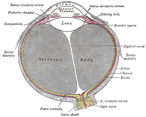 Horizontal section of the eyeball.
 -
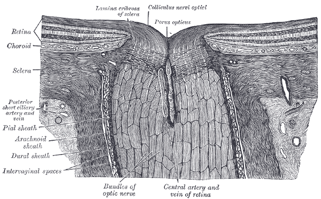 The terminal portion of the optic nerve and its entrance into the eyeball, in horizontal section.


External links
- Diagram at suncoastretina.com
- emerg/777 at eMedicine ("Retinal artery occlusion")
- Template:EMedicineDictionary