Aneurysm of sinus of valsalva pathophysiology
|
Aneurysm of Sinus of Valsalva Microchapters |
|
Differentiating Aneurysm of sinus of valsalva from other Diseases |
|---|
|
Diagnosis |
|
Treatment |
|
Aneurysm of sinus of valsalva pathophysiology On the Web |
|
American Roentgen Ray Society Images of Aneurysm of sinus of valsalva pathophysiology |
|
Risk calculators and risk factors for Aneurysm of sinus of valsalva pathophysiology |
Editor-In-Chief: C. Michael Gibson, M.S., M.D. [1]; Associate Editor(s)-In-Chief: Priyamvada Singh, M.B.B.S.[2]; Assistant Editor(s)-In-Chief: Kristin Feeney, B.S.[3]
Overview
In a normal heart, the typical path of blood flow is to shunt from right-to-left allowing for blood to leave the deoxygenated right system and become oxygenated in the left system. In patients with an aneurysm of the sinus of valsalva, the typical path of blood flow is disrupted by the presence of the aneurysm. This can result in blood shunting from left-to-right. The severity of circulatory complications depends largely on the size of the aneurysm and any accompanying defects. The larger the defect, the greater the amount of mixing between deoxygenated and oxygenated blood.
Pathophysiology
-
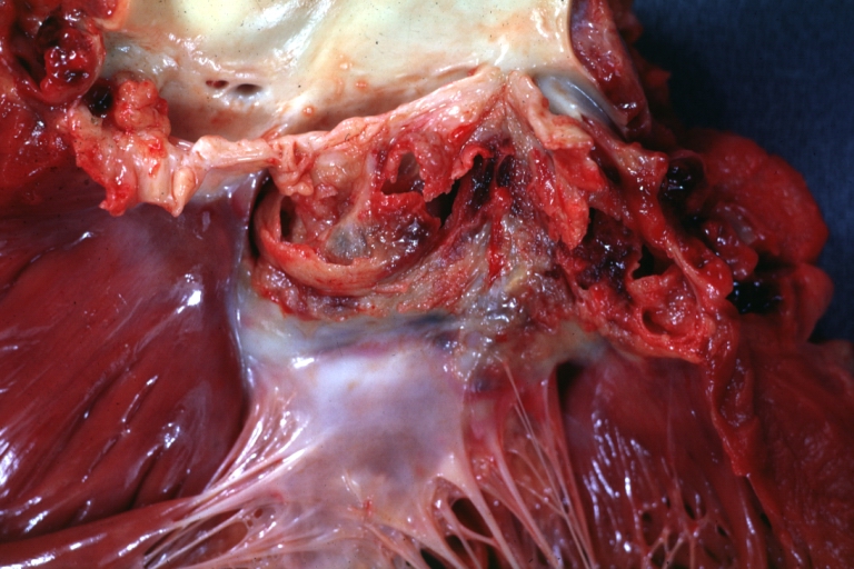
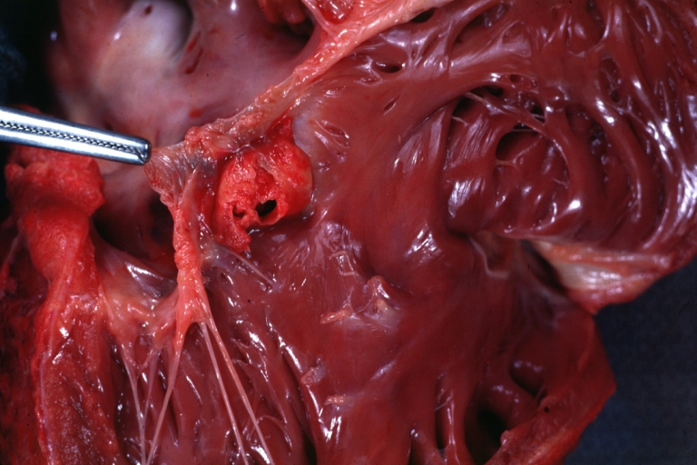 Sinus of Valsalva Aneurysm: Gross; a beautiful picture of windsock opening of aneurysm beneath anterior leaflet of tricuspid valve into right ventricle
 -
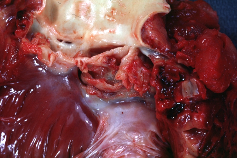
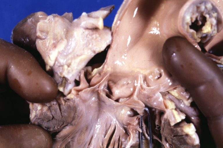
 Sinus of Valsalva Aneurysm: Gross; an excellent picture; cut through aneurysm.
 -
 Sinus of Valsalva Aneurysm: Gross; An infected aneurysm with a ruptured aortic cusp due to endocarditis.
 -
 Sinus of Valsalva Aneurysm: Gross; difficult to orient but aneurysm is visible.
 -
 Sinus of Valsalva Aneurysm Infected: Gross; natural color, close-up view of coronary cusps with extensive ulceration and fibrinous material. Staphylococcus infection of a bicuspid valve.
 -
 Sinus of Valsalva Aneurysm Infected: Gross; natural color fibrin exudate and some valve scarring are easily seen but aneurysm not seen. The bicuspid valve requires careful look to appreciate Staphylococcus caused lesion.
 -
 Ruptured Sinus of Valsalva Aneurysm: Gross; natural color view from left atrium of left sinus aneurysm that is pointing into left atrium through mitral ring at anterior mitral commissure (the most unusual lesion).
 -
 Ruptured Sinus of Valsalva Aneurysm: Gross; natural color view of left ventricular outlet with hemorrhagic lesion in left coronary sinus.
 -
 Ruptured Sinus of Valsalva Aneurysm: Gross; natural color close-up of aneurysm in aortic valve.
 -
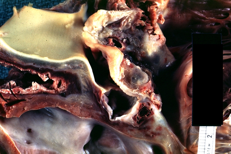
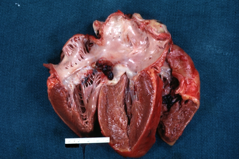
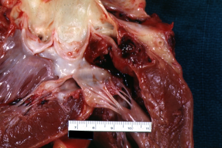
 Sinus of Valsalva Aneurysm: Gross fixed tissue shows aneurysm in noncoronary cusp about as well as possible see for picture with probe in aneurysm
 -
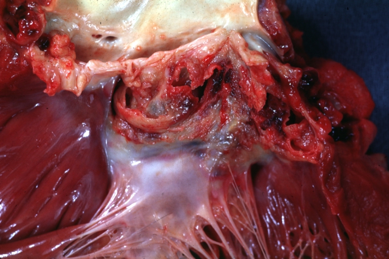
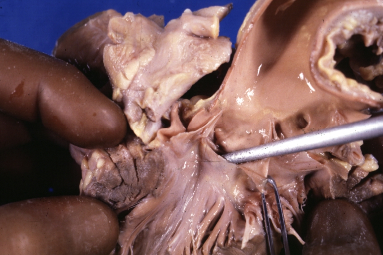 Sinus of Valsalva Aneurysm: Gross fixed tissue close-up view with probe in non coronary sinus aneurysm see for photo without probe
 -
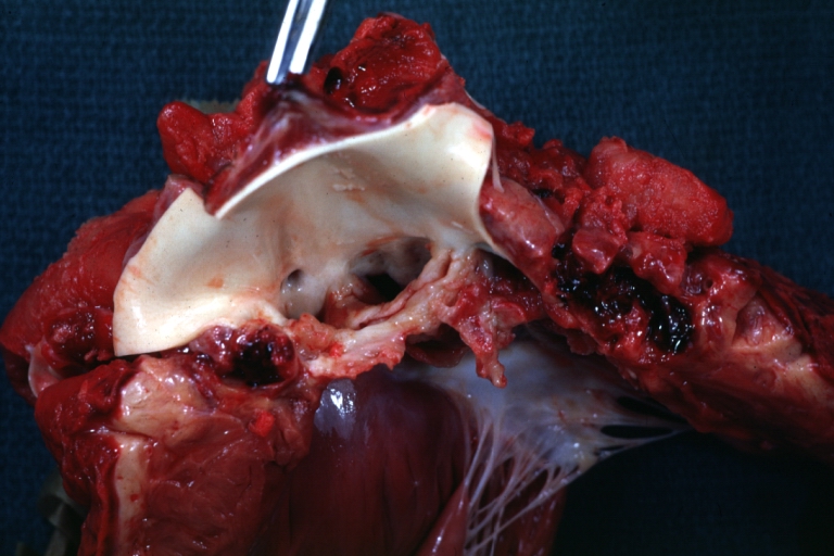
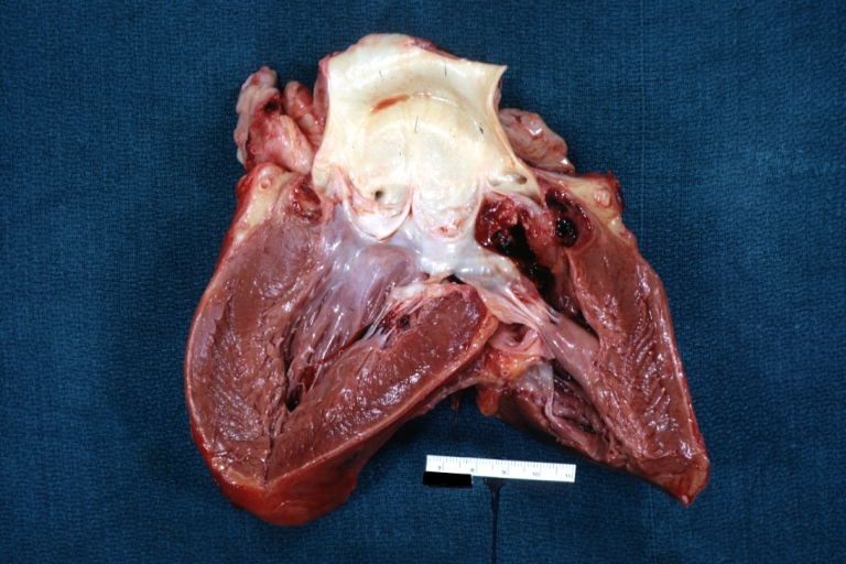
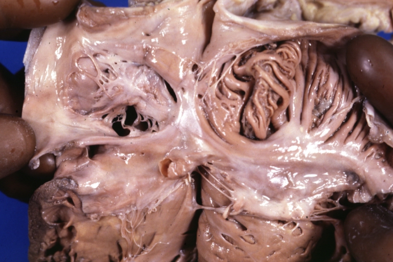 Sinus of Valsalva Aneurysm Ruptured: Gross fixed tissue view of right side of heart showing site of rupture below tricuspid ring also patent foramen ovale


