Gallery of new files
Jump to navigation
Jump to search
This special page shows the last uploaded files.
-
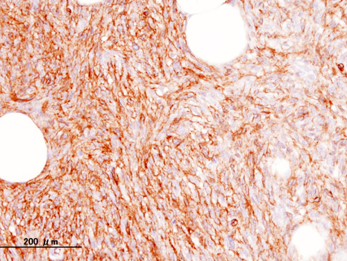
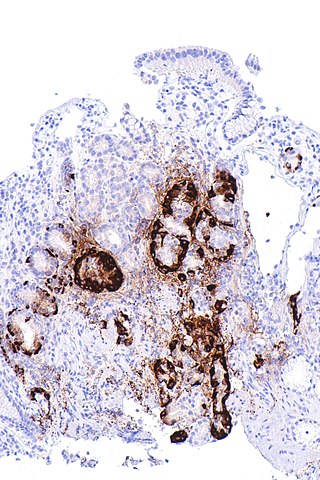 320px-Metaplastic atrophic gastritis - body - chromogranin A -- intermed mag.jpg Sanajan
18:01, 24 June 2020
320 × 480; 66 KB
-
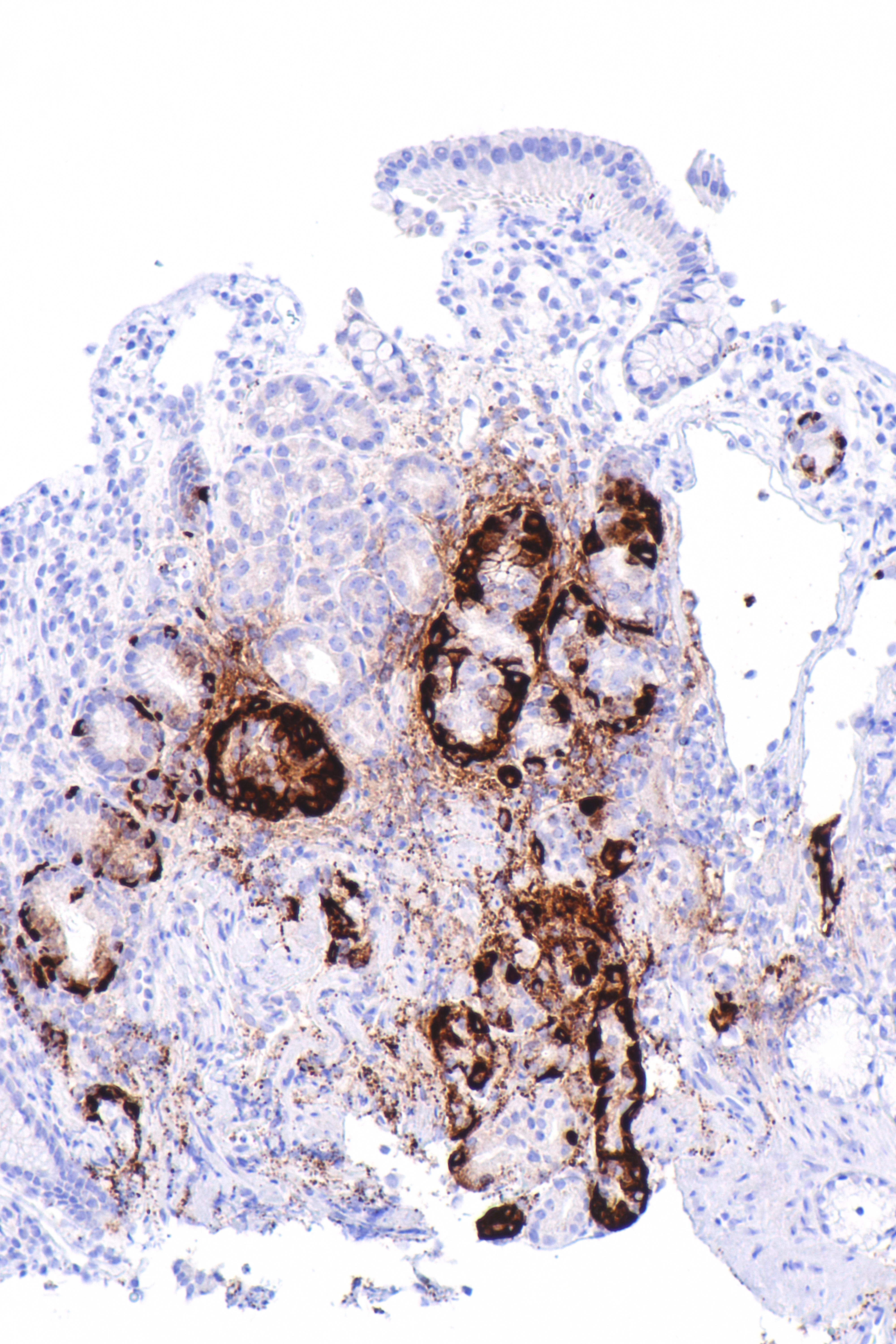
 Metaplastic atrophic gastritis - body - chromogranin A -- intermed mag.jpg Sanajan
17:26, 24 June 2020
4,000 × 6,000; 8.77 MB
-
-
-

-
-
 Acute Phosphate Nephropathy - Calcium Phosphate crystal deposition.jpg AFarheen
22:34, 22 June 2020
640 × 480; 91 KB
-

-

-
-
-
-
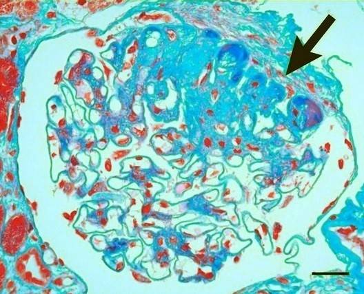 Histopathology of secondary segmental glomerular sclerosis of hypertensive nephropathy.jpg NNikravangolsefid
23:52, 19 June 2020
528 × 427; 128 KB
-
-
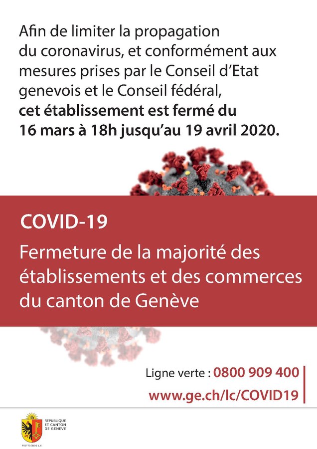
 Page1-636px-COVID-19 Key-Message A3-Posters Stop.pdf.jpg Sara Mohsin
16:11, 19 June 2020
636 × 899; 79 KB
-
-

-

-
-
 Complete AV Block with Sinus slowing during carotid Sinus massage Case report.jpg Akash Daswaney
11:27, 18 June 2020
1,168 × 534; 119 KB
-
-
-
-
 79px-Life cycle of Rustic Butterfly (2663558308).jpg Maneesha Nandimandalam
15:18, 15 June 2020
79 × 119; 3 KB
-

-
-
-
-
-
-
-
-
-
-
-
-
-
-

-
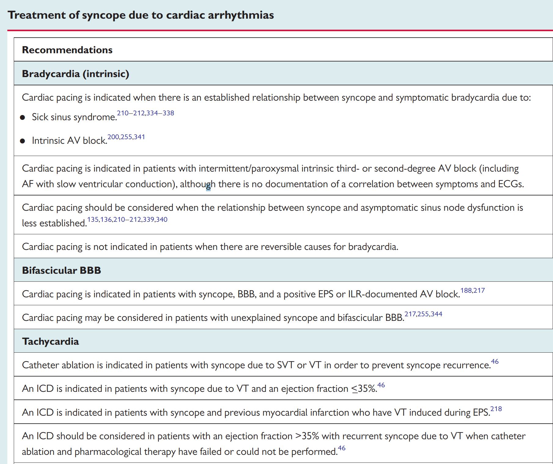
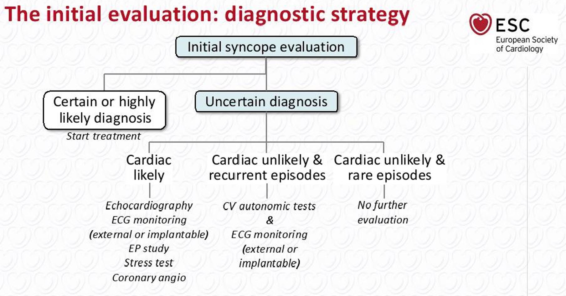 Initial Strategy Syncope or Paroxysmal AV Block.JPG Akash Daswaney
10:56, 13 June 2020
1,808 × 946; 188 KB
-
-
-
-
-
-
-
-
-
-

-

-
-
-

-

-

-

-

-

-
-
-
-
-
-
-
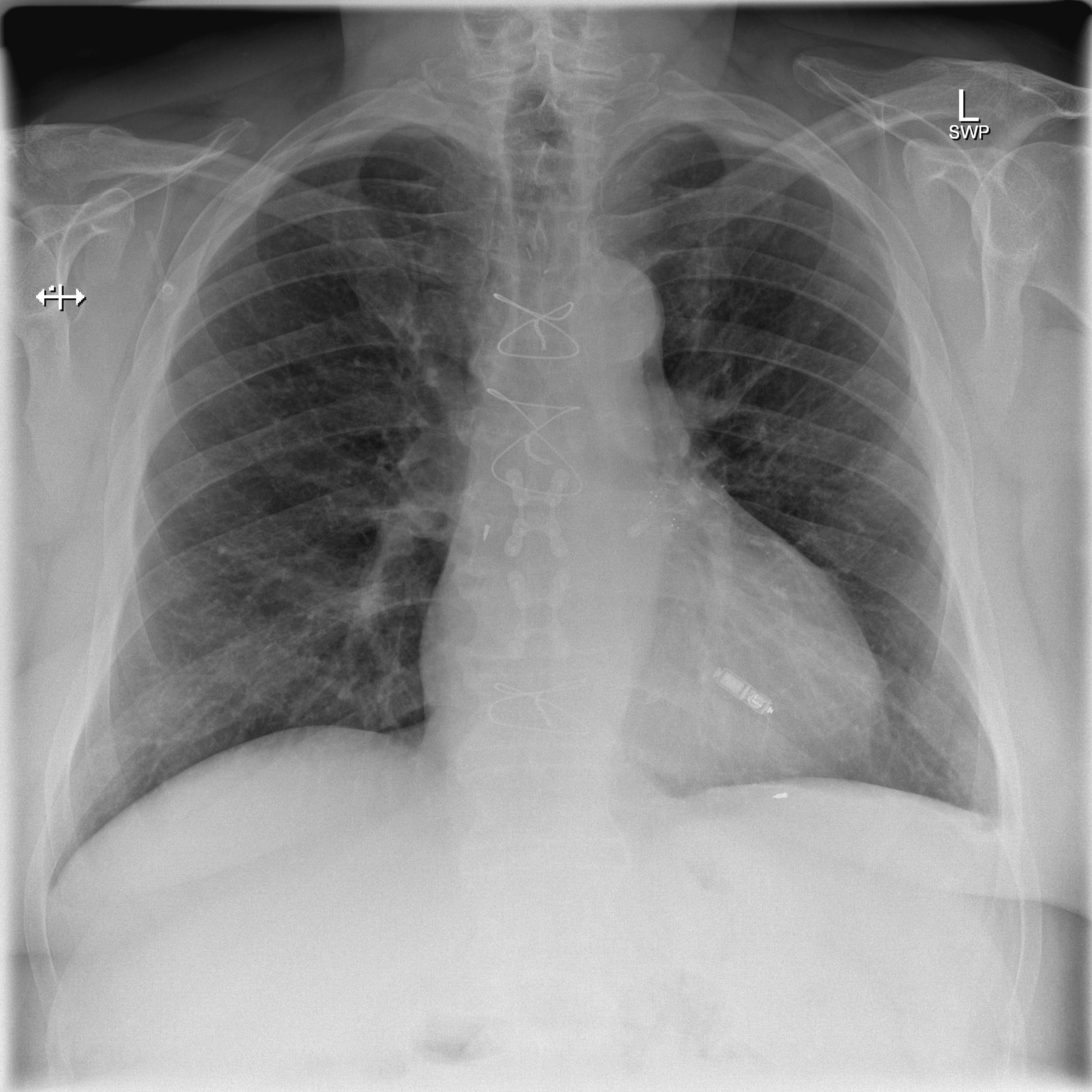
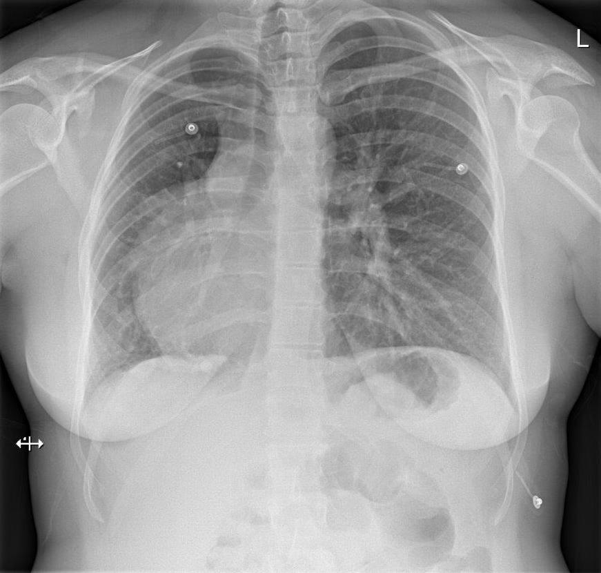
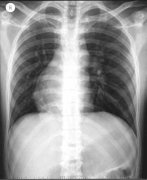
 X-ray of pacemaker with right atrial and ventricular lead.jpg Javaria Anwer
09:40, 9 June 2020
725 × 361; 46 KB
-

-

-

-
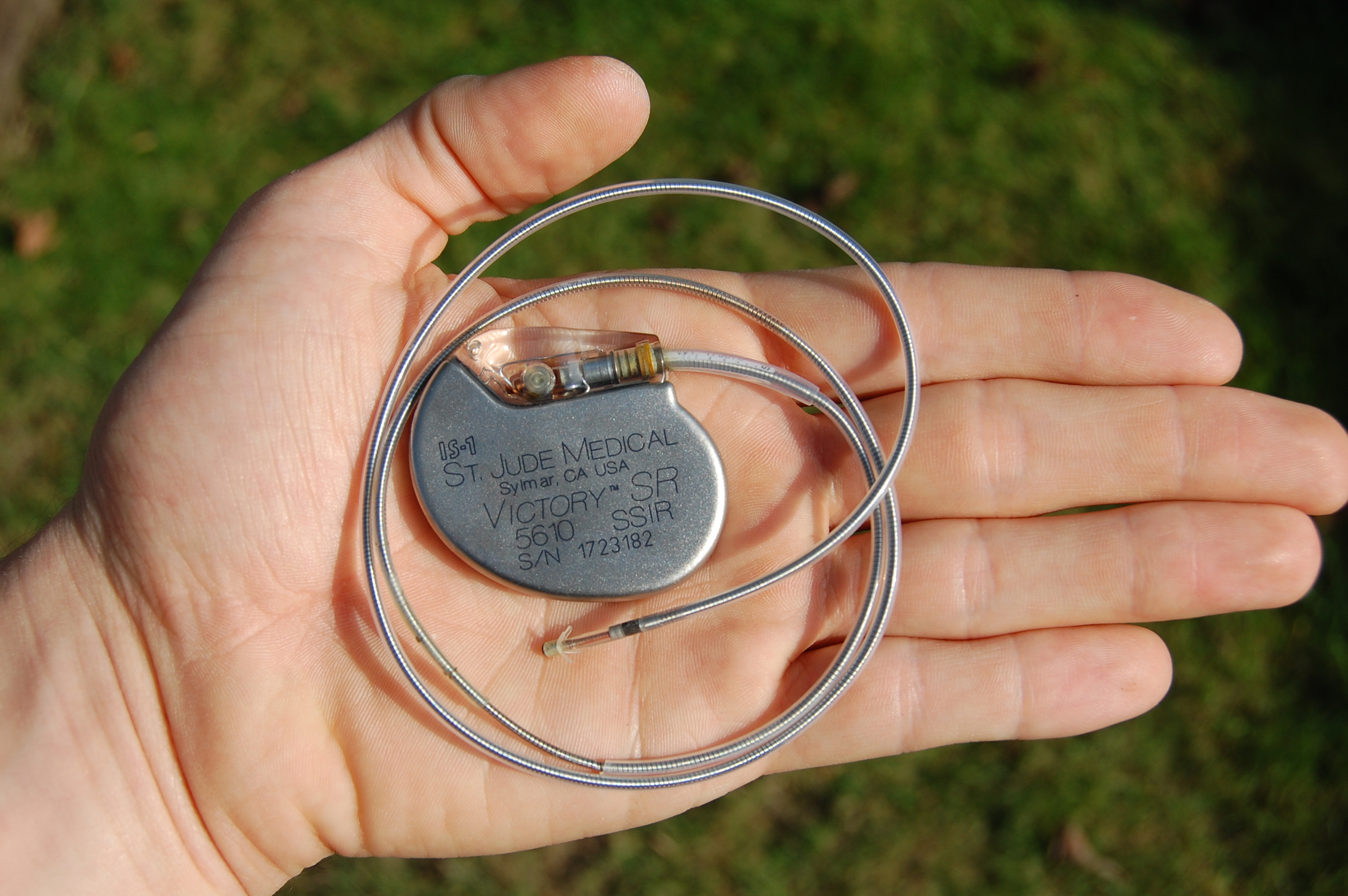 An artificial pacemaker shown in hand with electrode and lead (from St Jude medical).jpg Javaria Anwer
05:26, 8 June 2020
3,008 × 2,000; 2.83 MB
-
 Renal arterial hyalinosis - pas - very high mag.jpg NNikravangolsefid
22:51, 7 June 2020
4,272 × 2,848; 3.91 MB
-

-
-
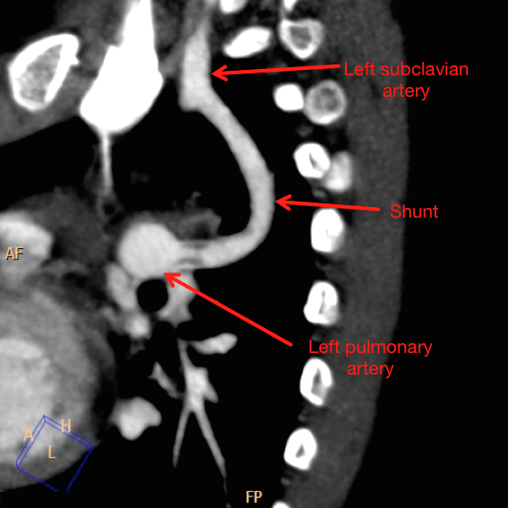
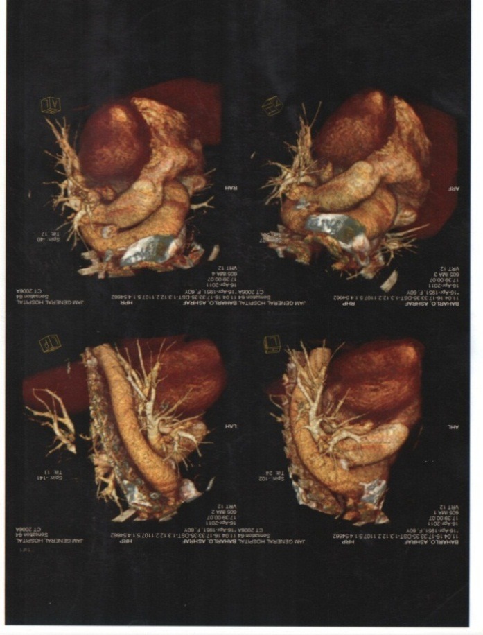
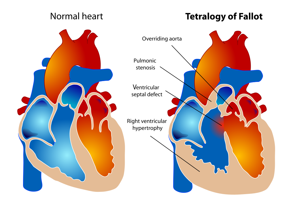
 Unilateral-pulmonary-oedema-blalock-taussig-shunt-in-pulmonary-atresia-with-ventricular-septal-defect-1.jpg Usmanaliakbar
03:49, 7 June 2020
2,037 × 2,642; 3 MB
-
 Histopathology of hypertensive glomerular lesion of hypertensive nephropathy.jpg NNikravangolsefid
21:34, 5 June 2020
558 × 452; 114 KB
-
 Fibrous intimal thickening in hypertensive nephropathy.jpg NNikravangolsefid
20:37, 5 June 2020
251 × 317; 41 KB
-
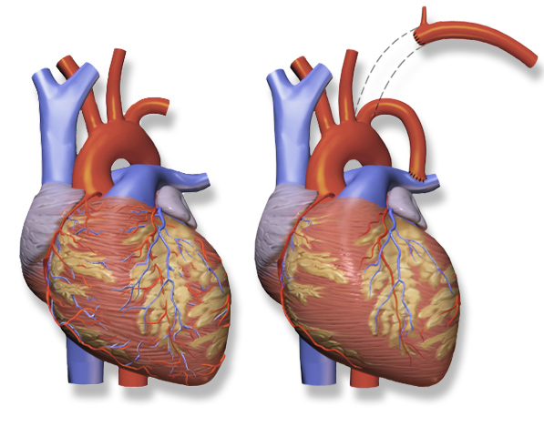
 Modified-blalock-taussig-shunt-creative-commons.png Usmanaliakbar
18:50, 5 June 2020
359 × 476; 165 KB
-
 Original-blalock-taussig-shunt-creative-commons.png Usmanaliakbar
15:55, 5 June 2020
597 × 469; 282 KB
-

-
 Sbo-secondary-to-ileal-stricture-from-crohns-disease.jpg Bosky Soni
03:11, 5 June 2020
766 × 857; 203 KB
-

-

-

-

-

-

-

-

-

-

-

-
 Monosodium-urate-crystals-in-tophaceous-gout.jpeg Jsoujanya
14:06, 31 May 2020
2,048 × 2,048; 2.5 MB
-

-
-

-

-

-

-
 C0438160-Life Cycle of the Black-legged Tick and Lyme, Illustration.jpg Rina Ghorpade
20:51, 29 May 2020
800 × 518; 104 KB
-

-

-
-
-
-
-

-

-

-
-
-
-
-
-
 Overheating is one of the chief risk factors for SIDS.jpg Gunnam
14:41, 15 May 2020
800 × 1,200; 385 KB
-
-
-

-
-

-

-
-
-
-
-

-
-

-

-
-

-
 WhatsApp Image 2020-05-07 at 1.36.56 AM.jpeg Abdulkareem Opeoluwalukan
11:41, 7 May 2020
640 × 640; 21 KB
-

-
-
-

-
-

-
-
-

-
-
-
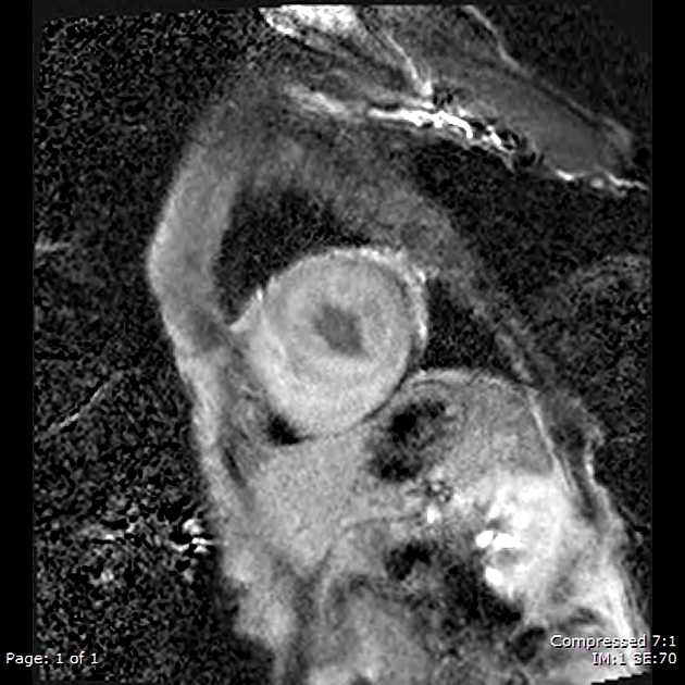
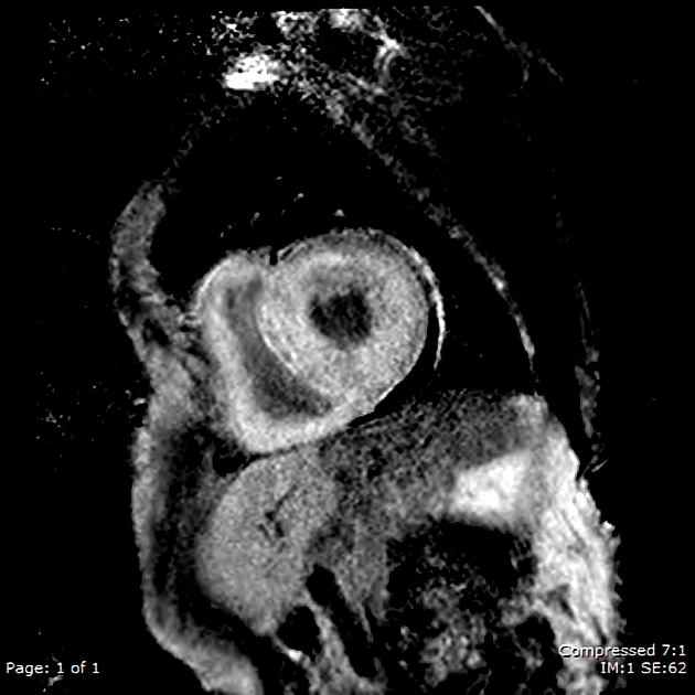
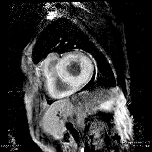
 CMR four-chamber cine view. Seen here is the grossly dilated right heart, with an atrialized RV and dilated tricuspid annulus.jpg Gunnam
20:36, 22 April 2020
771 × 322; 68 KB
-
-
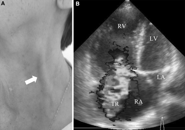
 Right-atrial-enlargement in Tricuspid regurgitation .jpg Gunnam
16:31, 20 April 2020
2,645 × 2,791; 396 KB
-

-
-
 Severe tricuspid regurgitation E00572 (CardioNetworks ECHOpedia).jpg Gunnam
16:29, 16 April 2020
660 × 526; 202 KB
-
-
-
-

-
-

-
-
-
-

-
-
 Normal echocardiographic appearance of tricuspid valve.jpg Gunnam
16:47, 19 March 2020
709 × 299; 65 KB
-

-
-

-
-
-

-
-
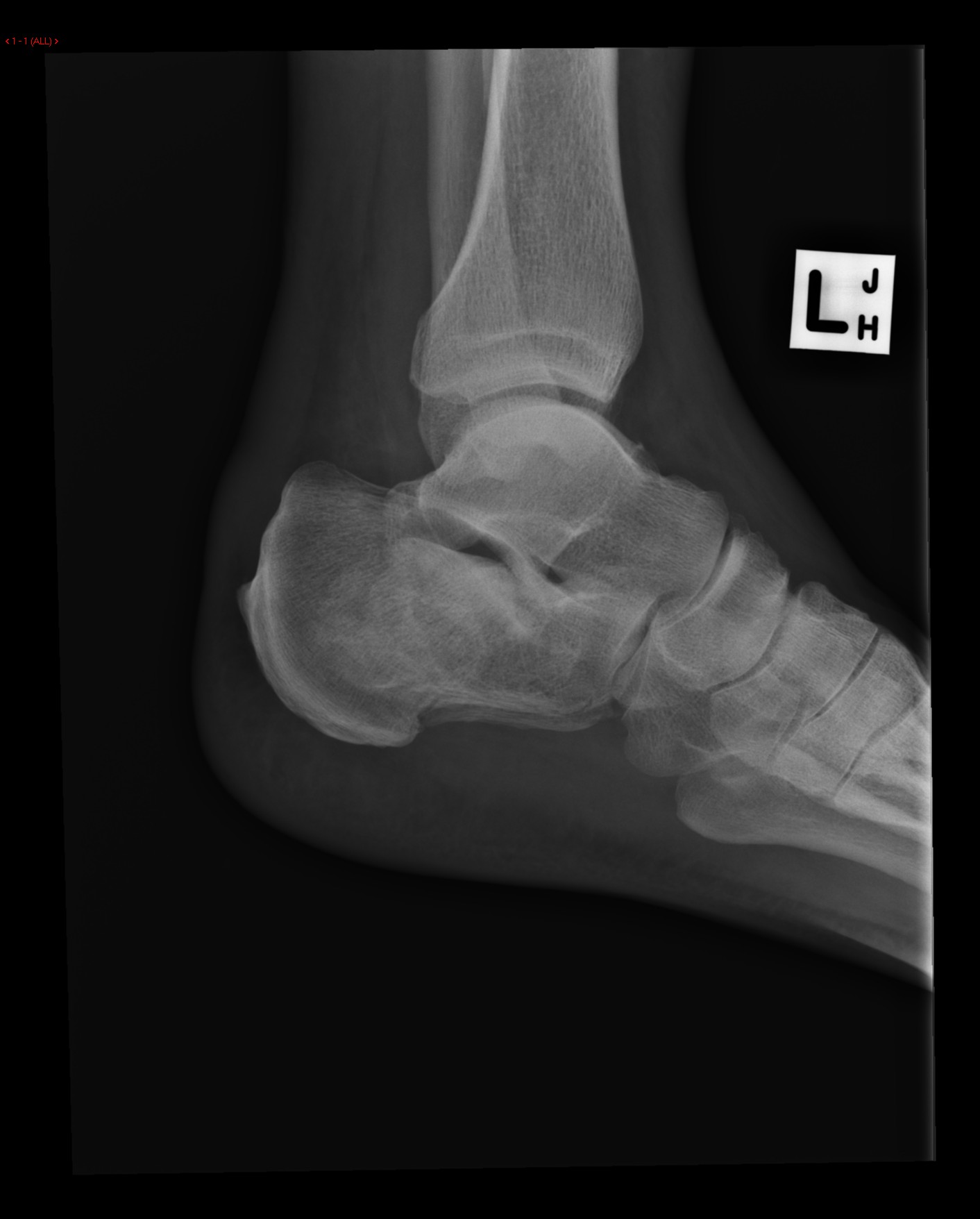
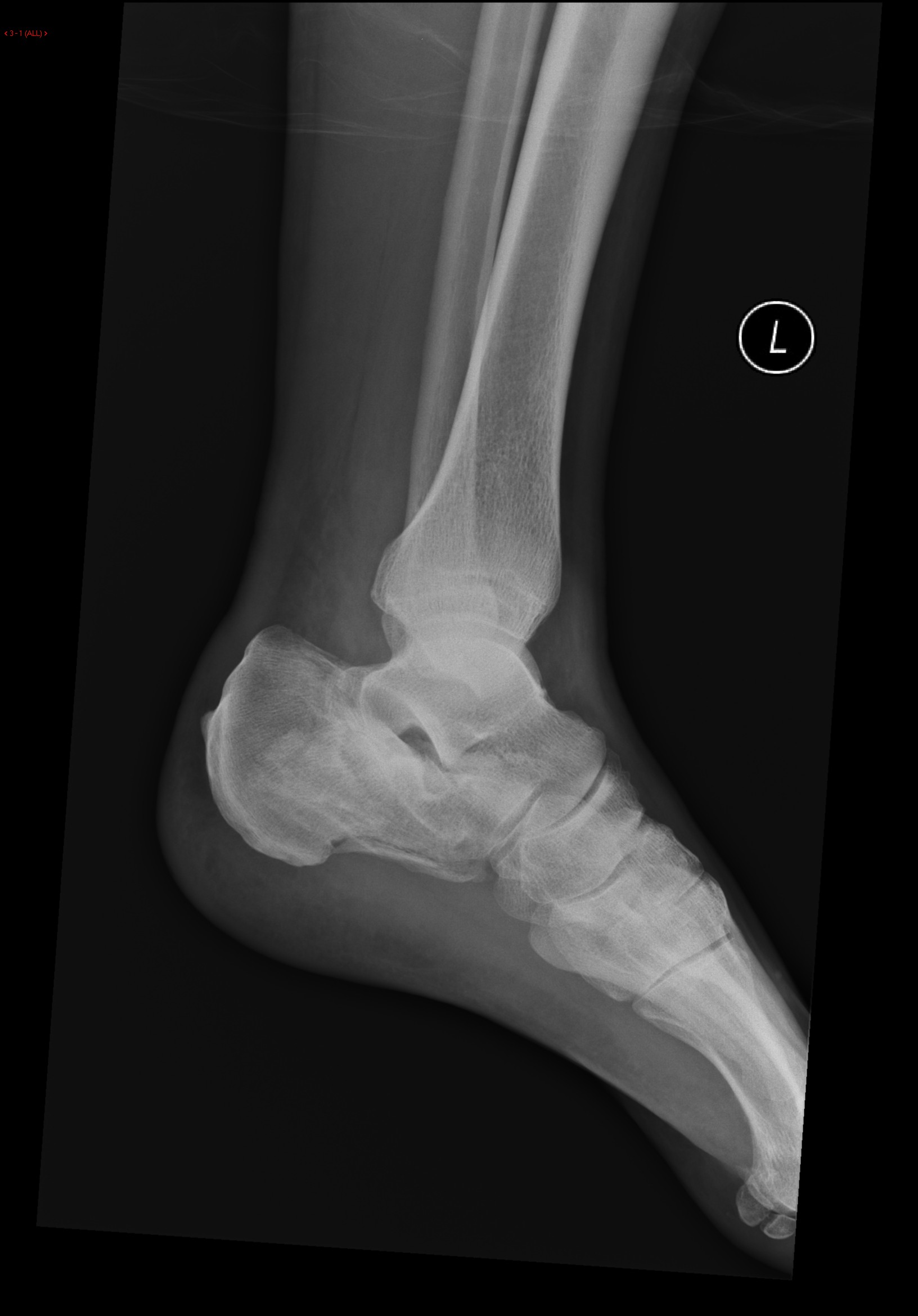
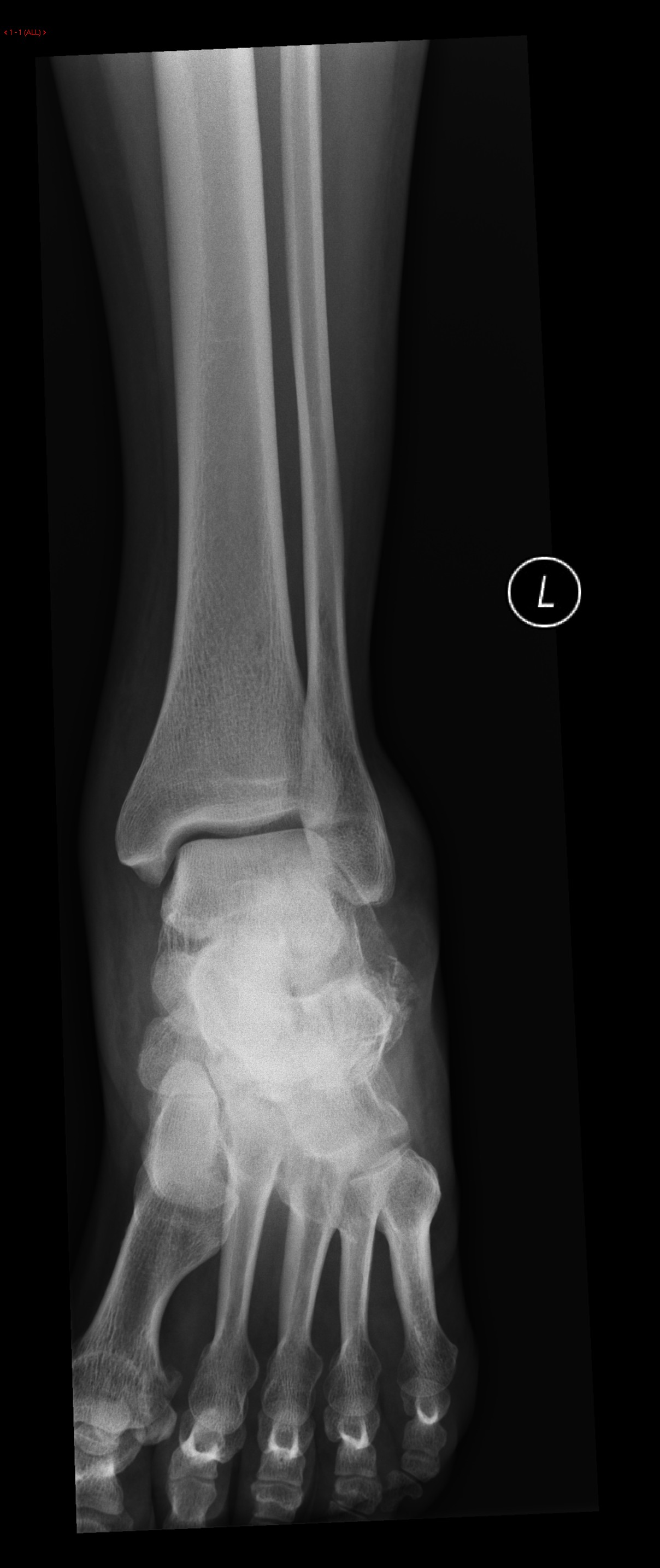
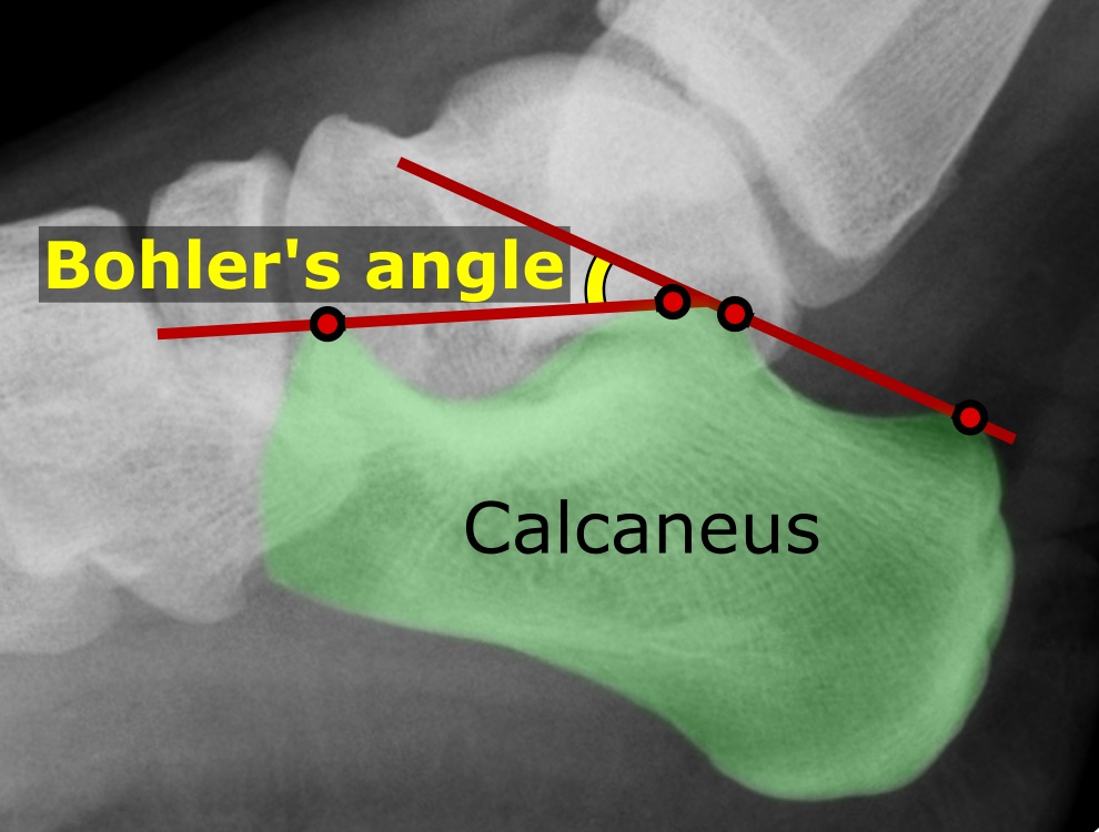
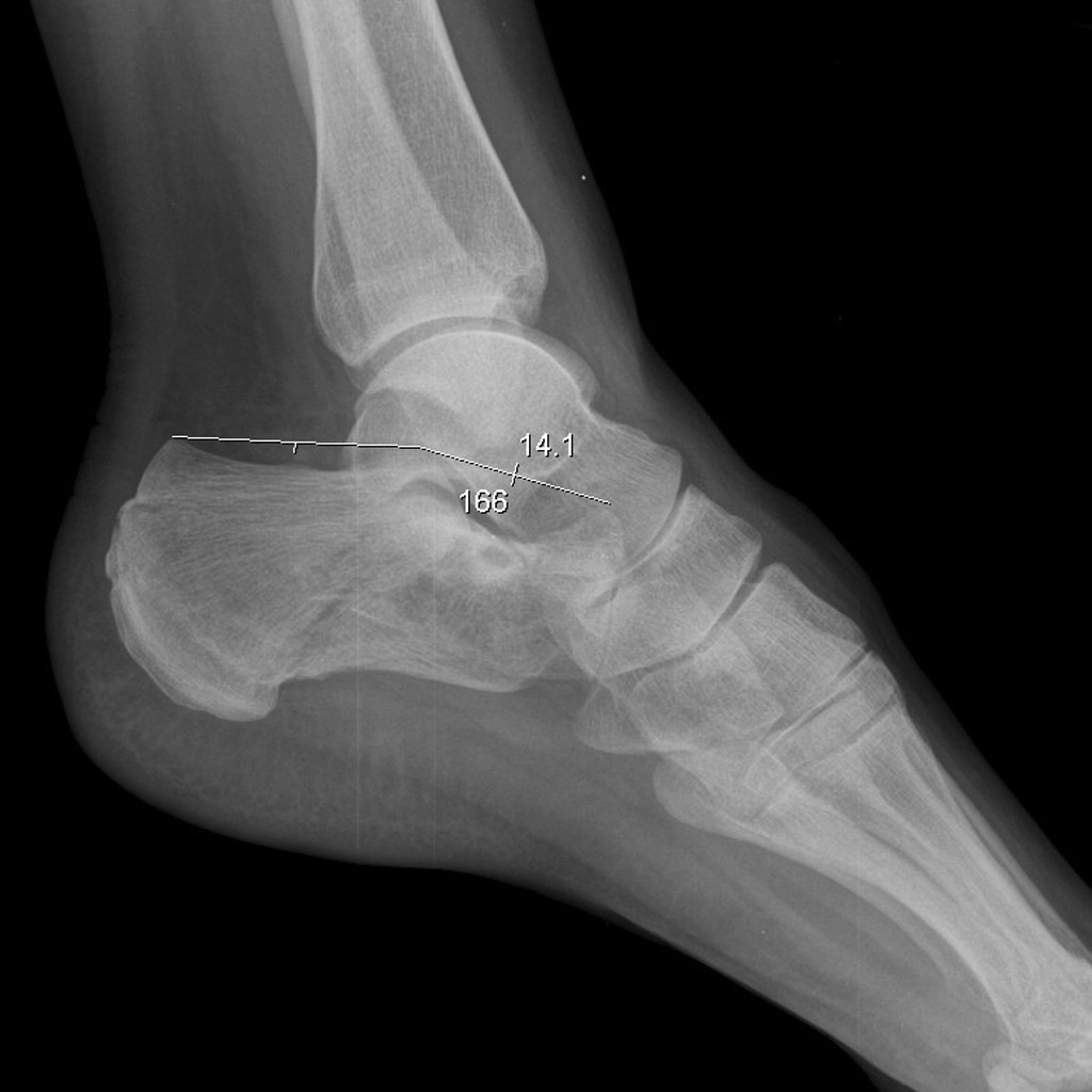
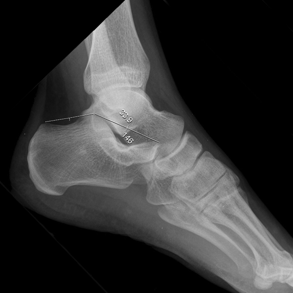
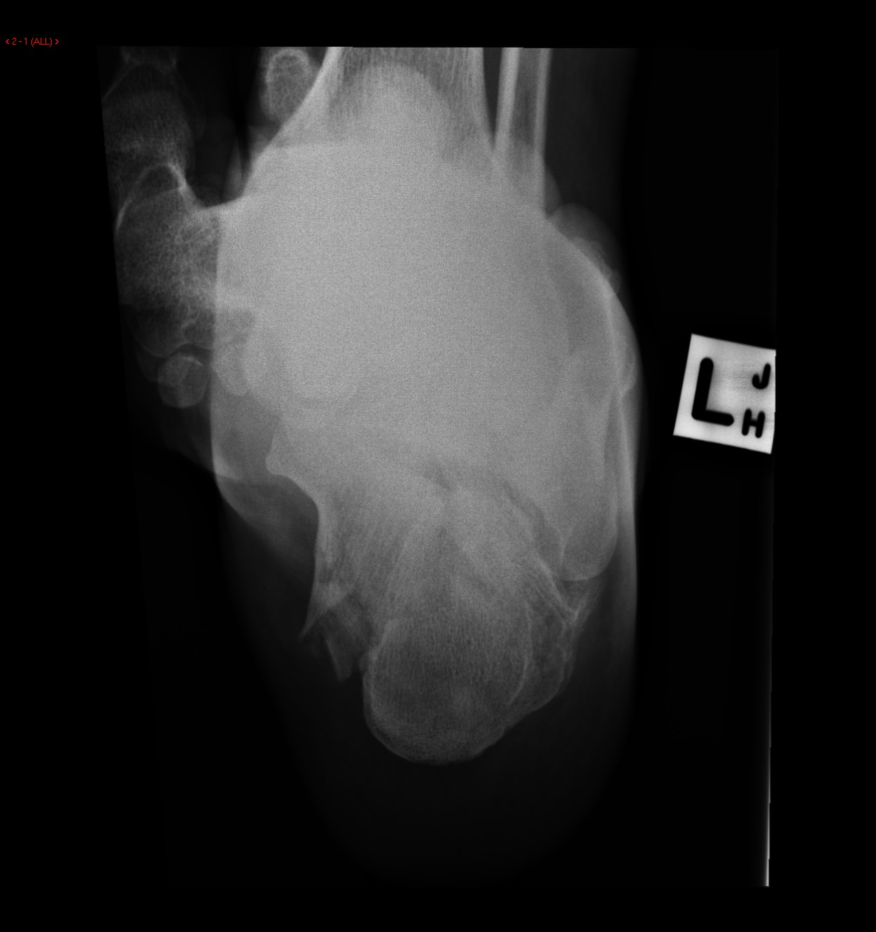 Calcaneal-fracture-and-associated-spinal-injury (4).jpg DrMars
14:53, 14 March 2020
1,252 × 1,332; 150 KB
-
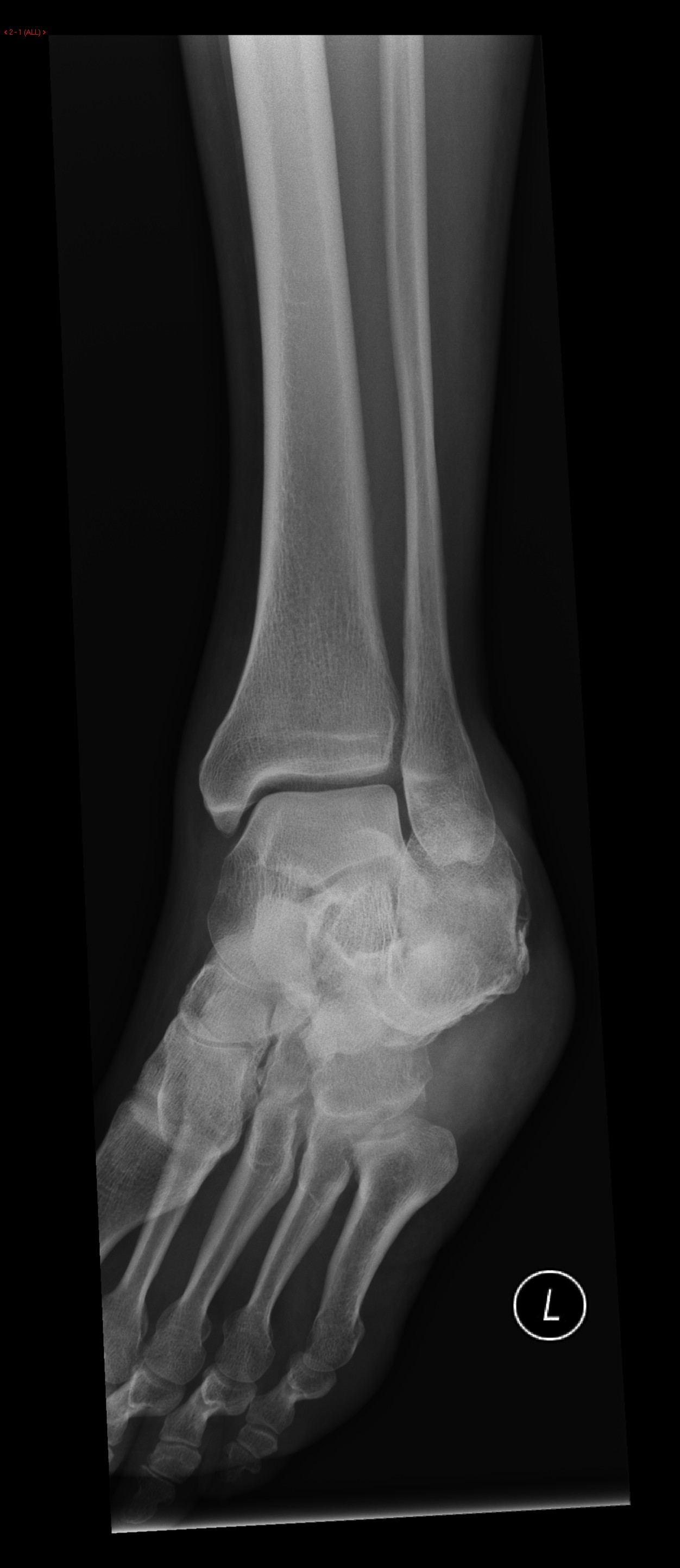 Calcaneal-fracture-and-associated-spinal-injury (3).jpg DrMars
14:53, 14 March 2020
1,252 × 2,884; 282 KB
-
 Calcaneal-fracture-and-associated-spinal-injury (2).jpg DrMars
14:53, 14 March 2020
1,412 × 1,756; 157 KB
-
 Calcaneal-fracture-and-associated-spinal-injury (1).jpg DrMars
14:52, 14 March 2020
1,632 × 2,340; 250 KB
-
 Calcaneal-fracture-and-associated-spinal-injury.jpg DrMars
14:52, 14 March 2020
1,208 × 2,868; 297 KB
-

-
-
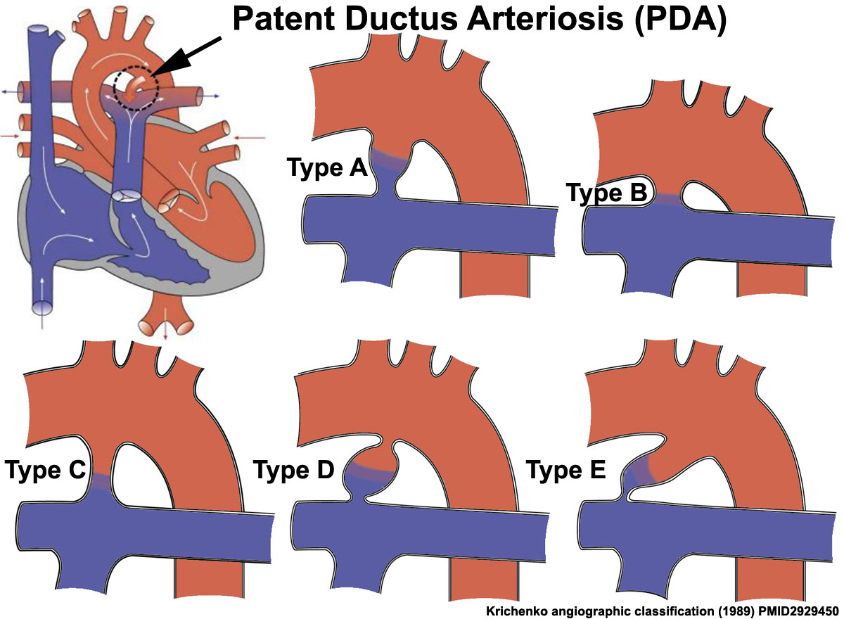
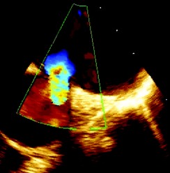 Patent ductus arteriosus (PDA) in Transesophageal echocardiography.jpg Gunnam
15:50, 13 March 2020
244 × 248; 16 KB
-
 Transesophageal echocardiogram of a patent ductus arteriosus.jpg Gunnam
14:28, 13 March 2020
654 × 250; 39 KB
-
-
-
-
-
-
-
-
-

-
-

-
-
-
-

-
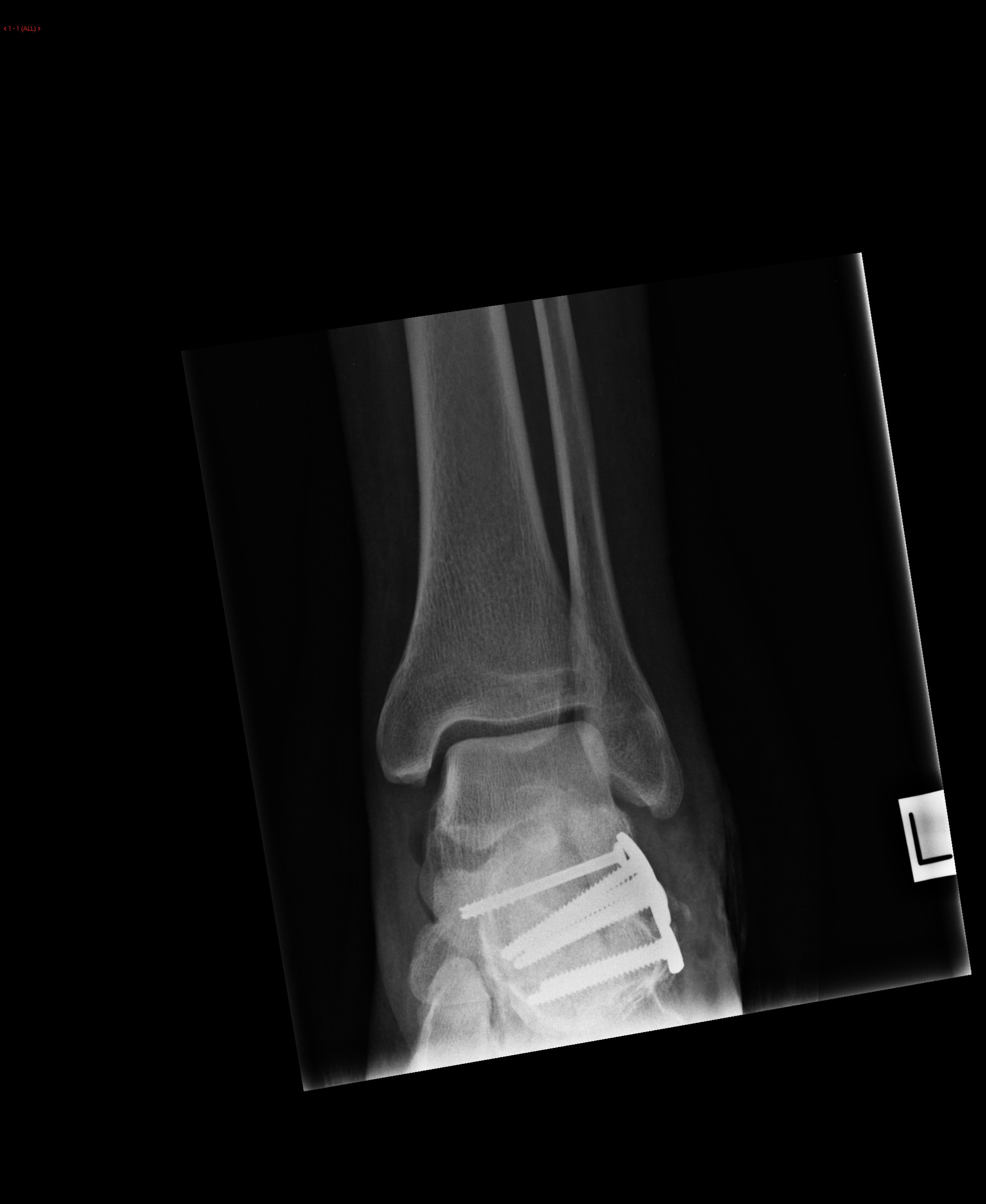
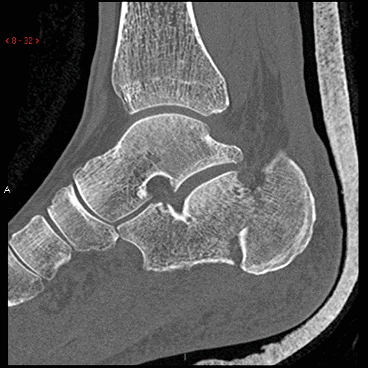
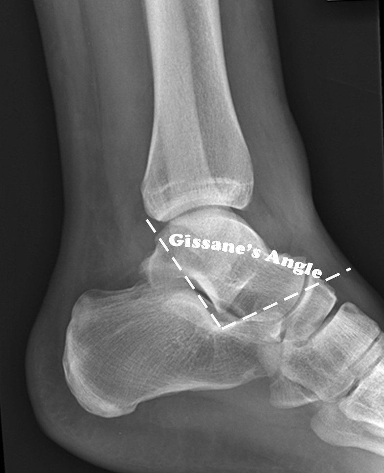
 Sanders-classification-of-calcaneal-fractures-1.jpg DrMars
00:16, 7 March 2020
9,000 × 9,000; 12.6 MB
-

-

-

-
-

-

-
-

-
-
-
-
-
-
-
-

-
 Focal granule cell dispersion in the dentate gyrus (DG).jpg Gunnam
15:36, 29 February 2020
708 × 533; 221 KB
-
 Total-anomalous-pulmonary-venous-return-type-iv.jpg Sahar Memar Montazerin
18:27, 26 February 2020
386 × 386; 36 KB
-
-
-
-
-
-

-
-

-
-

-
-
 Libman-Sacks-Endocarditis-The-presence-of-vegetations-predisposes-patients-to-bacterial.png Sara Mohsin
22:05, 25 February 2020
623 × 389; 365 KB
-
-

-

-
 Total-anomalous-pulmonary-venous-return-illustration.png Sahar Memar Montazerin
19:22, 19 February 2020
803 × 800; 288 KB
-
-

-
-
-
-
-
-

-

-

-
-

-
-
-
-
-
-
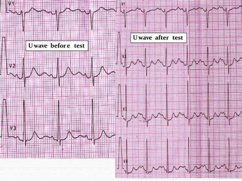 U-wave amplitude after “adrenaline test” in an ATS1 patient.jpg Gunnam
23:29, 11 February 2020
772 × 579; 179 KB
-
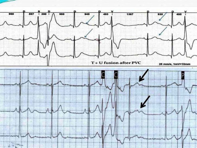 LQTS pattern in Andersen - Tawil syndrome (ATS).jpg Gunnam
23:21, 11 February 2020
793 × 595; 165 KB
-
-
-
-
-
-

-

-

-

-
-
-
-
-
-

-

-

-

-
-

-
 Grab von August Eisenmenger auf dem Wiener Zentralfriedhof.jpg AbdelrahmanAbushouk
12:44, 26 January 2020
512 × 683; 174 KB
-
-

-

-

-
-

-
-
 Aortic-intramural-haematoma-6 (1).jpg Sahar Memar Montazerin
20:10, 17 January 2020
509 × 439; 39 KB
-

-
 Aortic-intramural-haematoma-1 (1).jpg Sahar Memar Montazerin
19:23, 17 January 2020
512 × 512; 52 KB
-
-
 Pathogenesis-of-aortic-intramural-haematoma-illustration.png Sahar Memar Montazerin
18:16, 17 January 2020
1,128 × 1,128; 875 KB
-
-
-

-
-
 Penetrating-aortic-atherosclerotic-ulcer-with-false-aneurysm (2).jpg Sahar Memar Montazerin
21:18, 3 January 2020
1,024 × 1,024; 156 KB
-
 Penetrating-aortic-atherosclerotic-ulcer-with-false-aneurysm (1).jpg Sahar Memar Montazerin
21:07, 3 January 2020
1,024 × 1,024; 196 KB
-
 Penetrating-aortic-atherosclerotic-ulcer-with-false-aneurysm.jpg Sahar Memar Montazerin
20:56, 3 January 2020
1,024 × 1,024; 177 KB
-
 Pathogenesis-of-penetrating-atherosclerotic-ulcer-illustration (1).png Sahar Memar Montazerin
18:38, 3 January 2020
1,128 × 1,128; 877 KB
-
-
 Cardiac amyloidosis very high mag he.jpg Maneesha Nandimandalam
15:23, 3 January 2020
4,272 × 2,848; 3.16 MB
-

-
-
-
-
-
-

-

-
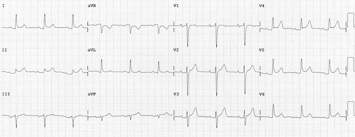
 ECG-Idiopathic-dilated-cardiomyopathy-Biatrial-hypertrophy.jpg AbdelrahmanAbushouk
18:56, 29 December 2019
1,200 × 507; 142 KB
-
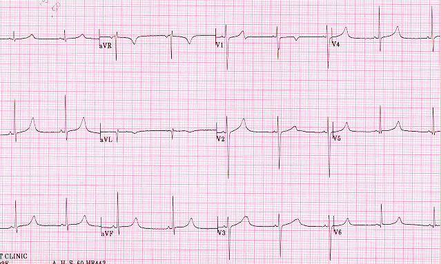
 ECG-Ischaemic-dilated-cardiomyopathy-1.jpg AbdelrahmanAbushouk
18:50, 29 December 2019
1,200 × 533; 192 KB
-

-
-
-

-

-
-
 438a79613ec103c0a40e06b1cb6402 big gallery.jpg Fahimeh Shojaei
15:23, 16 December 2019
520 × 630; 27 KB
-
 Fa018be29edc63c9ab2c136c0ee43b big gallery.jpeg Fahimeh Shojaei
15:22, 16 December 2019
630 × 630; 35 KB
-
 D435dff9c313eed96624ff2361b64d big gallery.jpeg Fahimeh Shojaei
15:19, 16 December 2019
630 × 429; 30 KB
-

-

-
 1feebfb356e0ad4f722c1640566332 big gallery.jpg.bmp Fahimeh Shojaei
15:34, 10 December 2019
630 × 376; 695 KB
-

-
 A6ab844024ae1ae5f996fc6dd7fcb2 jumbo.jpeg Fahimeh Shojaei
15:15, 10 December 2019
1,024 × 1,023; 130 KB
-
 A4c19436ff054f7414e2af1c30067a jumbo.jpeg Fahimeh Shojaei
15:14, 10 December 2019
1,013 × 1,024; 146 KB
-

-
 DNA chemical structure with repositioned labels.png Marshallsumter
01:41, 1 December 2019
1,500 × 1,750; 545 KB
-
 Iatrogenic-abdominal-aortic-dissection.jpg Sahar Memar Montazerin
18:29, 27 November 2019
400 × 400; 26 KB
-
 Aortic-dissection-debakey-type-1.jpg Sahar Memar Montazerin
18:21, 27 November 2019
512 × 512; 73 KB
-
-

-

-
-

-
-
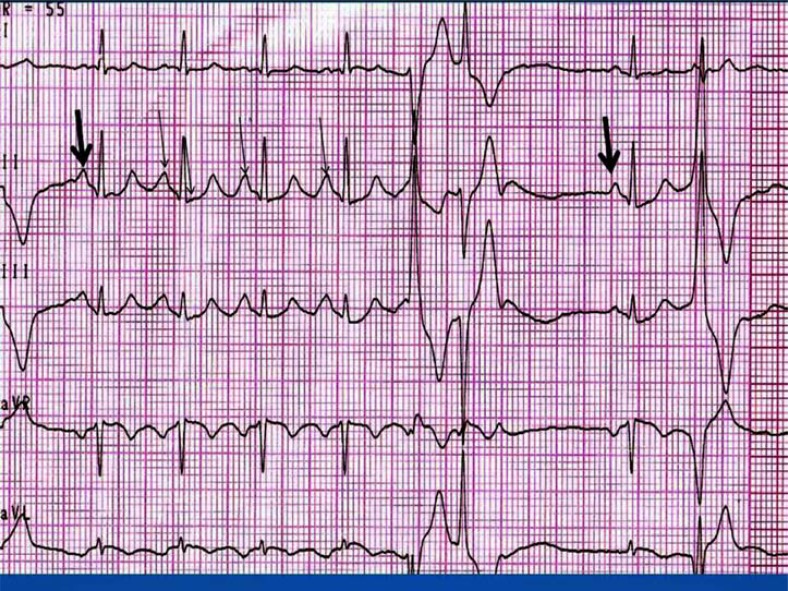
 Electrocardiograms (ECG) from members of a family with LQTS.gif Gunnam
13:59, 26 November 2019
220 × 299; 231 KB
-
-

-

-

-
-
 GEO Profile of A1BG tissue expression.jpg Marshallsumter
00:08, 20 November 2019
1,250 × 419; 346 KB
-
-
-
-
-
-
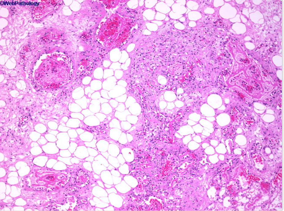
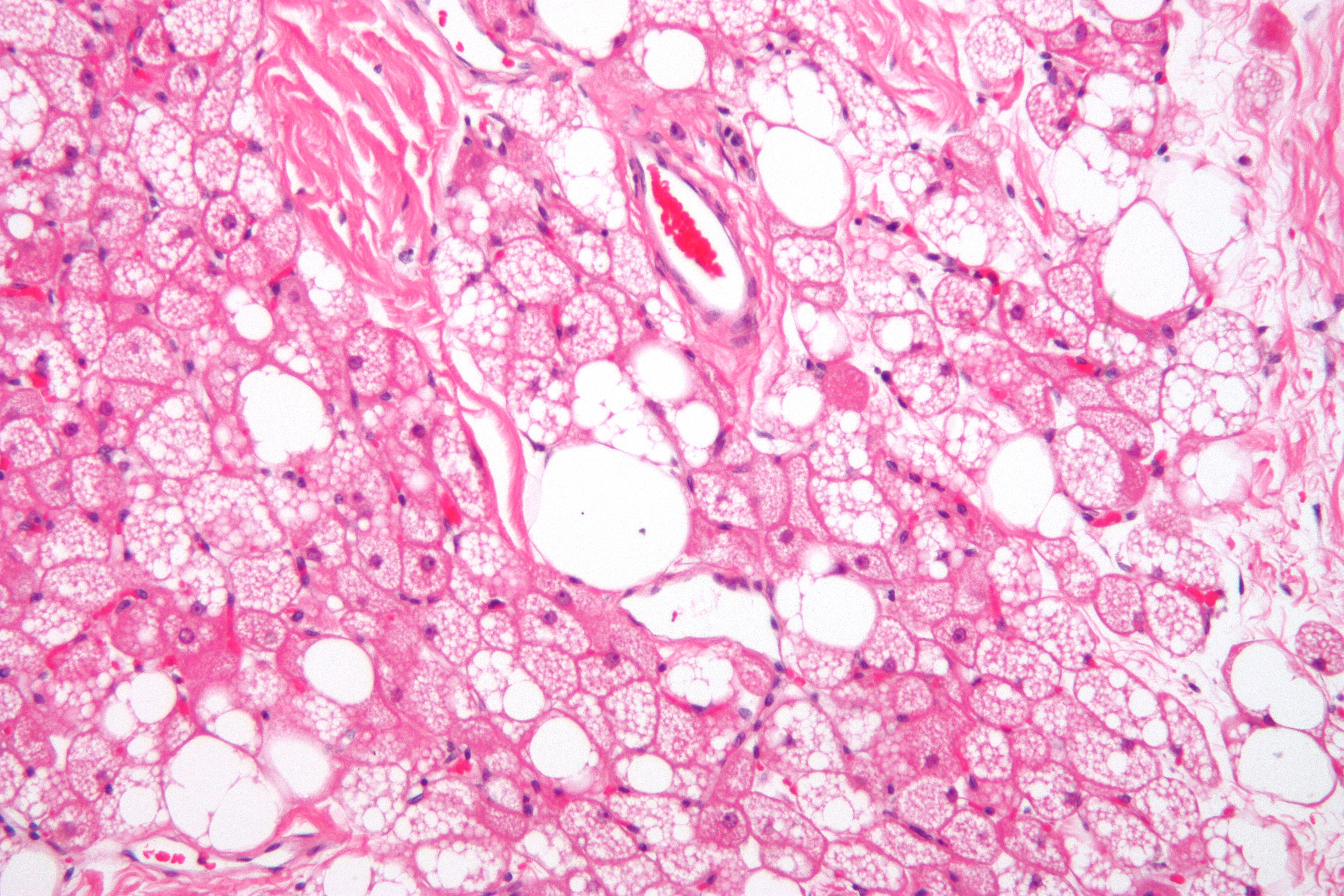
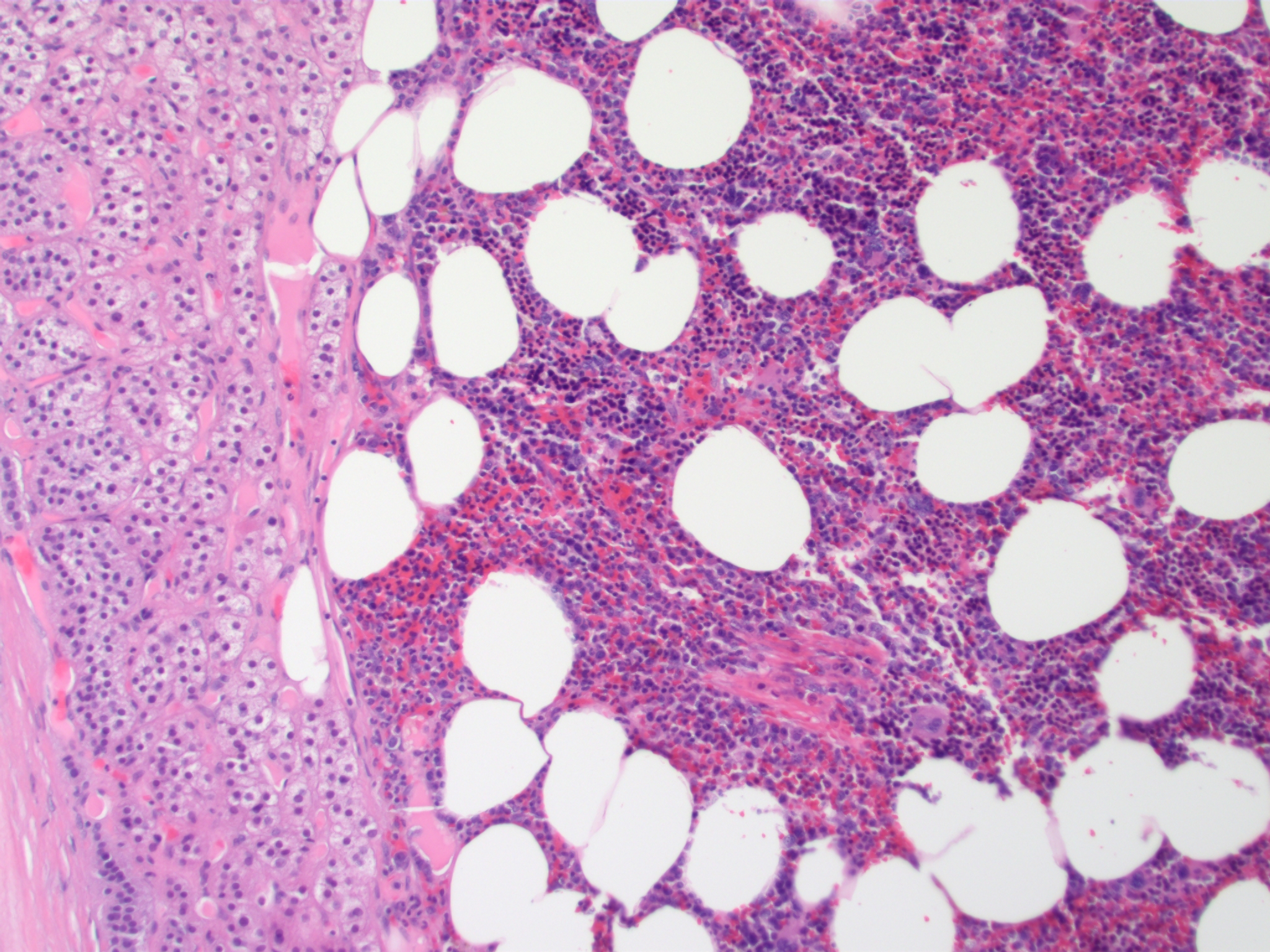 Adrenal Myelolipoma MP CTR.jpg Sahar Memar Montazerin
22:11, 13 November 2019
2,048 × 1,536; 2.29 MB
-
-
 MRI FLAIR sequence showing hyperintense signal in the periaqueducal grey matter in Wernicke Encephalopathy.jpg AbdelrahmanAbushouk
19:35, 12 November 2019
1,004 × 1,050; 72 KB
-
 Tachycardia-mediated-cardiomyopathy.jpg AbdelrahmanAbushouk
18:52, 12 November 2019
4,280 × 3,520; 2.27 MB
-
-
 Tear drop poikilocytes in Myelofibrosis.jpg Sabawoon Mirwais
15:02, 12 November 2019
1,280 × 1,024; 133 KB
-
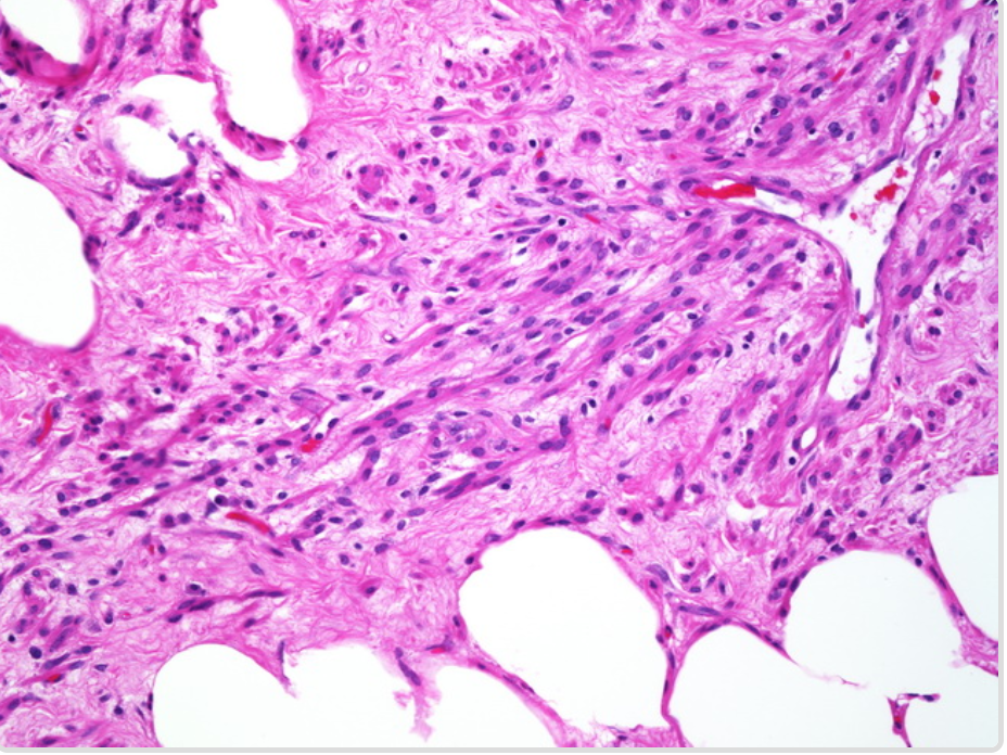
 Spindle cell lipoma -- intermed mag.jpg Sahar Memar Montazerin
21:37, 11 November 2019
4,000 × 6,000; 11.18 MB
-
 Spindle Cell Lipoma (2275051876).jpg Sahar Memar Montazerin
21:23, 11 November 2019
1,634 × 1,332; 1.08 MB
-

-
 MRI FLAIR sequence Wernicke Encephalopathy.jpg AbdelrahmanAbushouk
17:21, 11 November 2019
800 × 837; 62 KB
-
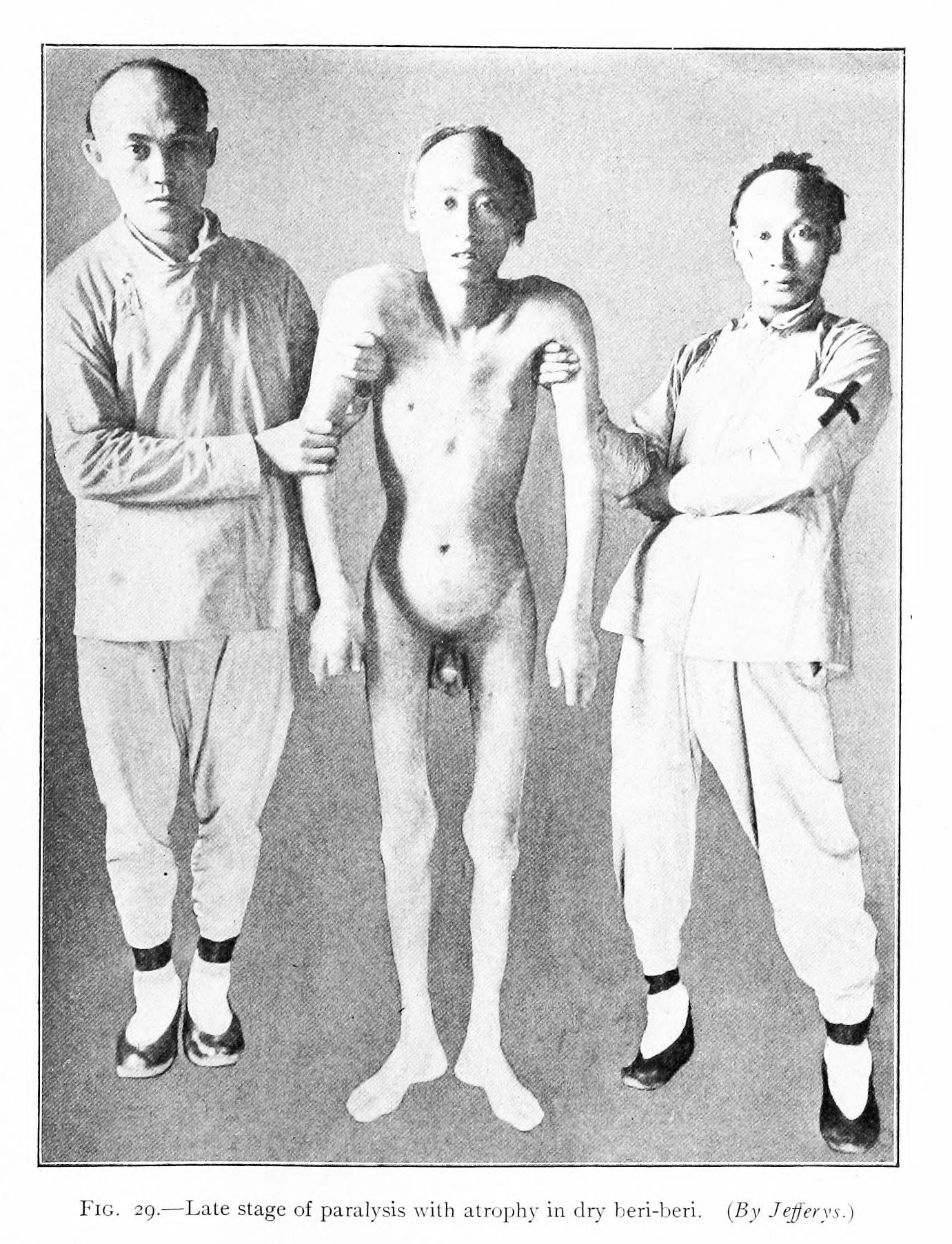 Late stage of paralysis with atrophy in dry beriberi.jpg AbdelrahmanAbushouk
14:56, 11 November 2019
1,381 × 1,805; 883 KB
-

-

-
-

-
-

-
-
 Cardiac-amyloidosis-Nuclear-posterior.jpg Mandana Chitsazan
23:47, 3 November 2019
512 × 1,024; 157 KB
-
 Cardiac-amyloidosis-Nuclear-anterior.jpg Mandana Chitsazan
23:46, 3 November 2019
512 × 1,024; 134 KB
-
 Nodular-pulmonary-amyloidosis-3 (1).jpg Sahar Memar Montazerin
17:54, 1 November 2019
3,056 × 3,056; 2.3 MB
-
 Nodular-pulmonary-amyloidosis-3.jpg Sahar Memar Montazerin
17:47, 1 November 2019
2,816 × 2,364; 1.47 MB
-
-

-

-
-

-
-
-
-
-
-
-
-
-
-
-

-

-

-

-

-
-
-

-

-
 Primitive-neuroectodermal-tumour-of-the-cns-1.jpg Sahar Memar Montazerin
15:43, 28 October 2019
512 × 512; 29 KB
-
-
-
-
-
-

-

-
-

-

-

-

-
-
-
-
-

-

-
 Amyloidosis-bronchial-and-diffuse-nodular-pulmonary-involvement.jpg Sabawoon Mirwais
01:23, 26 October 2019
512 × 512; 58 KB
-
-
-
-
-
-
-
-
-
-
-
 Amyloidosis, Node, Lugol's Reaction.jpg Sabawoon Mirwais
19:45, 25 October 2019
4,006 × 2,242; 4.23 MB
-
-
-

-

-
 Glioblastoma-nos-butterfly-morphologyyy.jpg Fahimeh Shojaei
15:41, 25 October 2019
819 × 1,024; 106 KB
-
 Glioblastoma-nos-butterfly-morphology.jpg Fahimeh Shojaei
15:38, 25 October 2019
819 × 1,024; 106 KB
-

-

-
 2169e7e3234fa8afb3a89e0a0bab37 big gallery.jpg Fahimeh Shojaei
15:29, 25 October 2019
630 × 630; 88 KB
-

-

-
-

-

-
-
-

-
-
-
-
 Follicular-thyroid-cancer-metastasis.jpg Sahar Memar Montazerin
19:27, 17 October 2019
2,328 × 2,930; 243 KB
-
 800px-Macrophages in bronchial wash specimen -- very high mag.jpg Ramyar
15:46, 17 October 2019
800 × 533; 38 KB
-

-
 Angiosarcoma (5617087462) (1).jpg Sahar Memar Montazerin
15:13, 15 October 2019
1,976 × 1,415; 945 KB
-
-

-

-
-
-

-

-
-

-
 Bundles of wavy spindle cells with serpentine nuclei .jpg Gunnam
13:50, 27 September 2019
779 × 502; 89 KB
-
-
-
 Forest plot of target HbA1c for Diabetes Type II.jpg Badgettrg
13:28, 24 September 2019
734 × 728; 97 KB
-
 Intravascular lymphoma - very high mag.jpg Syed Musadiq Ali
12:07, 19 September 2019
4,272 × 2,848; 5.33 MB
-
 Hypertrophic plexus chiari II intermed mag.jpg Fahimeh Shojaei
21:58, 18 September 2019
2,080 × 1,542; 668 KB
-
 Hypertrophic plexus chiari II low mag.jpg Fahimeh Shojaei
21:53, 18 September 2019
2,080 × 1,542; 658 KB
-

-
 Chiari-ii-malformation-ct-morphology.jpg Fahimeh Shojaei
14:27, 18 September 2019
761 × 1,253; 392 KB
-

-
-
 Atypical-teratoid-rhabdoid-tumour-atrt v2.jpg Gerald Chi
14:59, 16 September 2019
1,024 × 1,024; 197 KB
-
-
-

-
-
-
-
-
-
-

-

-
-
-
-

-

-
-
-
-
-
-
-
 Spindle-shaped fibroblasts, arranged in a storiform pattern.png Homa Najafi
03:47, 27 August 2019
512 × 385; 504 KB
-
 Proliferating histiocytic cells with foamy, granular cytoplasm.png Homa Najafi
03:44, 27 August 2019
512 × 384; 525 KB
-

-

-

-
-

-

-
 Hepatic Adenoma CT (portal venous phase).jpg Sabawoon Mirwais
23:13, 20 August 2019
800 × 800; 63 KB
-

-

-

-
-
-
 Dermatofibrosarcoma protuberans (3) recurrence.jpg Sara Mohsin
17:16, 16 August 2019
500 × 376; 99 KB
-
 Dermatofibrosarcoma protuberans (2) recurrence.jpg Sara Mohsin
17:13, 16 August 2019
500 × 376; 136 KB
-
 Dermatofibrosarcoma protuberans (1) recurrence.jpg Sara Mohsin
17:06, 16 August 2019
500 × 376; 149 KB
-
-
 Miliary-metastases-from-papillary-thyroid-cancer.png Sahar Memar Montazerin
13:39, 16 August 2019
624 × 556; 297 KB
-
-


.gif)


.jpg)


.jpg)


.jpg)


.jpg)


.jpg)
.jpg)


.jpg)


.jpg)
.jpg)
.jpg)
.jpg)


.jpg)


.jpg)


.jpg)


.jpg)

.jpg)


.jpg)

.jpg)


.jpg)
.jpg)

.png)


_from_members_of_a_family_with_LQTS.gif)


.jpg)


.jpg)


.jpg)


.jpg)


.jpg)


_(1).jpg)


.jpg)


.jpg)
.jpg)


_CD34.jpg)
_recurrence.jpg)
_recurrence.jpg)
_recurrence.jpg)


_in_Transesophageal_echocardiography.jpg)
_.gif)