Gallery of new files
Jump to navigation
Jump to search
This special page shows the last uploaded files.
-
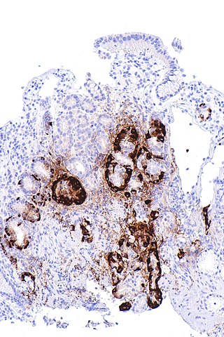 320px-Metaplastic atrophic gastritis - body - chromogranin A -- intermed mag.jpg Sanajan
18:01, 24 June 2020
320 × 480; 66 KB
-
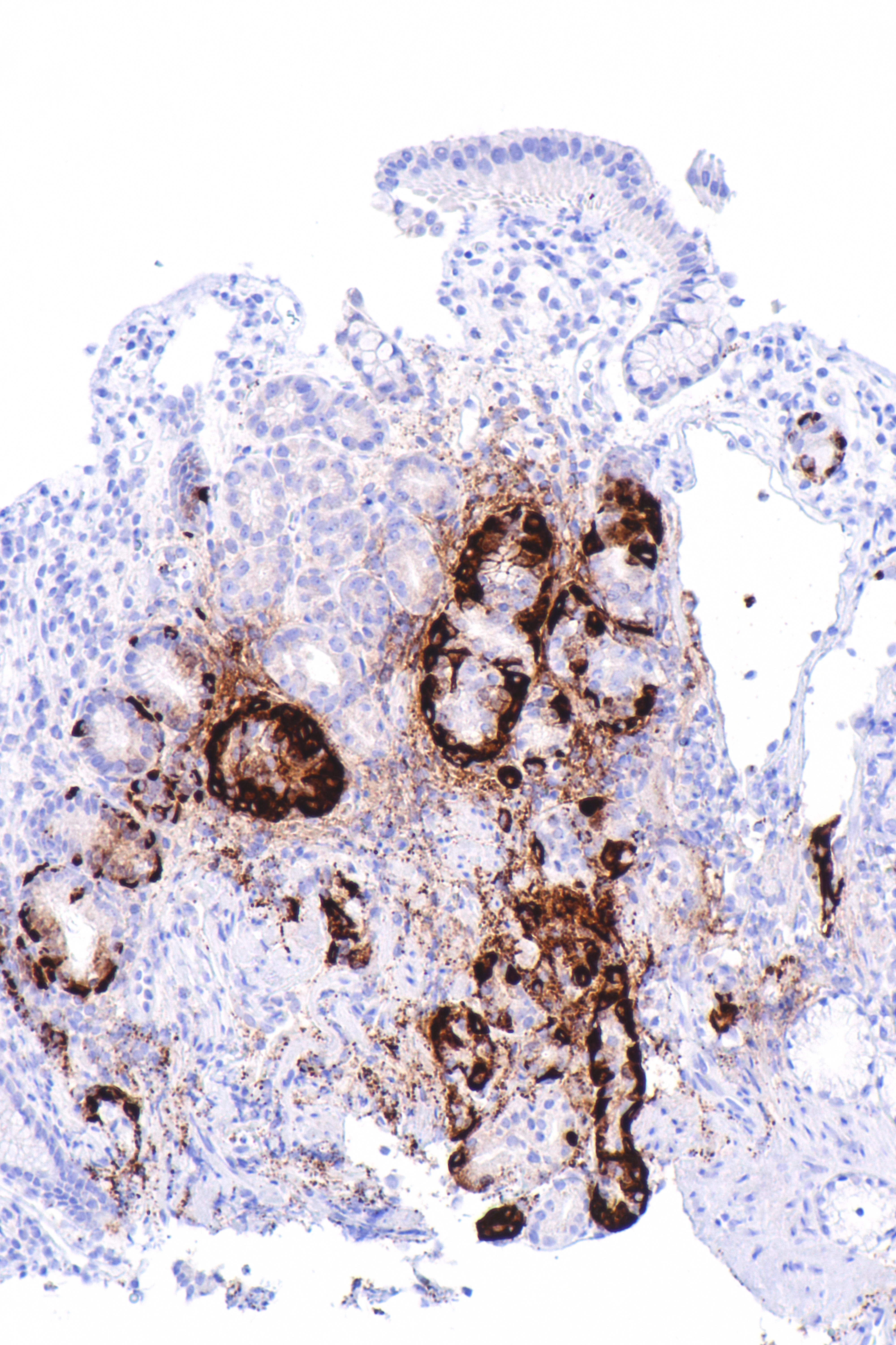
 Metaplastic atrophic gastritis - body - chromogranin A -- intermed mag.jpg Sanajan
17:26, 24 June 2020
4,000 × 6,000; 8.77 MB
-
-
-

-
-
 Acute Phosphate Nephropathy - Calcium Phosphate crystal deposition.jpg AFarheen
22:34, 22 June 2020
640 × 480; 91 KB
-

-

-
-
-
-
 Histopathology of secondary segmental glomerular sclerosis of hypertensive nephropathy.jpg NNikravangolsefid
23:52, 19 June 2020
528 × 427; 128 KB
-
-
 Page1-636px-COVID-19 Key-Message A3-Posters Stop.pdf.jpg Sara Mohsin
16:11, 19 June 2020
636 × 899; 79 KB
-
-

-

-
-
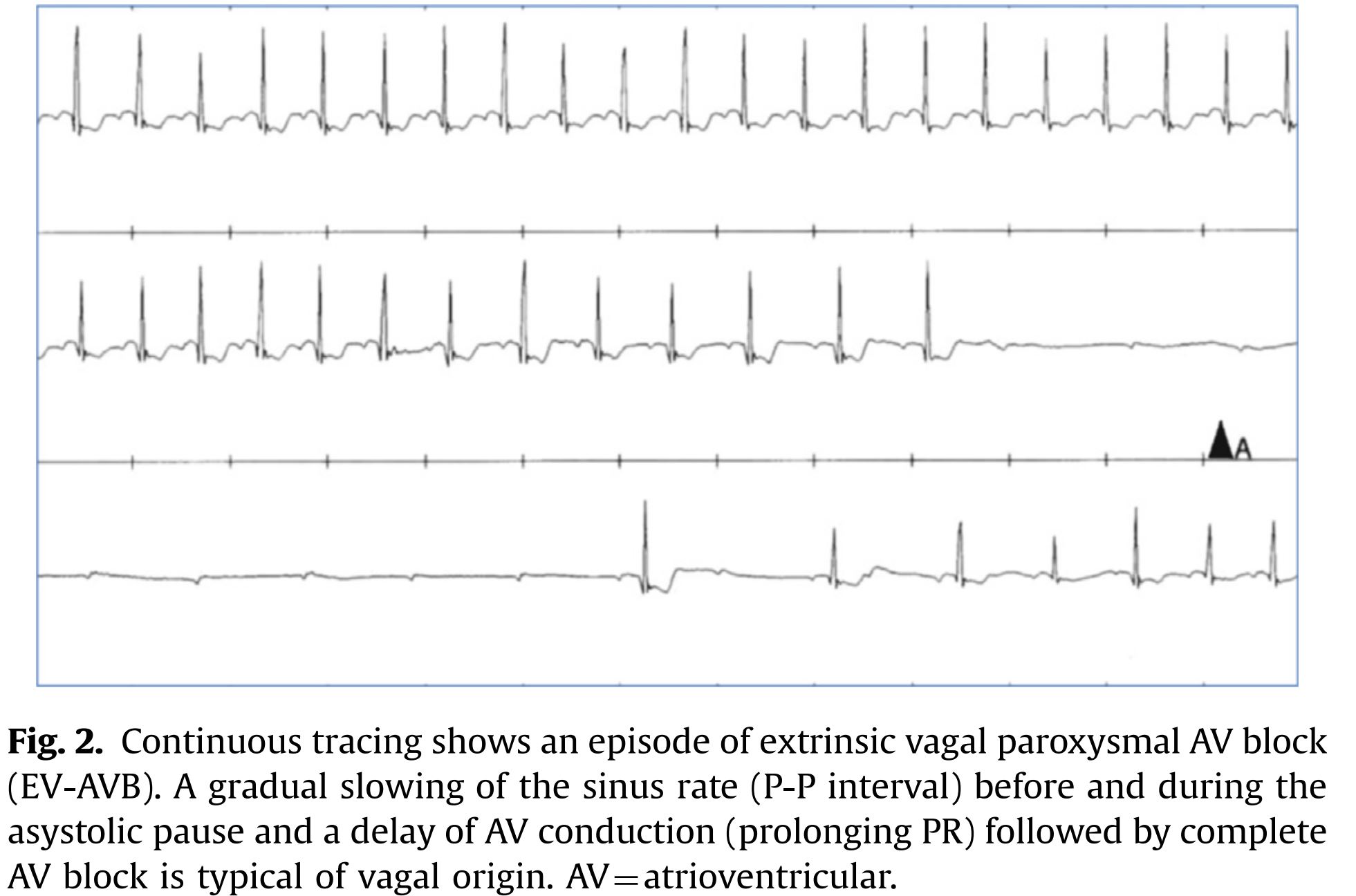
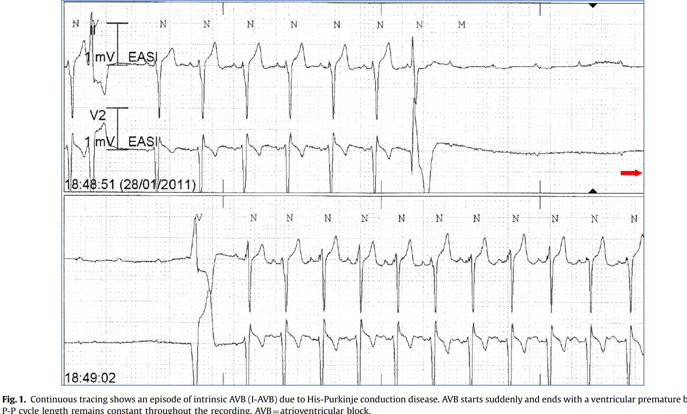
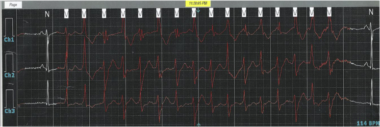
 Complete AV Block with Sinus slowing during carotid Sinus massage Case report.jpg Akash Daswaney
11:27, 18 June 2020
1,168 × 534; 119 KB
-
-
-
-
 79px-Life cycle of Rustic Butterfly (2663558308).jpg Maneesha Nandimandalam
15:18, 15 June 2020
79 × 119; 3 KB
-

-
-
-
-
-
-
-
-
-
-
-
-
-
-

-
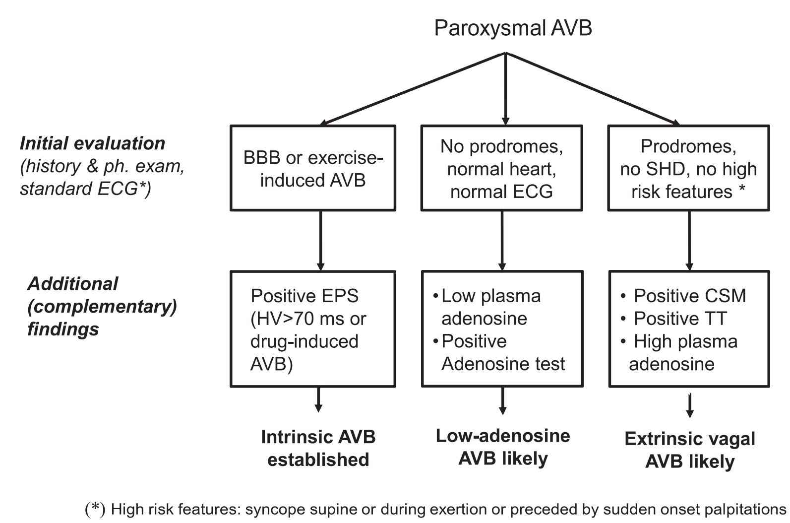
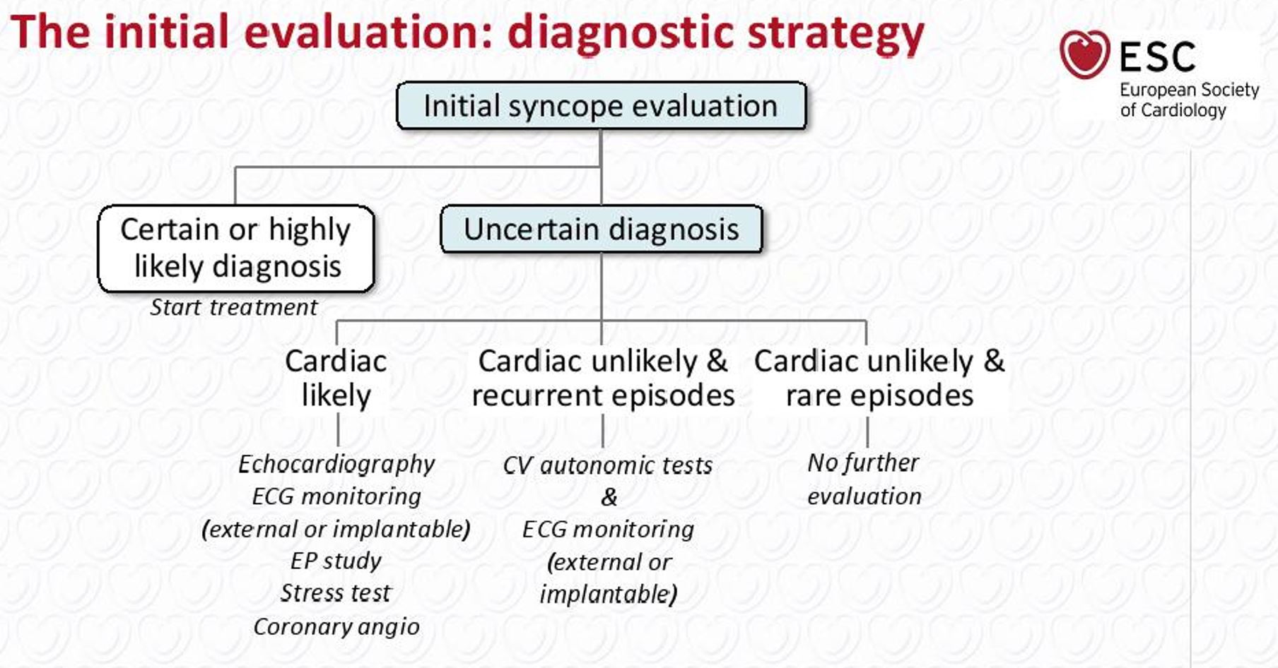 Initial Strategy Syncope or Paroxysmal AV Block.JPG Akash Daswaney
10:56, 13 June 2020
1,808 × 946; 188 KB
-
-
-
-
-
-
-
-
-
-

-

-
-
-

-

-

-

-

-

-
-
-
-
-
-
-
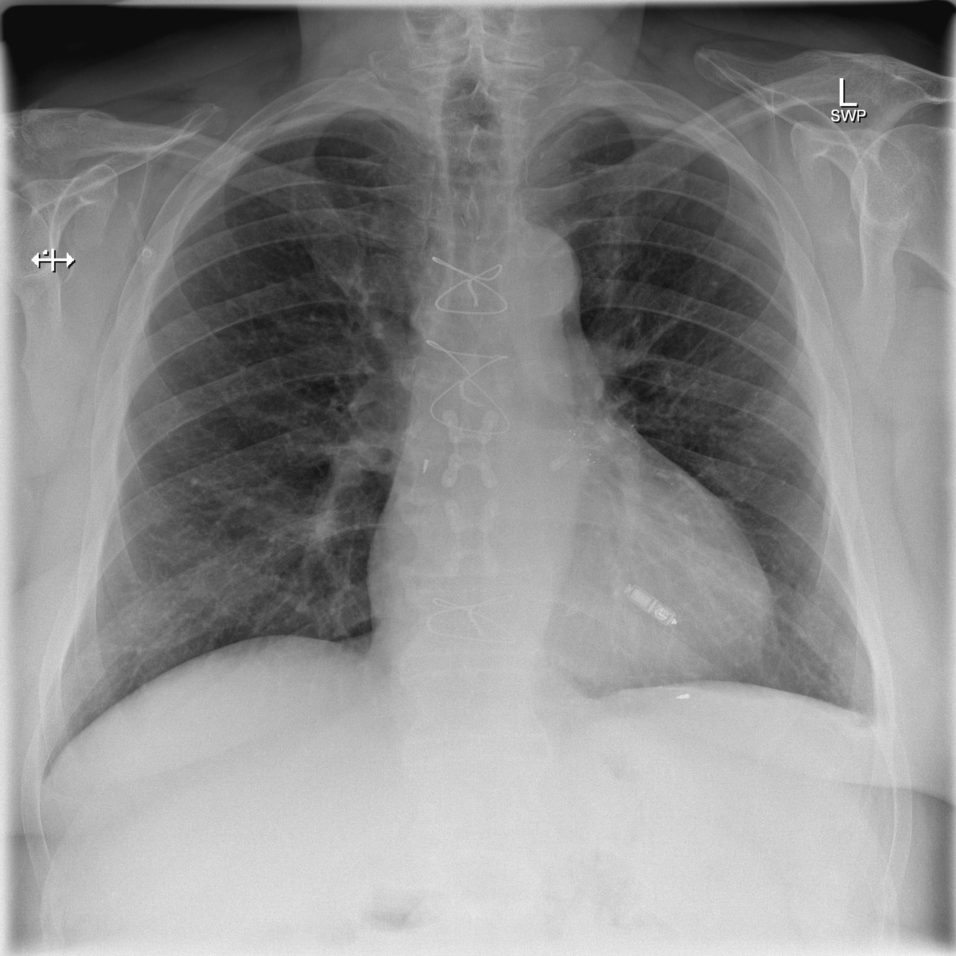
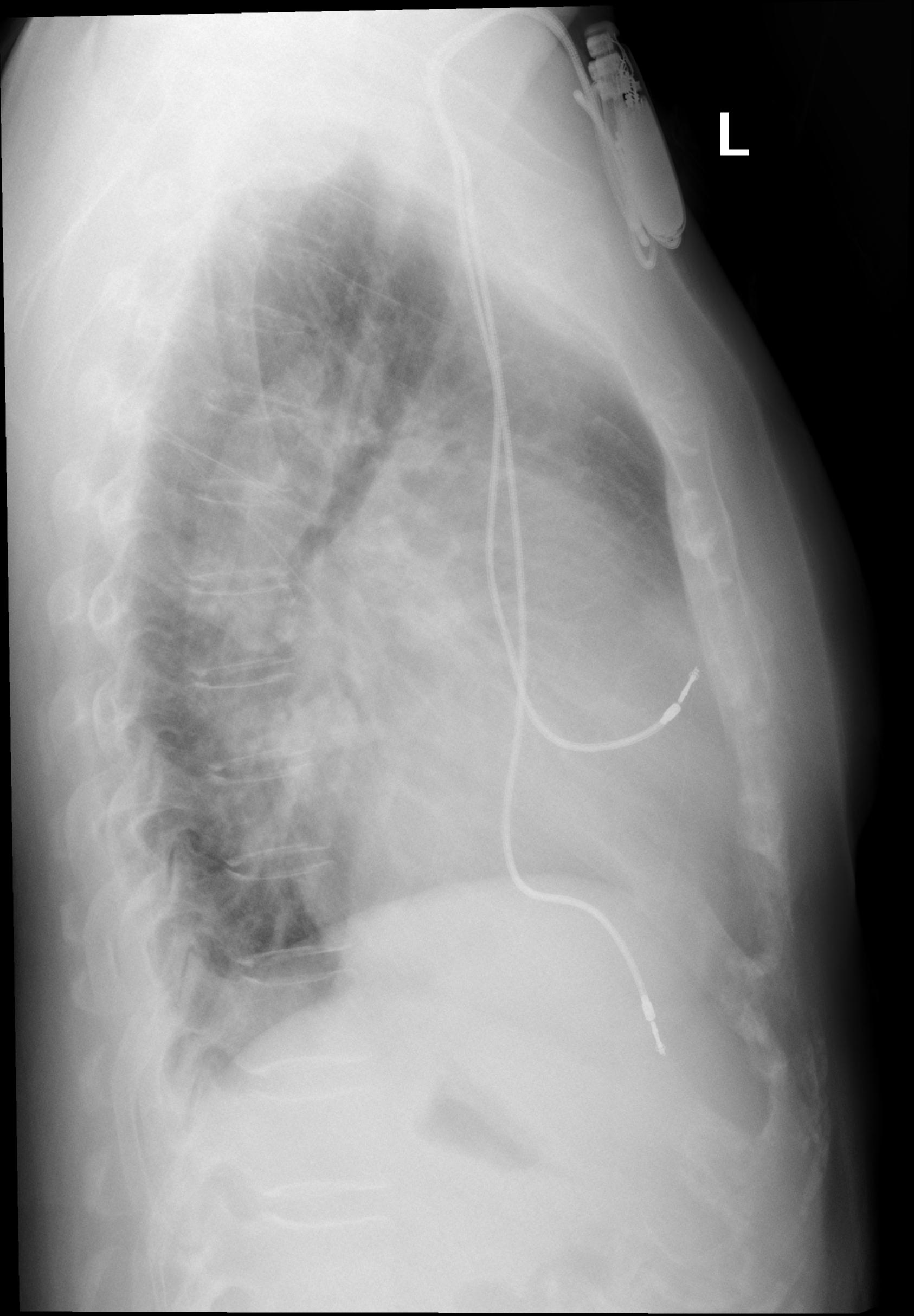
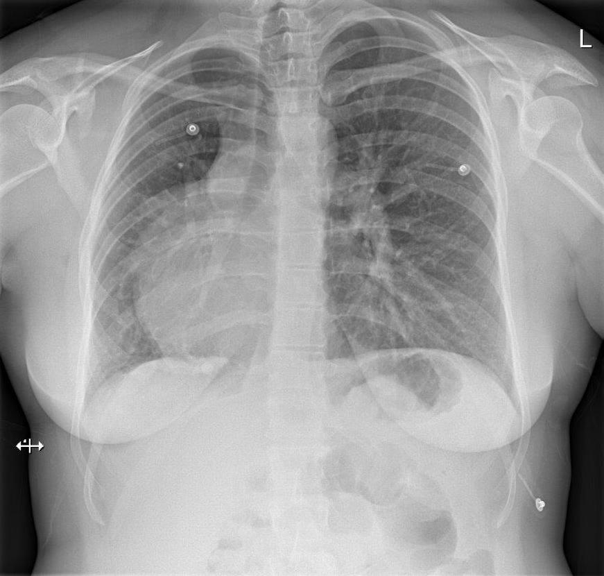
 X-ray of pacemaker with right atrial and ventricular lead.jpg Javaria Anwer
09:40, 9 June 2020
725 × 361; 46 KB
-

-

-

-
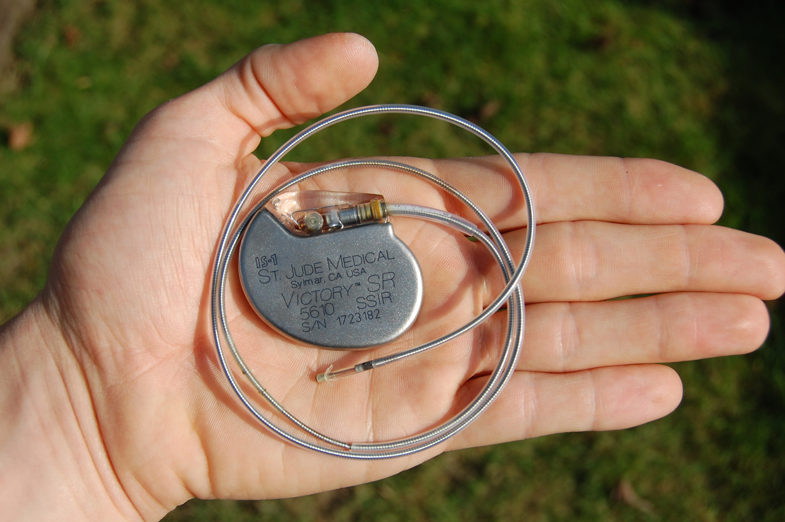 An artificial pacemaker shown in hand with electrode and lead (from St Jude medical).jpg Javaria Anwer
05:26, 8 June 2020
3,008 × 2,000; 2.83 MB
-
 Renal arterial hyalinosis - pas - very high mag.jpg NNikravangolsefid
22:51, 7 June 2020
4,272 × 2,848; 3.91 MB
-

-
-
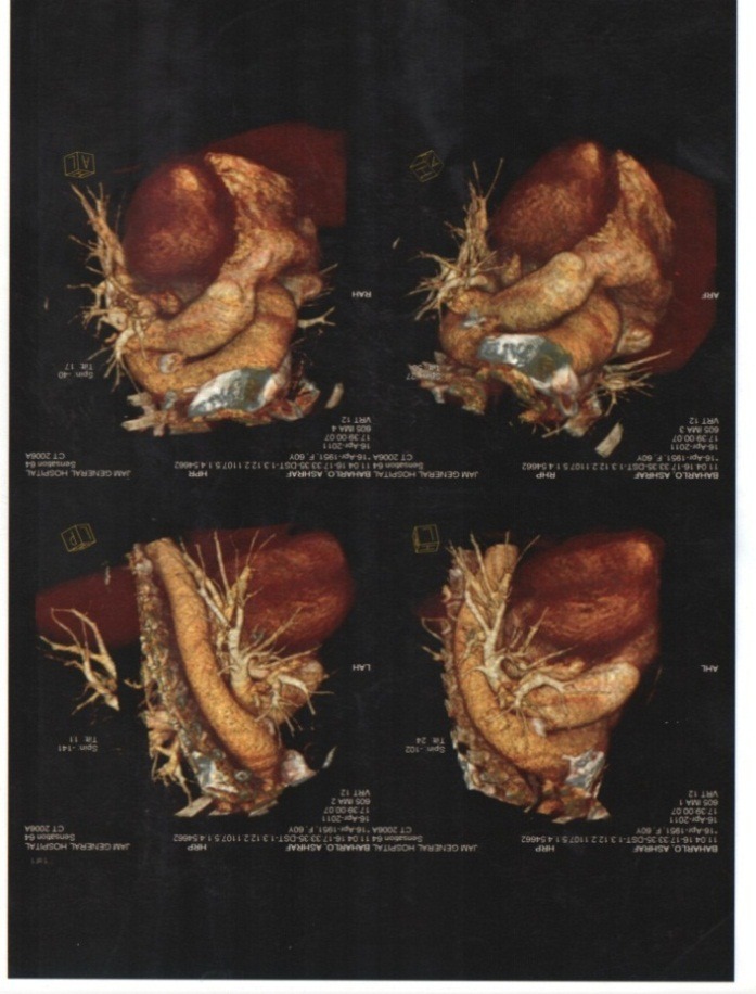
 Unilateral-pulmonary-oedema-blalock-taussig-shunt-in-pulmonary-atresia-with-ventricular-septal-defect-1.jpg Usmanaliakbar
03:49, 7 June 2020
2,037 × 2,642; 3 MB
-
 Histopathology of hypertensive glomerular lesion of hypertensive nephropathy.jpg NNikravangolsefid
21:34, 5 June 2020
558 × 452; 114 KB
-
 Fibrous intimal thickening in hypertensive nephropathy.jpg NNikravangolsefid
20:37, 5 June 2020
251 × 317; 41 KB
-
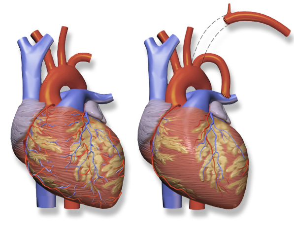
 Modified-blalock-taussig-shunt-creative-commons.png Usmanaliakbar
18:50, 5 June 2020
359 × 476; 165 KB
-
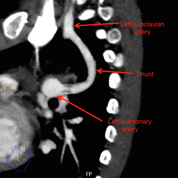
 Original-blalock-taussig-shunt-creative-commons.png Usmanaliakbar
15:55, 5 June 2020
597 × 469; 282 KB
-

-
 Sbo-secondary-to-ileal-stricture-from-crohns-disease.jpg Bosky Soni
03:11, 5 June 2020
766 × 857; 203 KB
-

-

-

-

-

-

-

-

-

-

-

-
 Monosodium-urate-crystals-in-tophaceous-gout.jpeg Jsoujanya
14:06, 31 May 2020
2,048 × 2,048; 2.5 MB
-

-
-

-

-

-

-
 C0438160-Life Cycle of the Black-legged Tick and Lyme, Illustration.jpg Rina Ghorpade
20:51, 29 May 2020
800 × 518; 104 KB
-

-

-
-
-
-
-

-

-

-
-
-
-
-
-
 Overheating is one of the chief risk factors for SIDS.jpg Gunnam
14:41, 15 May 2020
800 × 1,200; 385 KB
-
-
-

-
-

-

-
-
-
-
-

-
-

-

-
-

-
 WhatsApp Image 2020-05-07 at 1.36.56 AM.jpeg Abdulkareem Opeoluwalukan
11:41, 7 May 2020
640 × 640; 21 KB
-

-
-
-

-
-

-
-
-

-
-
-
 CMR four-chamber cine view. Seen here is the grossly dilated right heart, with an atrialized RV and dilated tricuspid annulus.jpg Gunnam
20:36, 22 April 2020
771 × 322; 68 KB
-
-
 Right-atrial-enlargement in Tricuspid regurgitation .jpg Gunnam
16:31, 20 April 2020
2,645 × 2,791; 396 KB
-

-
-
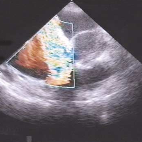
 Severe tricuspid regurgitation E00572 (CardioNetworks ECHOpedia).jpg Gunnam
16:29, 16 April 2020
660 × 526; 202 KB
-
-
-
-

-
-

-
-
-
-

-
-
 Normal echocardiographic appearance of tricuspid valve.jpg Gunnam
16:47, 19 March 2020
709 × 299; 65 KB
-

-
-

-
-
-

-
-
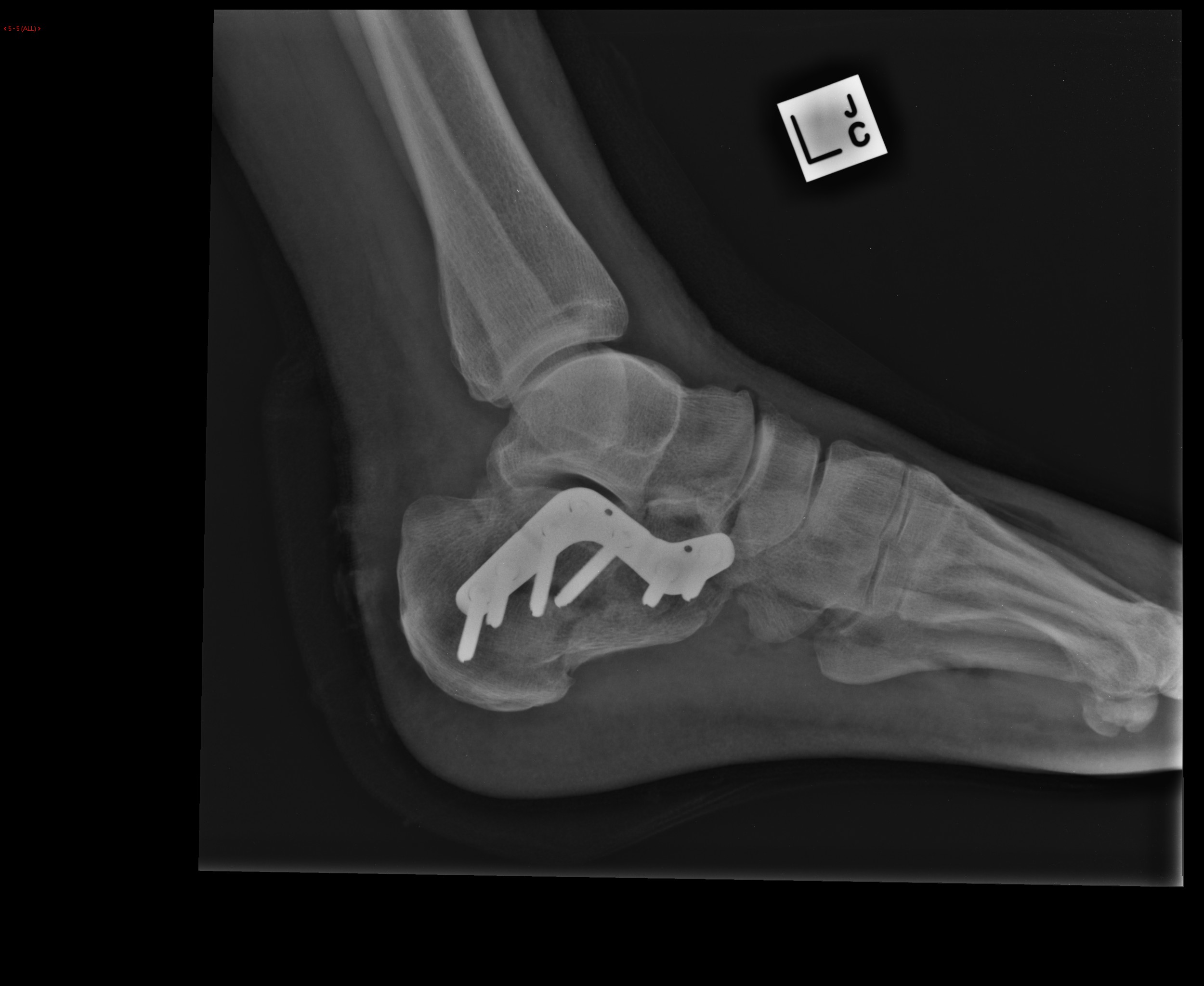
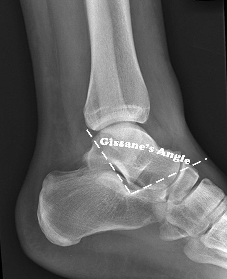
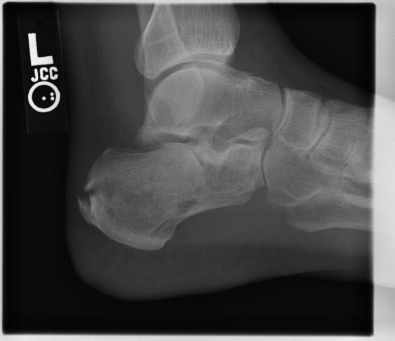
 Calcaneal-fracture-and-associated-spinal-injury (4).jpg DrMars
14:53, 14 March 2020
1,252 × 1,332; 150 KB
-
 Calcaneal-fracture-and-associated-spinal-injury (3).jpg DrMars
14:53, 14 March 2020
1,252 × 2,884; 282 KB
-
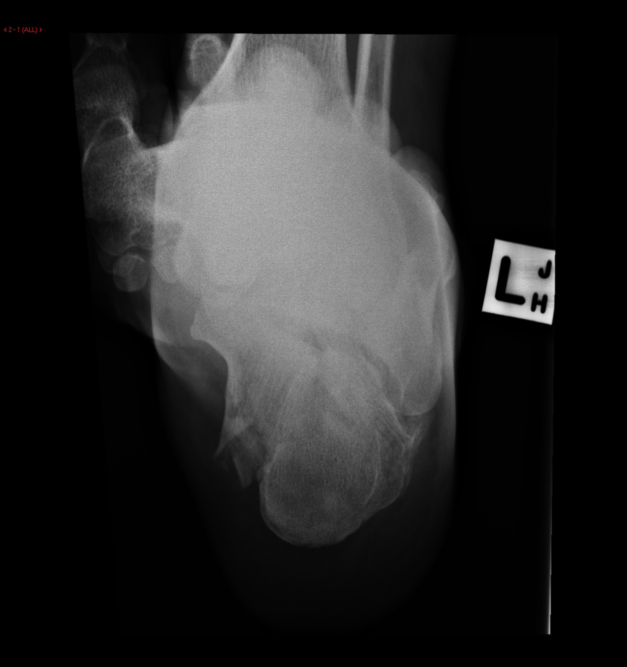
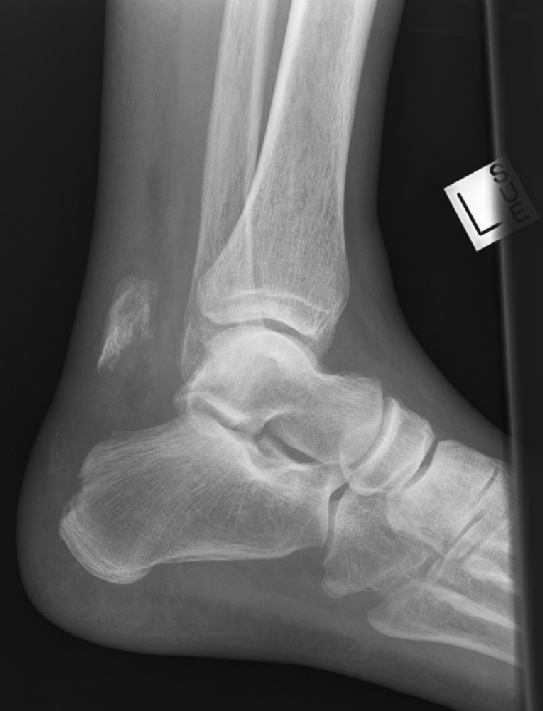
 Calcaneal-fracture-and-associated-spinal-injury (2).jpg DrMars
14:53, 14 March 2020
1,412 × 1,756; 157 KB
-
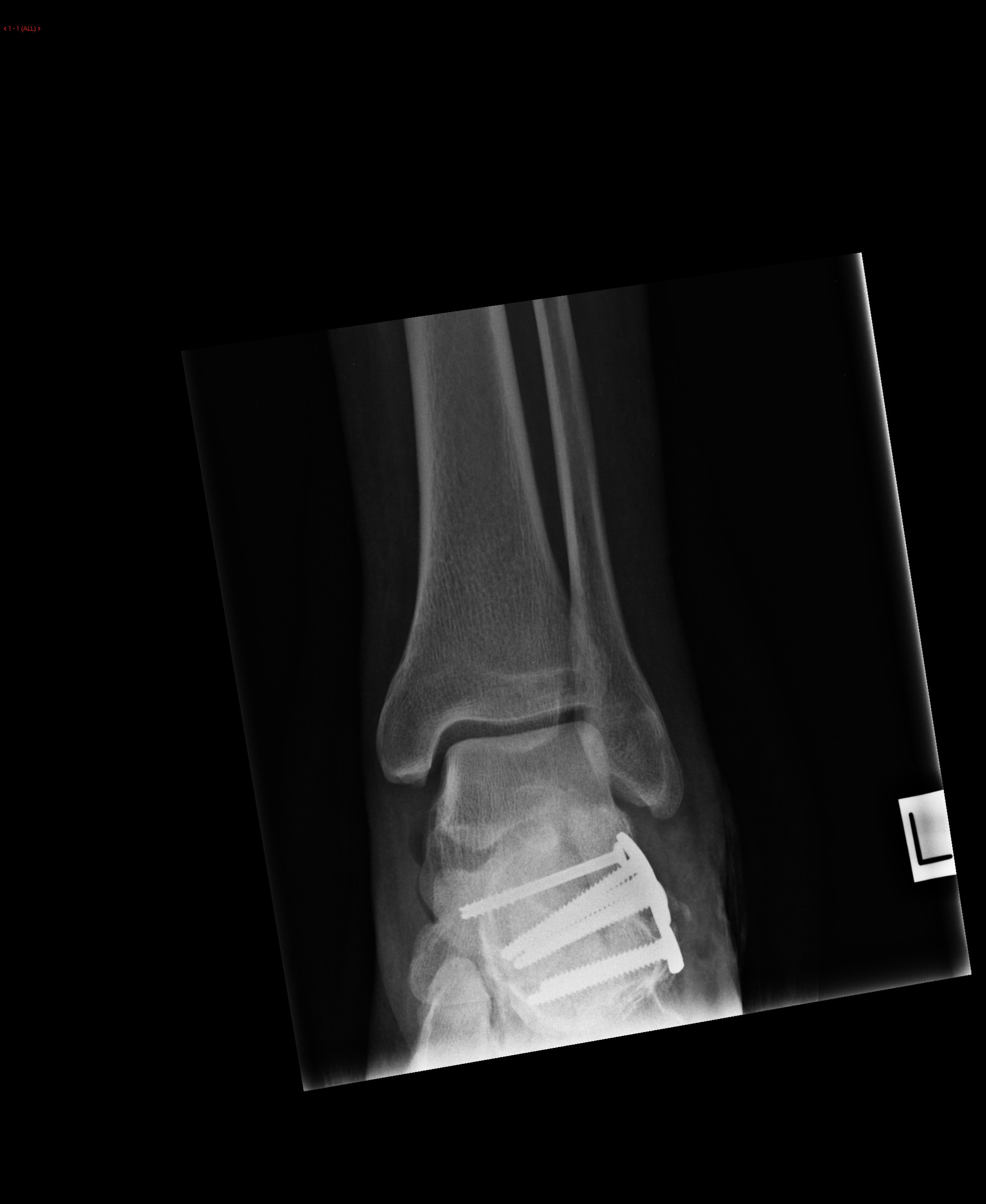
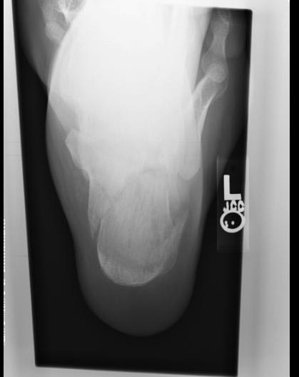
 Calcaneal-fracture-and-associated-spinal-injury (1).jpg DrMars
14:52, 14 March 2020
1,632 × 2,340; 250 KB
-
 Calcaneal-fracture-and-associated-spinal-injury.jpg DrMars
14:52, 14 March 2020
1,208 × 2,868; 297 KB
-

-
-
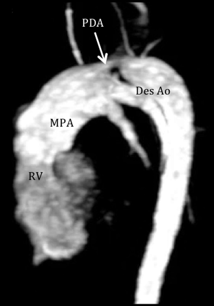
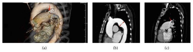
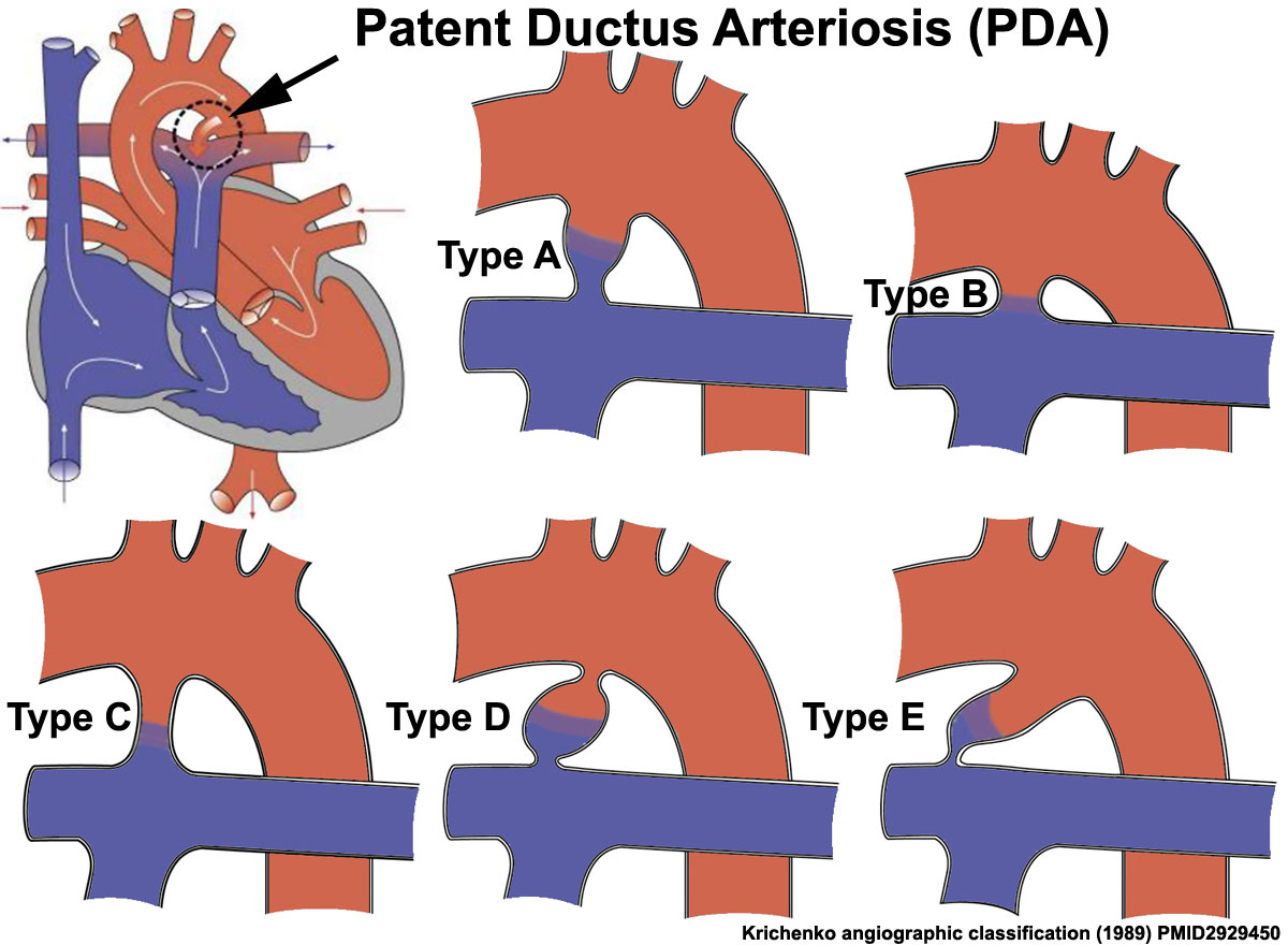
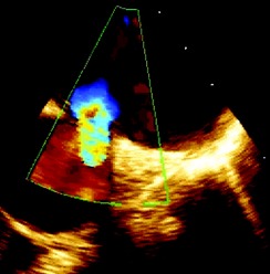 Patent ductus arteriosus (PDA) in Transesophageal echocardiography.jpg Gunnam
15:50, 13 March 2020
244 × 248; 16 KB
-
 Transesophageal echocardiogram of a patent ductus arteriosus.jpg Gunnam
14:28, 13 March 2020
654 × 250; 39 KB
-
-
-
-
-
-
-
-
-

-
-

-
-
-
-

-
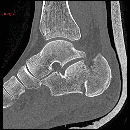
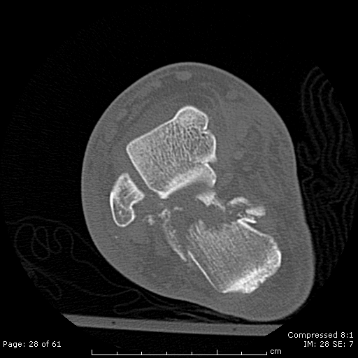
 Sanders-classification-of-calcaneal-fractures-1.jpg DrMars
00:16, 7 March 2020
9,000 × 9,000; 12.6 MB
-

-

-

-
-

-

-
-

-
-
-
-
-
-
-
-

-
 Focal granule cell dispersion in the dentate gyrus (DG).jpg Gunnam
15:36, 29 February 2020
708 × 533; 221 KB
-
 Total-anomalous-pulmonary-venous-return-type-iv.jpg Sahar Memar Montazerin
18:27, 26 February 2020
386 × 386; 36 KB
-
-
-
-
-
-

-
-

-
-

-
-
 Libman-Sacks-Endocarditis-The-presence-of-vegetations-predisposes-patients-to-bacterial.png Sara Mohsin
22:05, 25 February 2020
623 × 389; 365 KB
-
-

-

-
 Total-anomalous-pulmonary-venous-return-illustration.png Sahar Memar Montazerin
19:22, 19 February 2020
803 × 800; 288 KB
-
-

-
-
-
-
-
-

-

-

-
-

-
-
-
-
-
-
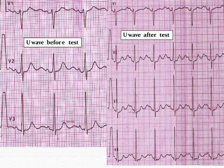 U-wave amplitude after “adrenaline test” in an ATS1 patient.jpg Gunnam
23:29, 11 February 2020
772 × 579; 179 KB
-
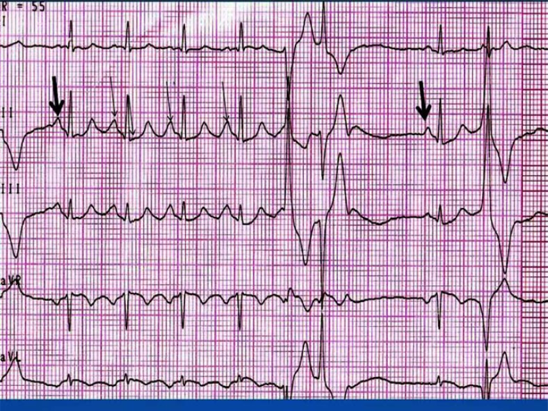
 LQTS pattern in Andersen - Tawil syndrome (ATS).jpg Gunnam
23:21, 11 February 2020
793 × 595; 165 KB
-
-
-
-
-


.gif)


.jpg)


.jpg)


.jpg)


.jpg)


.jpg)
.jpg)


.jpg)


.jpg)
.jpg)
.jpg)
.jpg)


.jpg)


.jpg)


.jpg)


_in_Transesophageal_echocardiography.jpg)