Gallery of new files
Jump to navigation
Jump to search
This special page shows the last uploaded files.
-

-

-

-

-
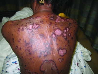
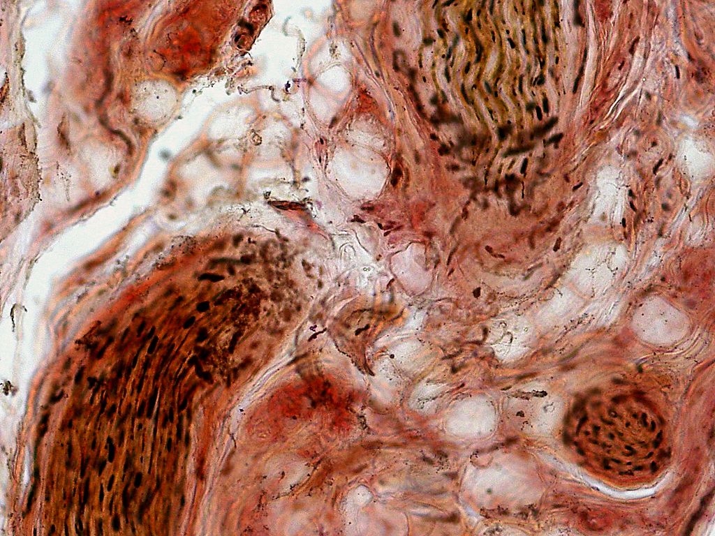
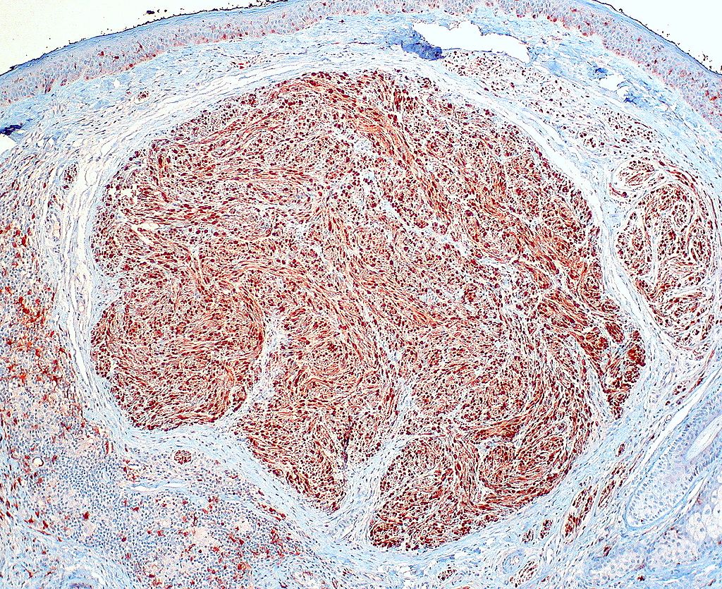
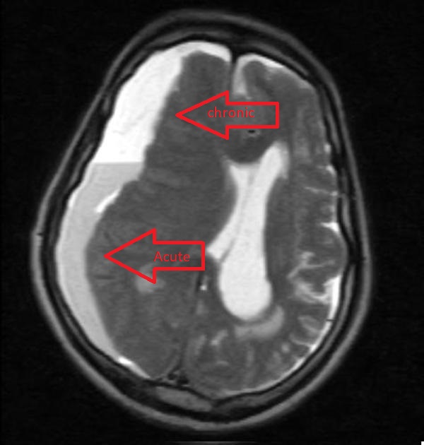
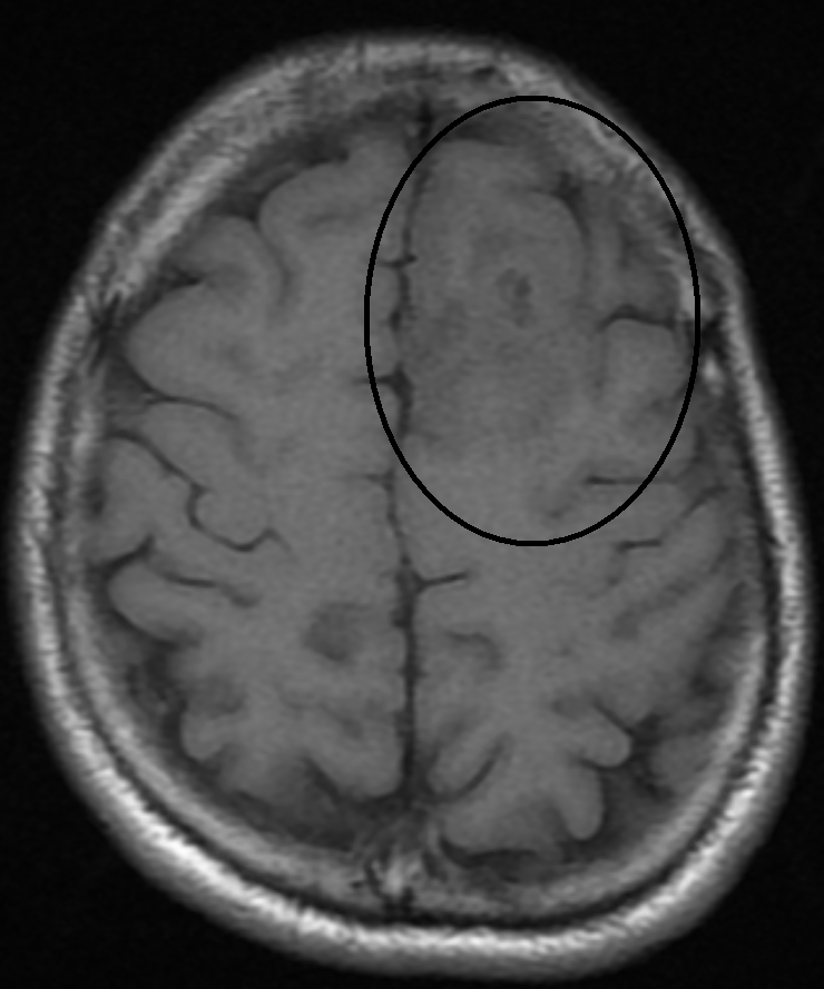
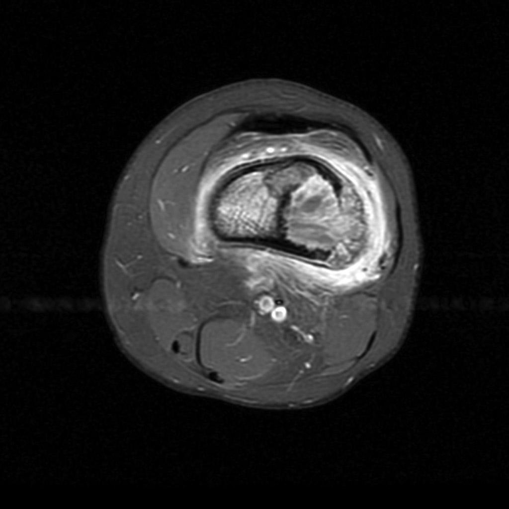
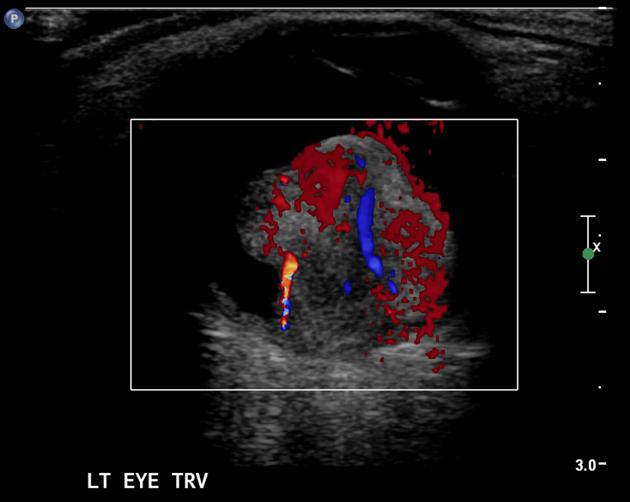 597ae8c0f532cd2564fbeab267ced3 big gallery.jpeg Fahimeh Shojaei
15:33, 24 June 2019
630 × 502; 35 KB
-

-

-
-
-

-
-

-
-

-
-

-
-
-
-

-

-
-

-
-
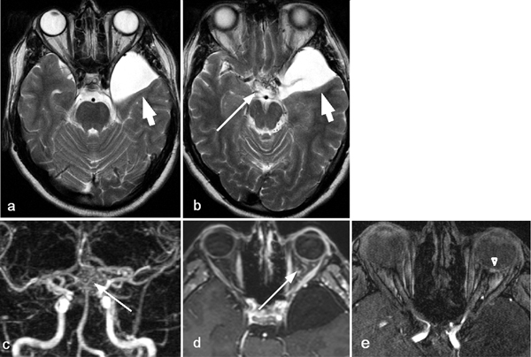
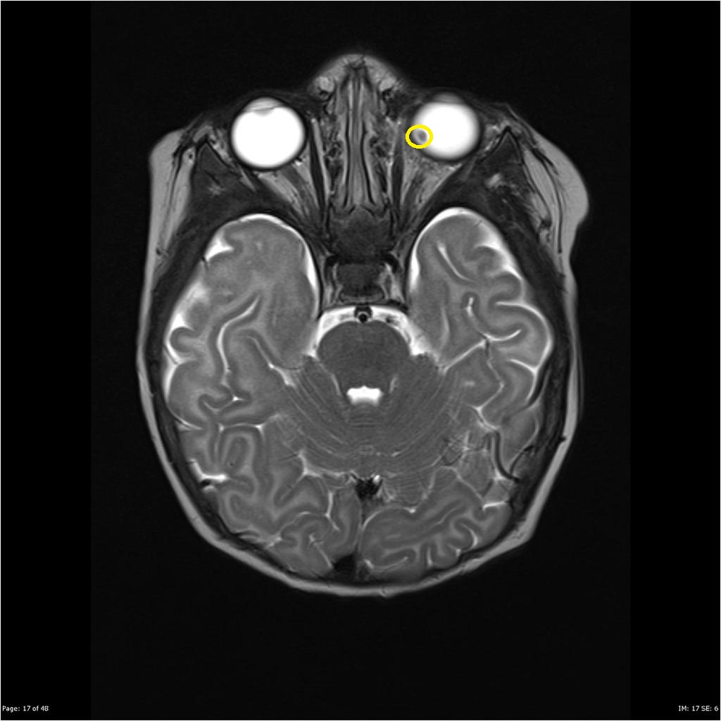
 Fundus photographs, fluorescein angiographic (FA) image, and enhanced depth imaging optical coherence tomographic (EDI- OCT).gif Gunnam
15:15, 21 June 2019
472 × 416; 385 KB
-
-

-

-
-

-
-

-

-
-
-

-

-
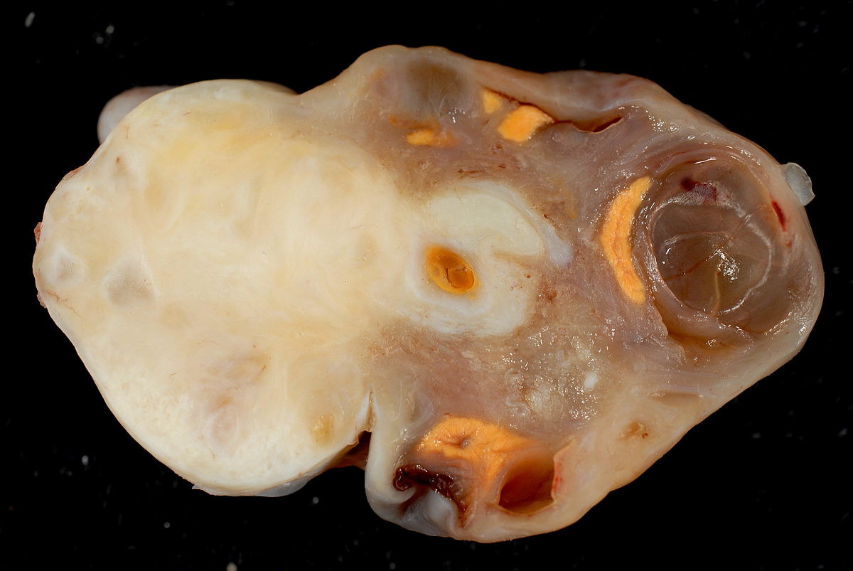
 Intraventricular-meningioma-third-ventricle-1.jpg Ifeoma odukwe
16:17, 20 June 2019
818 × 1,024; 74 KB
-

-

-

-

-
-

-
-
-
-
-

-

-
-
-
-
-
-
-
-
-
-
-
-
-
-
-

-

-

-
-
-
-
-
-
-

-
-
-
-
-
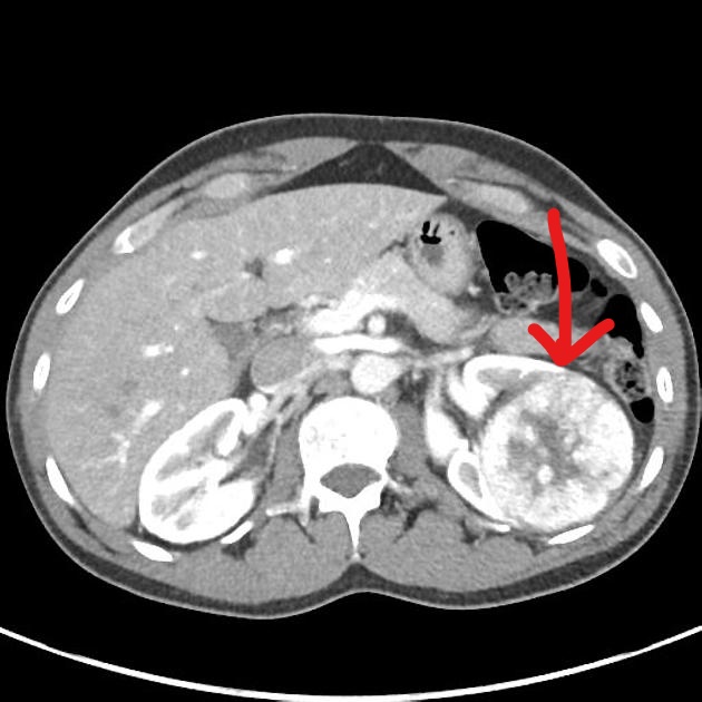
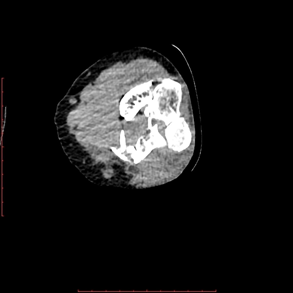
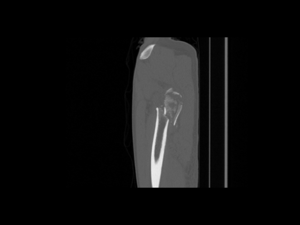
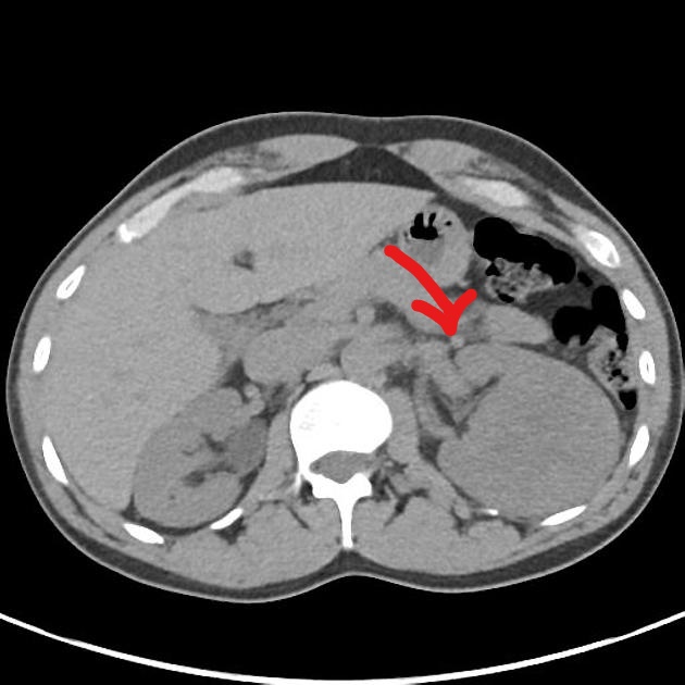 InkedAxial non-contrast CT of renal oncocytoma LI.jpg Homa Najafi
15:50, 17 June 2019
630 × 630; 82 KB
-
-
-
-
-
-
-
-

-
-
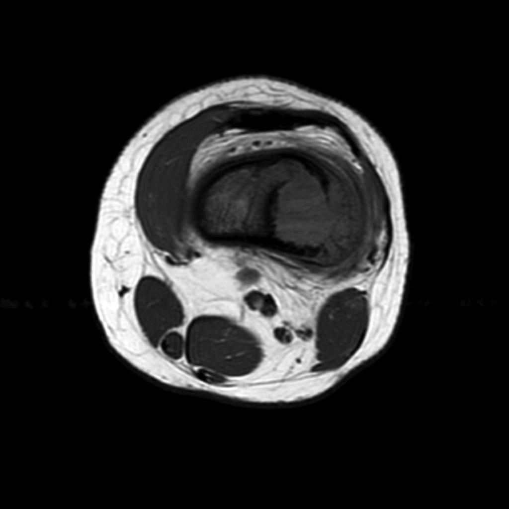
 D273cf22cccee8797c32f307cc1448 big gallery.jpg Sahar Memar Montazerin
19:02, 14 June 2019
630 × 630; 43 KB
-
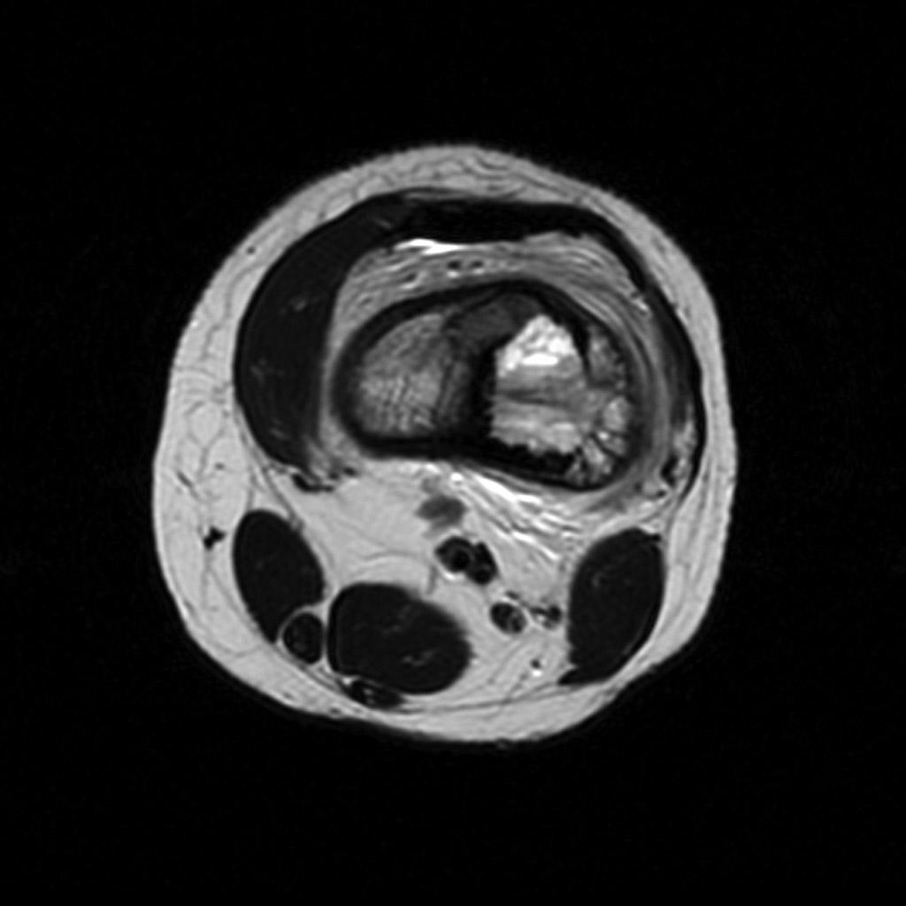
 487d9fd790cc79305d88478b100d61 big gallery.jpg Sahar Memar Montazerin
18:56, 14 June 2019
484 × 630; 34 KB
-
-
-
-
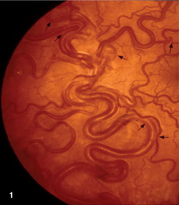
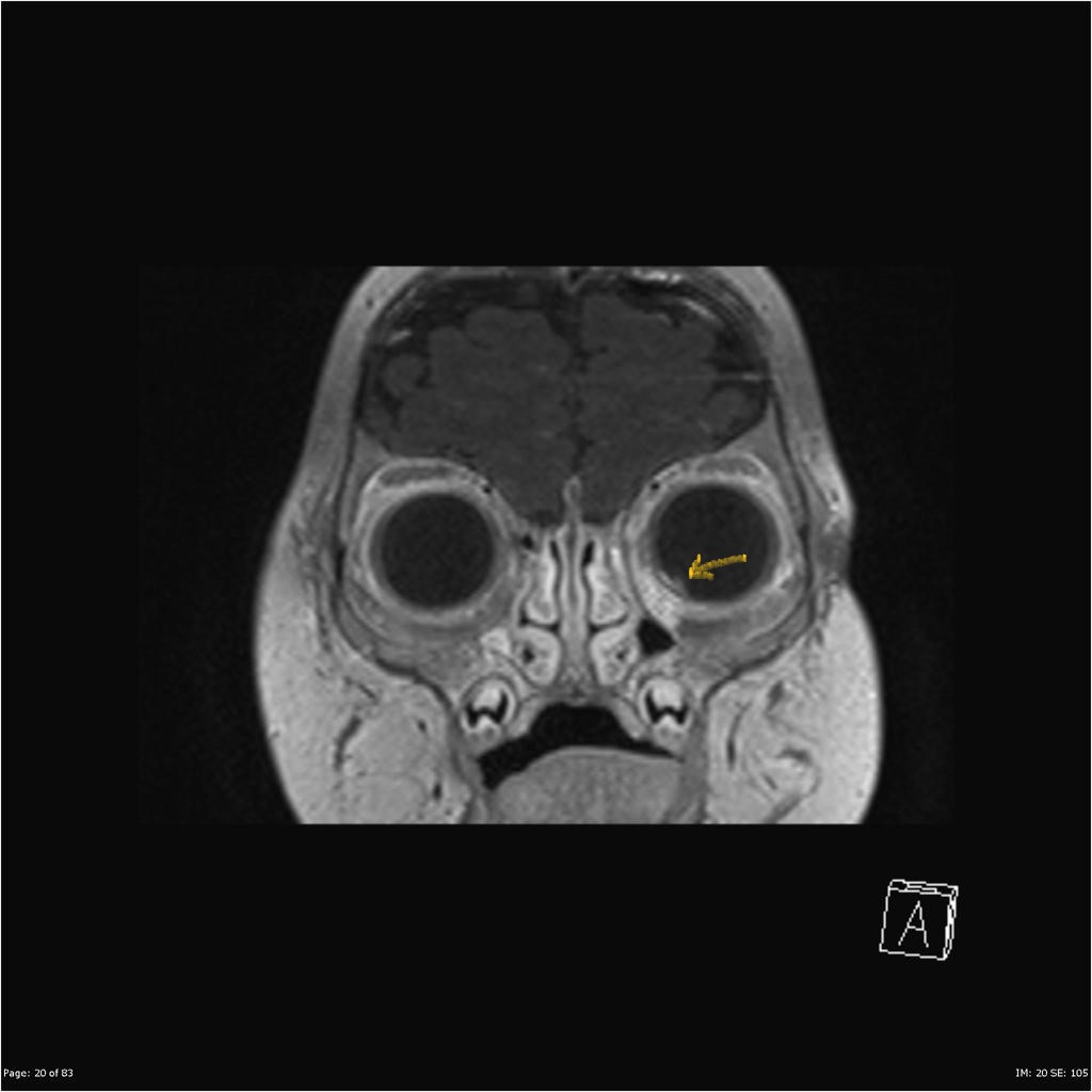
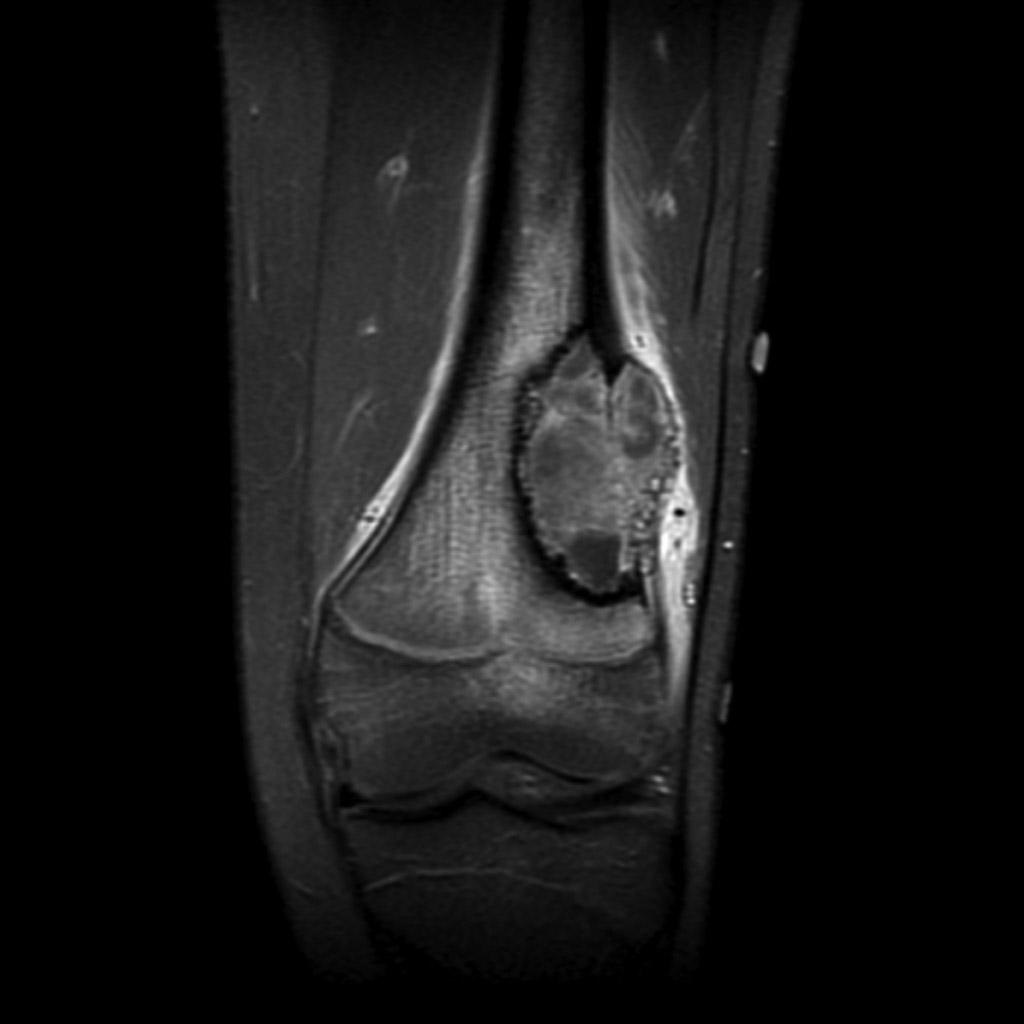
 Macular ischemia and extensive arteriovenous communications and dilated intertwined vessels.jpg Gunnam
20:01, 13 June 2019
256 × 256; 19 KB
-
-

-
-
-
-
-

-
 Breast-cancer-with-lymphangitis-carcinomatosa.jpg Swathi Venkatesan
13:17, 3 June 2019
1,024 × 1,024; 106 KB
-
 Metastatic gastric adenocarcinoma-lymphangitic carcinomatosis (7261944992).jpg Swathi Venkatesan
13:05, 3 June 2019
1,200 × 900; 376 KB
-

-

-

-
 1200px-IDH1 R132H in anaplastic ologodendroglioma.jpg Sara Mohsin
00:55, 1 June 2019
1,200 × 890; 255 KB
-
-
 1200px-Anaplastic oligodendroglioma minigemistocytes.jpg Sara Mohsin
00:49, 1 June 2019
1,200 × 890; 207 KB
-
 1200px-Oligodendroglioma discrete invasion HE.jpg Sara Mohsin
00:44, 1 June 2019
1,200 × 890; 411 KB
-

-

-

-

-

-

-
 Eosinophilic-granulomatosis-with-polyangiitis.jpg Soroush Seifirad
20:02, 31 May 2019
1,024 × 1,024; 147 KB
-
 Allergic-bronchopulmonary-aspergillosis-abpa-2.jpg Soroush Seifirad
19:59, 31 May 2019
1,024 × 1,024; 176 KB
-
 Allergic-bronchopulmonary-aspergillosis-6.jpg Soroush Seifirad
19:57, 31 May 2019
1,024 × 1,011; 87 KB
-

-

-

-

-

-

-

-
-

-

-

-

-

-

-

-
-

-

-

-

-
-
-

-
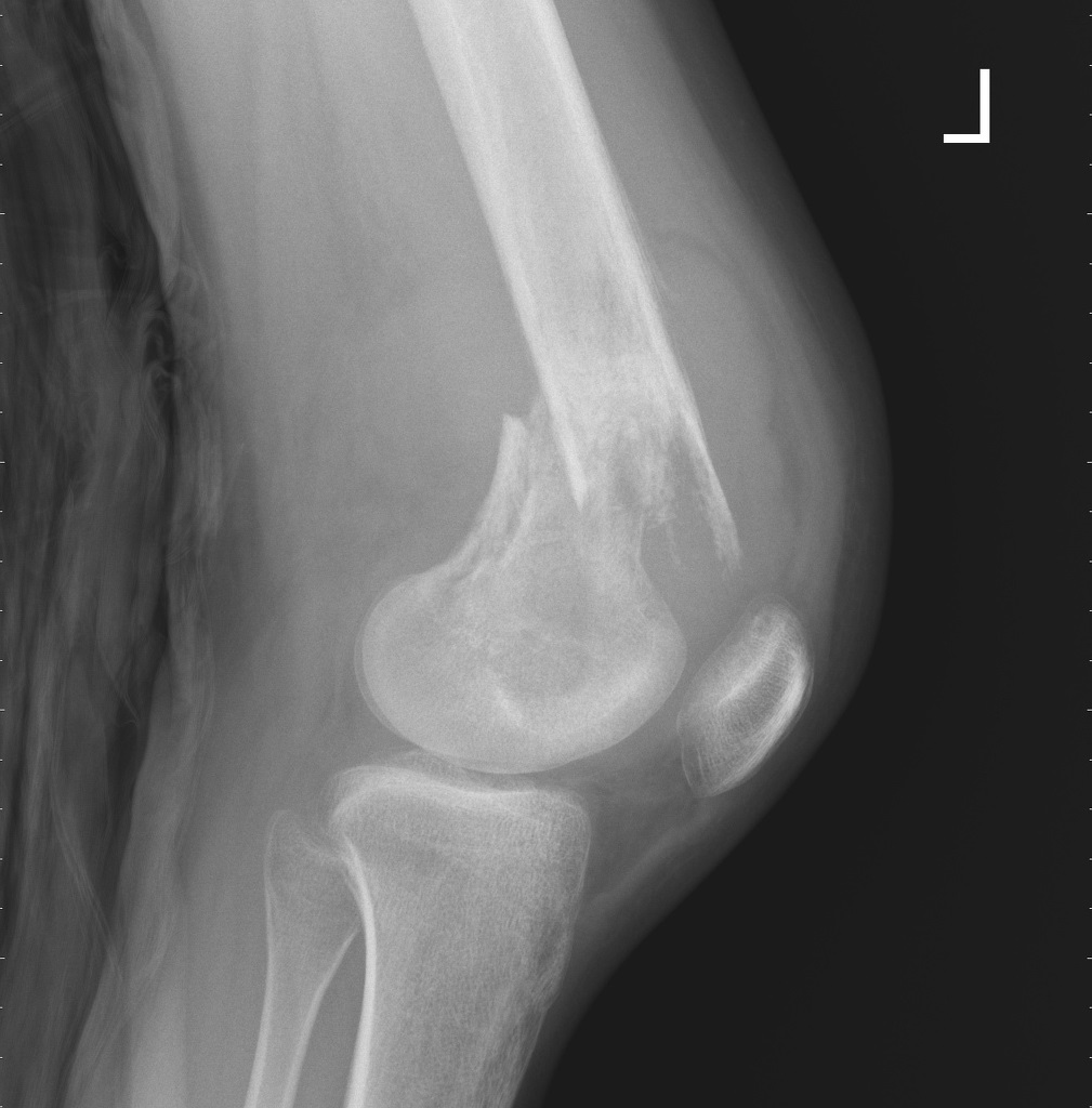
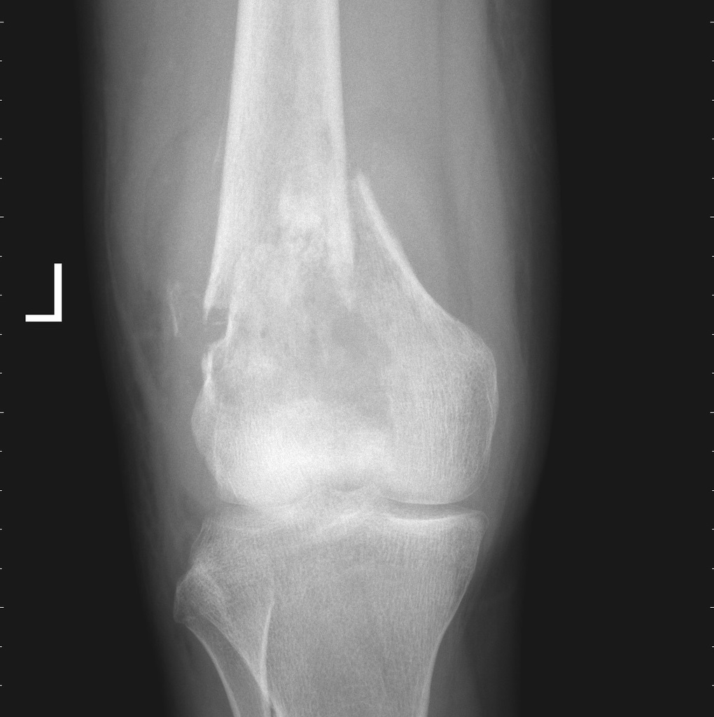
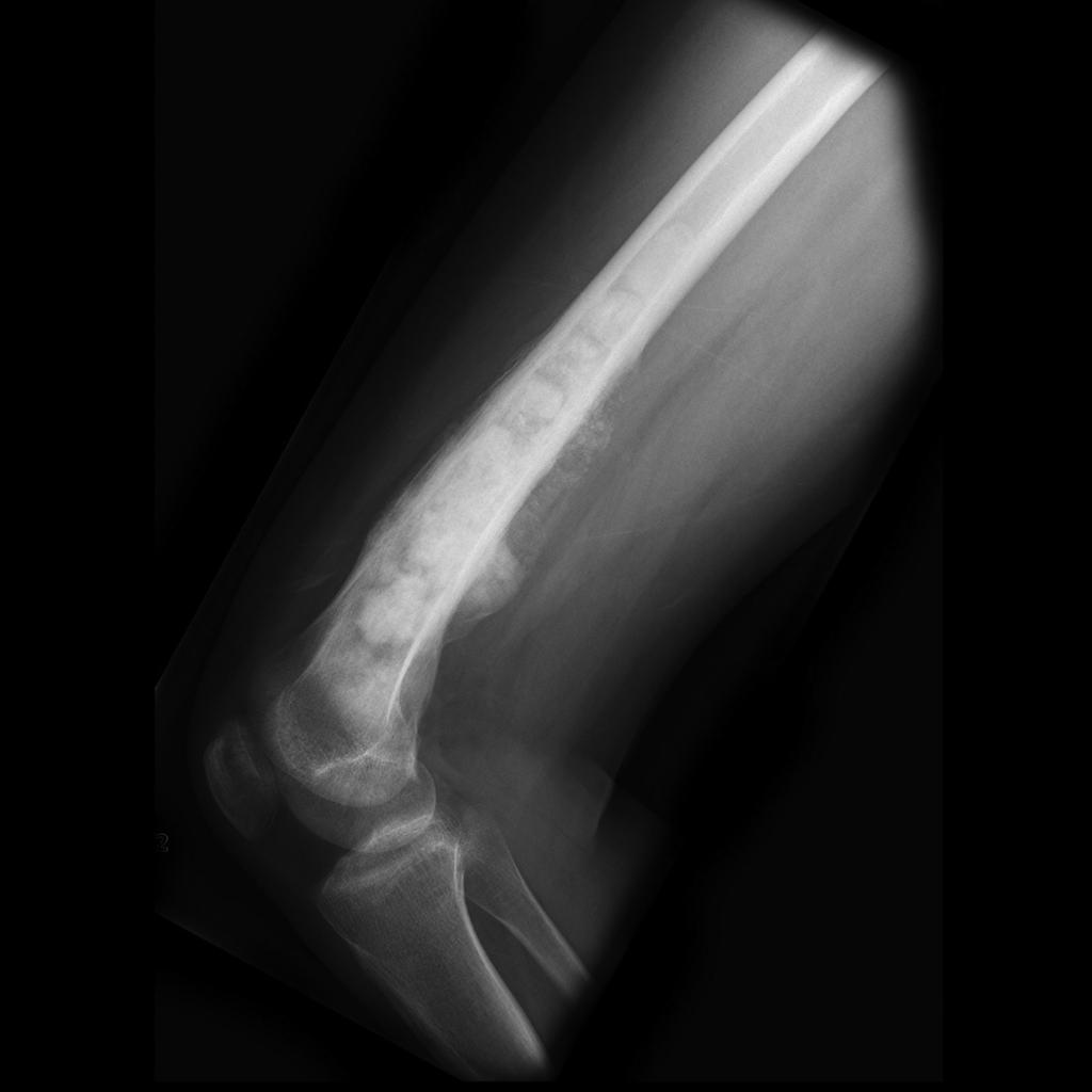
 Bone ChondromyxoidFibroma Calcium MP PA.JPG Maneesha Nandimandalam
17:24, 29 May 2019
1,076 × 899; 230 KB
-

-

-
-
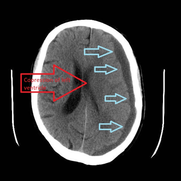
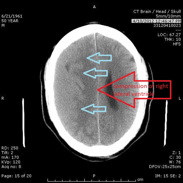
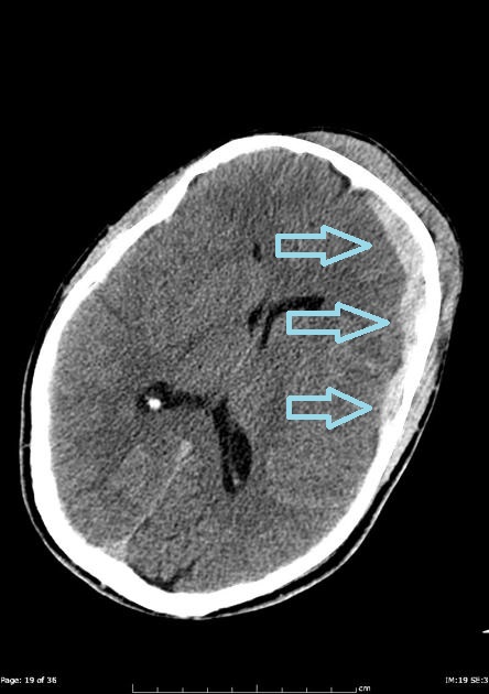
 1200px-Subdural hematoma - intermed low mag.jpg Fahimeh Shojaei
15:12, 29 May 2019
1,200 × 800; 389 KB
-
-
-

-

-

-
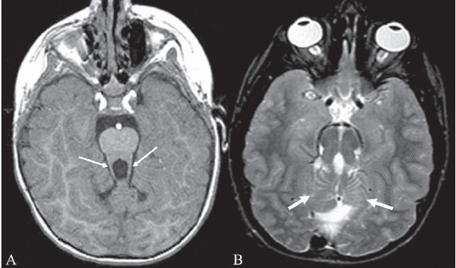
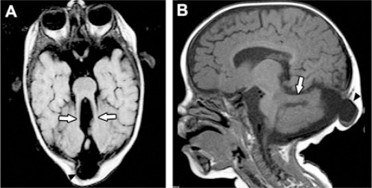 Joubert syndrome-molar tooth sign, occipital encephalocele.gif Gunnam
21:12, 28 May 2019
542 × 274; 324 KB
-
-
-
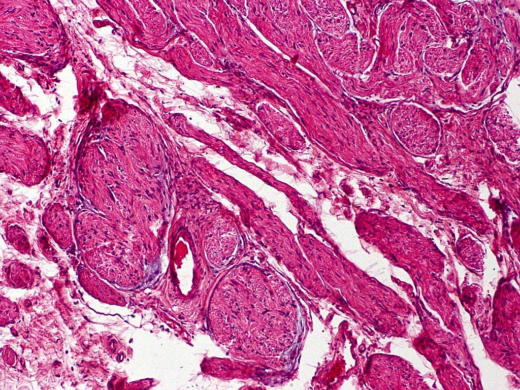
 Palisaded and Encapsulated Neuroma (3953412330).jpg Sara Mohsin
16:08, 28 May 2019
1,280 × 960; 527 KB
-
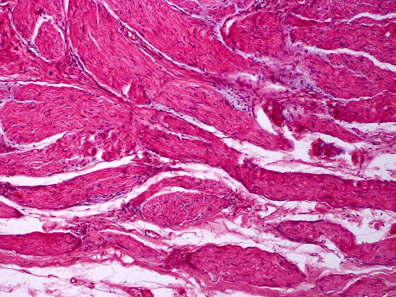
 Palisaded and Encapsulated Neuroma (3952635881).jpg Sara Mohsin
16:02, 28 May 2019
1,280 × 854; 343 KB
-
-
-
-
-
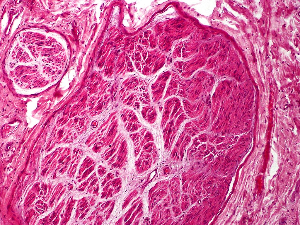
 Palisaded and Encapsulated Neuroma, S-100 Immunostain (3953412396).jpg Sara Mohsin
15:37, 28 May 2019
1,024 × 835; 460 KB
-

-

-

-

-

-

-

-
-
-
-

-
-
 81c080c7f013c25f665f1f49c03f56 big galleryy.BMP Fahimeh Shojaei
15:47, 23 May 2019
544 × 630; 1,004 KB
-

-

-

-

-
 81c080c7f013c25f665f1f49c03f56 big gallery.BMP Fahimeh Shojaei
14:44, 23 May 2019
544 × 630; 1.31 MB
-

-
 Molar tooth sign and hypoplasia of the cerebellar vermis in Joubert syndrome.gif Gunnam
01:49, 21 May 2019
651 × 382; 493 KB
-

-
-

-
-

-

-

-

-

-

-

-

-

-
-
-
-
-

-

-
-

-

-

-

-
-

-

-

-
-

-

-

-

-

-

-

-
-
-
-
-
-
-
-
-
-
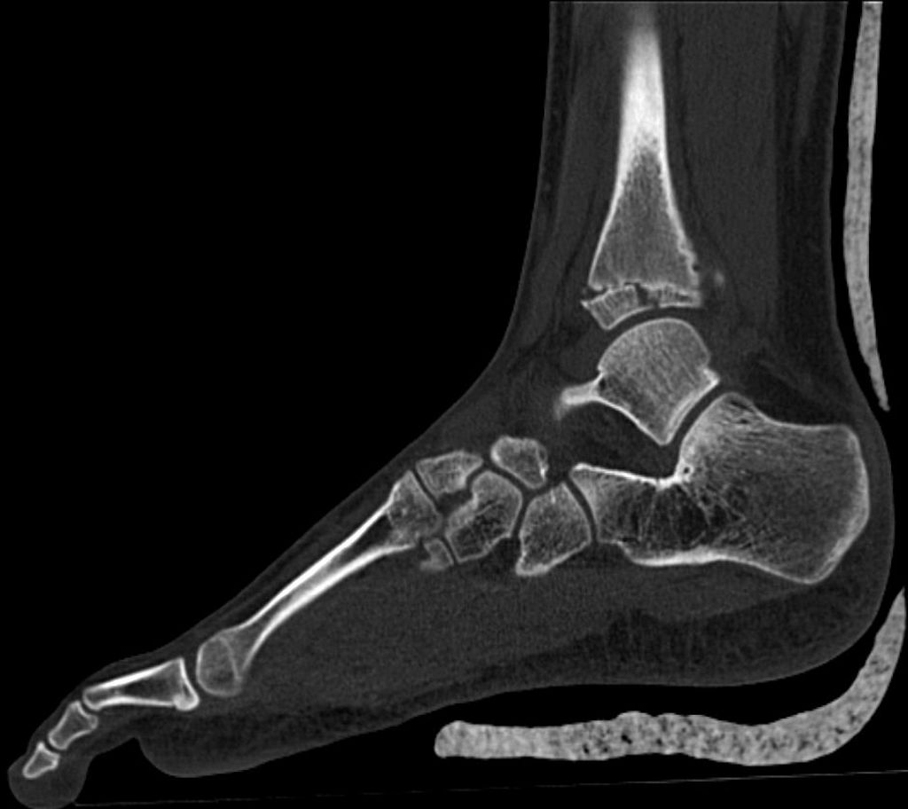
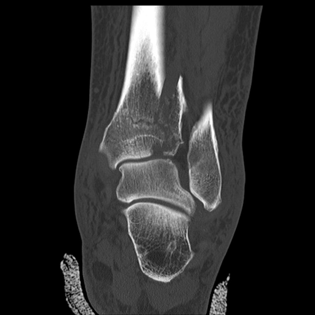
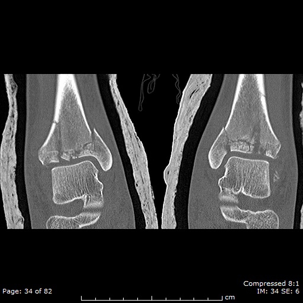
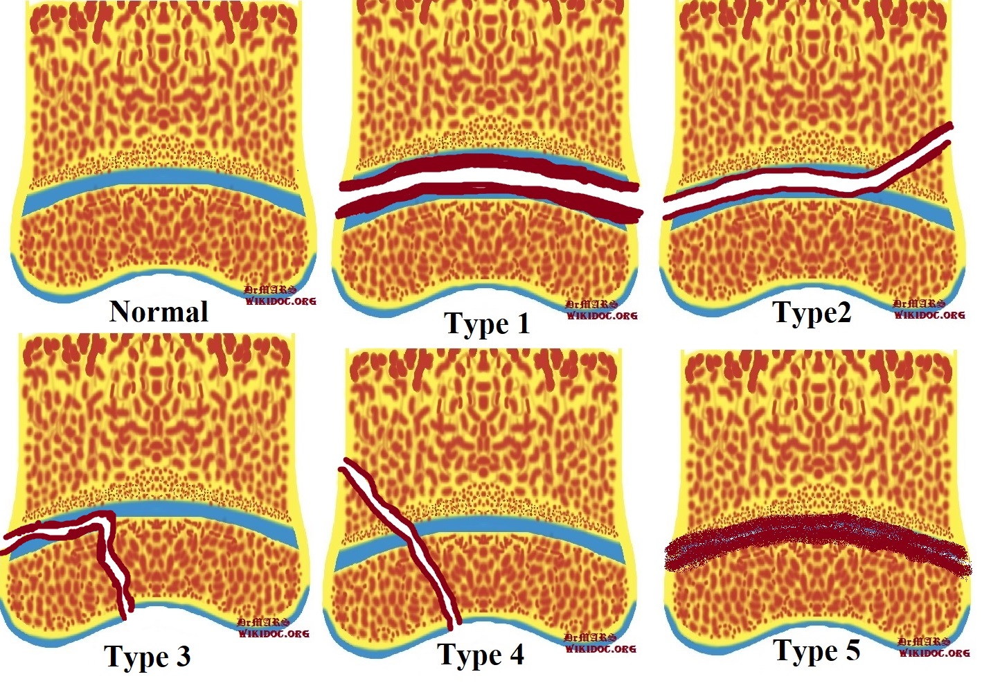
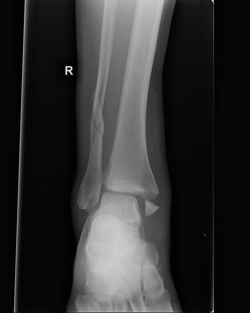 Ankle-fractures-pronation-external-rotation-mechanismmm.jpg DrMars
12:44, 12 May 2019
817 × 1,024; 90 KB
-
-
-
-

-
-

-

-

-
-
-
-

-

-

-

-

-

-

-

-
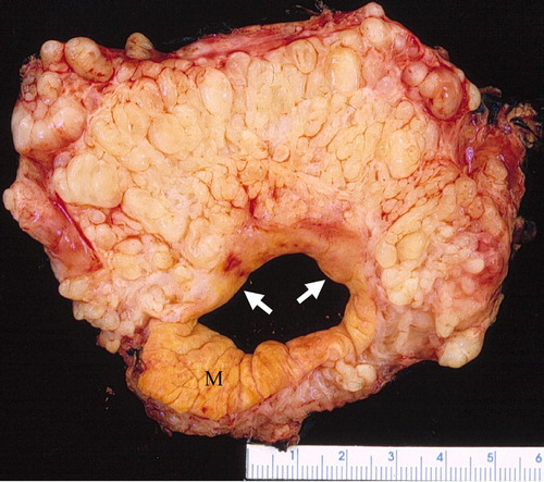
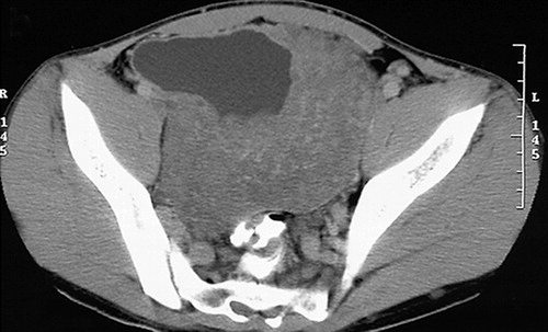
 CT image of ovarian granulosa cell tumor.jpg Maneesha Nandimandalam
19:39, 6 May 2019
473 × 205; 29 KB
-
-
-

-
-
 Chloroma histopathology micrograph (H&E stain).jpeg Parnian Jabbari
15:05, 2 May 2019
2,560 × 1,707; 1.08 MB
-

-

-

-
-

-

-

-

-
-
-

-
-
-

-
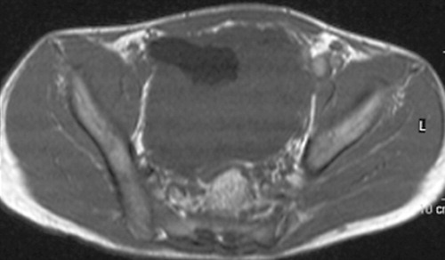
 Ultrasound imaging of ovarian thecofibroma.jpg Maneesha Nandimandalam
19:21, 19 April 2019
567 × 394; 88 KB
-
 3D Dual Color Super Resolution Microscopy Cremer 2010 .png Soroush Seifirad
14:47, 19 April 2019
3,486 × 1,280; 3 MB
-

-

-
-

-
-
-
-
-

-
-
-
-
 Sclerosing-stromal-tumour-of-the-ovary.jpg Maneesha Nandimandalam
19:19, 17 April 2019
709 × 583; 117 KB
-

-

-
-
-

-
 Breast carcinoma in a lymph node biopsy.jpg Soroush Seifirad
14:05, 17 April 2019
1,024 × 819; 215 KB
-

-
-

-

-
-
-
-
-
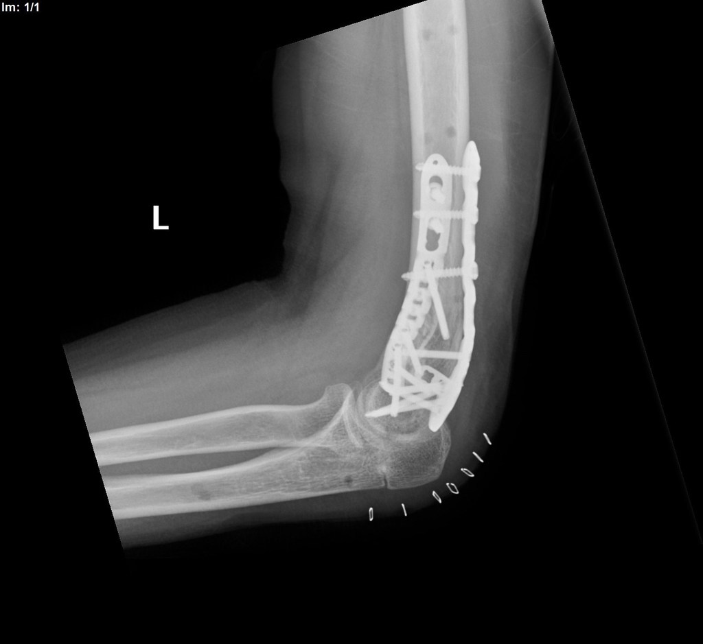
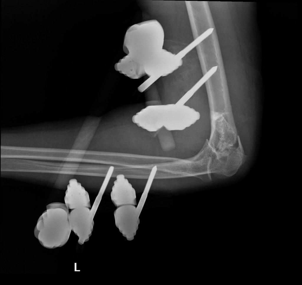
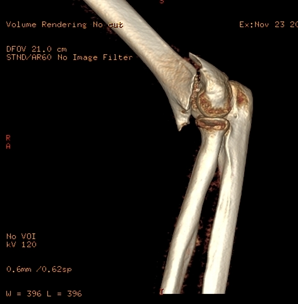
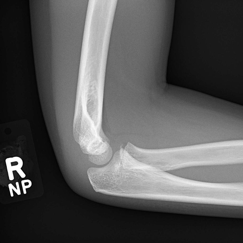
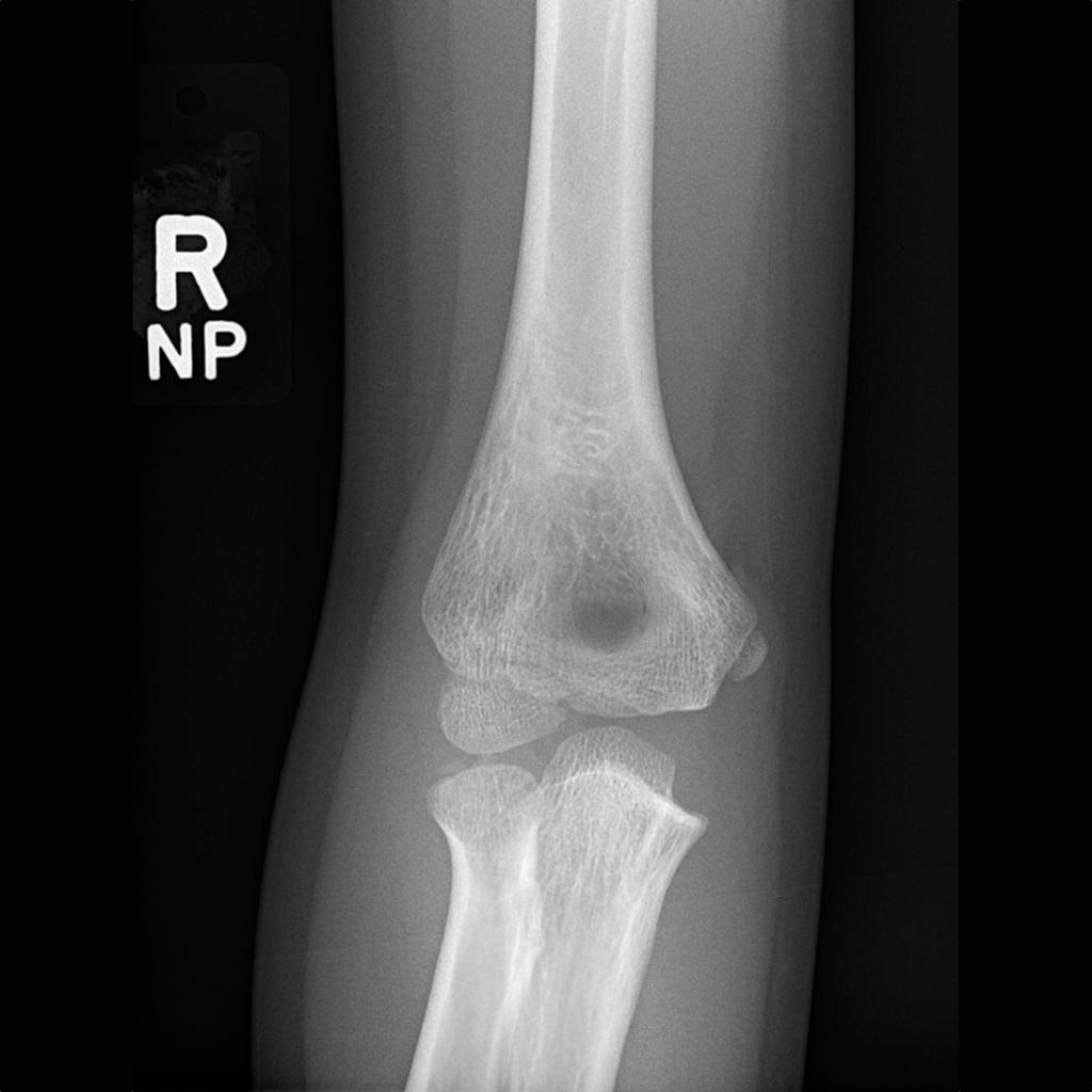
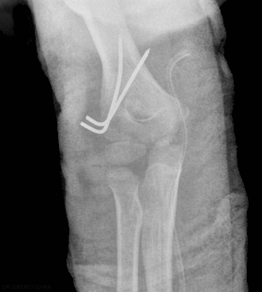
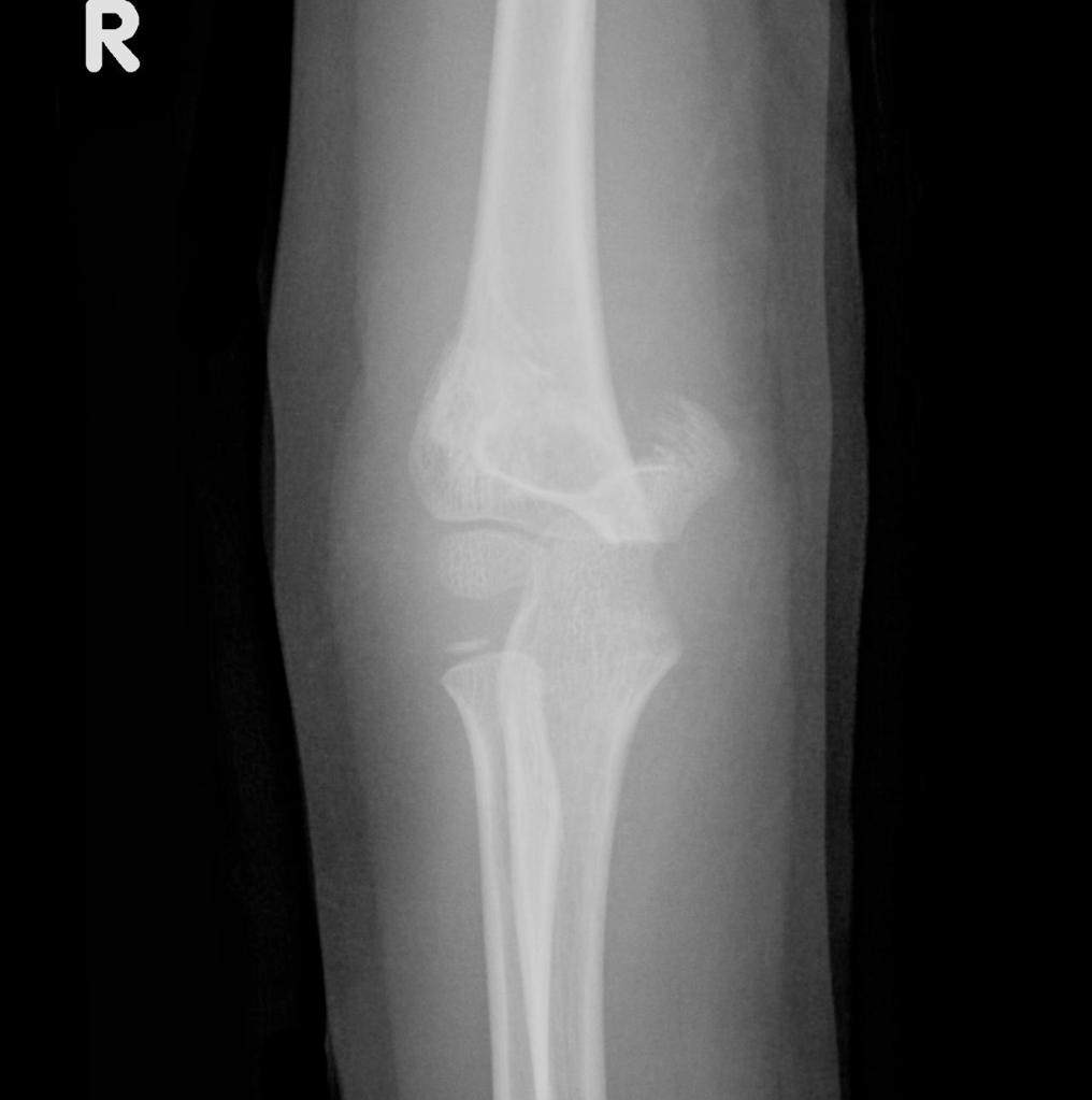
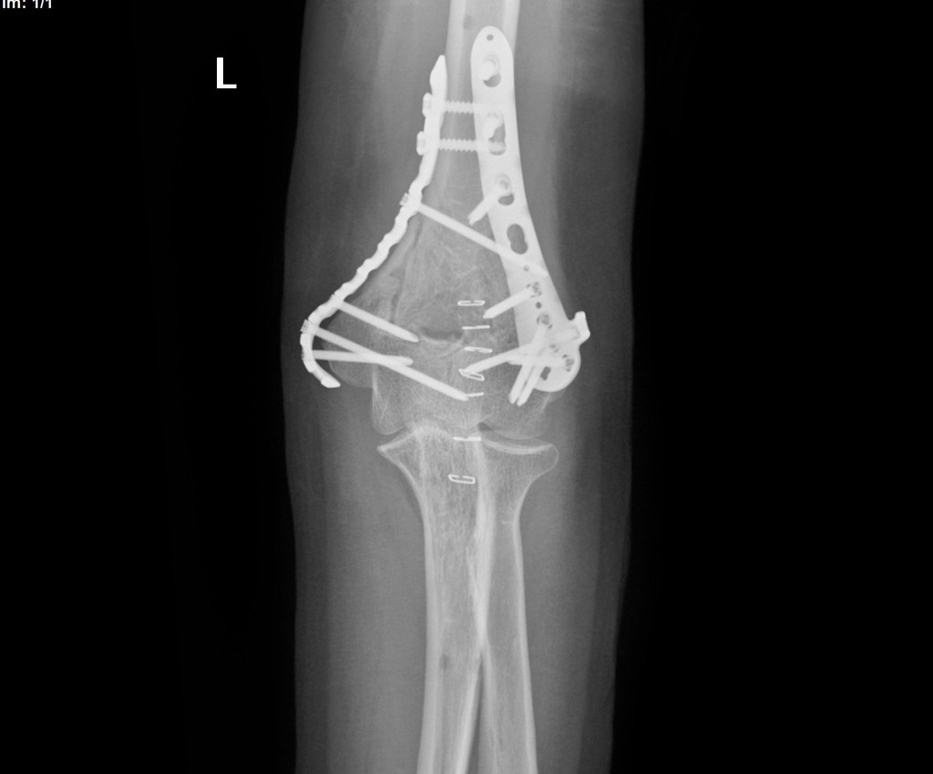 Displaced-t-condylar-and-supracondylar-fracture-of-the-distal-humerus (11).jpg DrMars
10:53, 17 April 2019
1,024 × 850; 53 KB
-
 Displaced-t-condylar-and-supracondylar-fracture-of-the-distal-humerus (10).jpg DrMars
10:53, 17 April 2019
1,024 × 939; 61 KB
-
 Displaced-t-condylar-and-supracondylar-fracture-of-the-distal-humerus (9).jpg DrMars
10:53, 17 April 2019
1,024 × 965; 72 KB
-
 Displaced-t-condylar-and-supracondylar-fracture-of-the-distal-humerus (8).jpg DrMars
10:49, 17 April 2019
1,003 × 1,024; 188 KB
-
 Displaced-t-condylar-and-supracondylar-fracture-of-the-distal-humerus (7).jpg DrMars
10:49, 17 April 2019
997 × 1,024; 169 KB
-
 Displaced-t-condylar-and-supracondylar-fracture-of-the-distal-humerus (6).jpg DrMars
10:49, 17 April 2019
846 × 1,024; 177 KB
-
 Displaced-t-condylar-and-supracondylar-fracture-of-the-distal-humerus (5).jpg DrMars
10:49, 17 April 2019
1,024 × 1,024; 86 KB
-
 Displaced-t-condylar-and-supracondylar-fracture-of-the-distal-humerus (4).jpg DrMars
10:49, 17 April 2019
1,024 × 1,024; 92 KB
-
 Displaced-t-condylar-and-supracondylar-fracture-of-the-distal-humerus (3).jpg DrMars
10:49, 17 April 2019
1,024 × 1,024; 104 KB
-
 Displaced-t-condylar-and-supracondylar-fracture-of-the-distal-humerus (2).jpg DrMars
10:48, 17 April 2019
1,024 × 1,024; 118 KB
-
 Displaced-t-condylar-and-supracondylar-fracture-of-the-distal-humerus (1).jpg DrMars
10:44, 17 April 2019
1,024 × 825; 225 KB
-
 Displaced-t-condylar-and-supracondylar-fracture-of-the-distal-humerus.jpg DrMars
10:44, 17 April 2019
1,024 × 877; 219 KB
-
-
-
-
-
-
-
-
-
-

-

-

-

-

-

-
 Supracondylar-fractures-gartland-classification-1.jpg DrMars
08:42, 17 April 2019
1,024 × 493; 67 KB
-
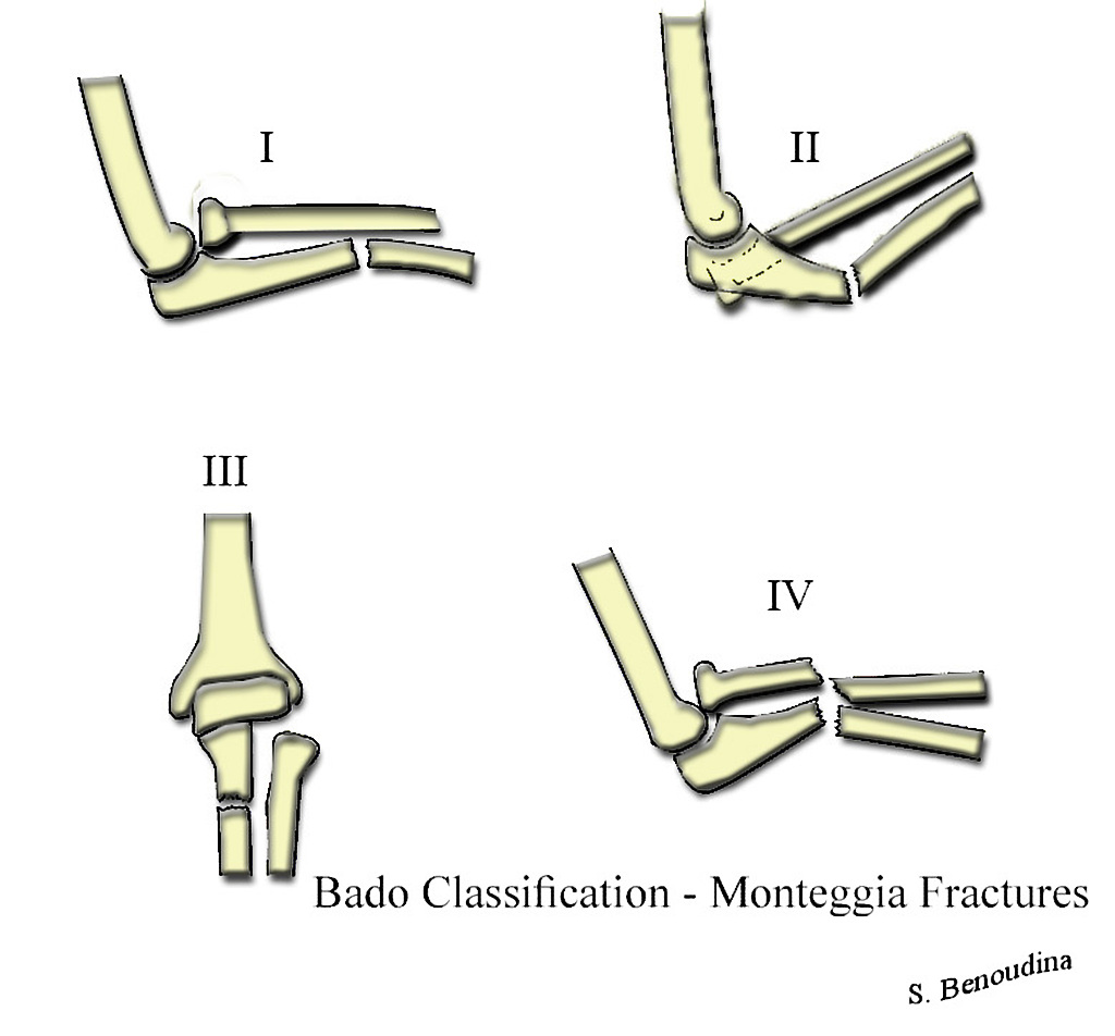
 Intercondylar-fractures-of-the-humerus-riseborough-and-radin-classification.jpg DrMars
08:21, 17 April 2019
917 × 1,024; 237 KB
-
-

-
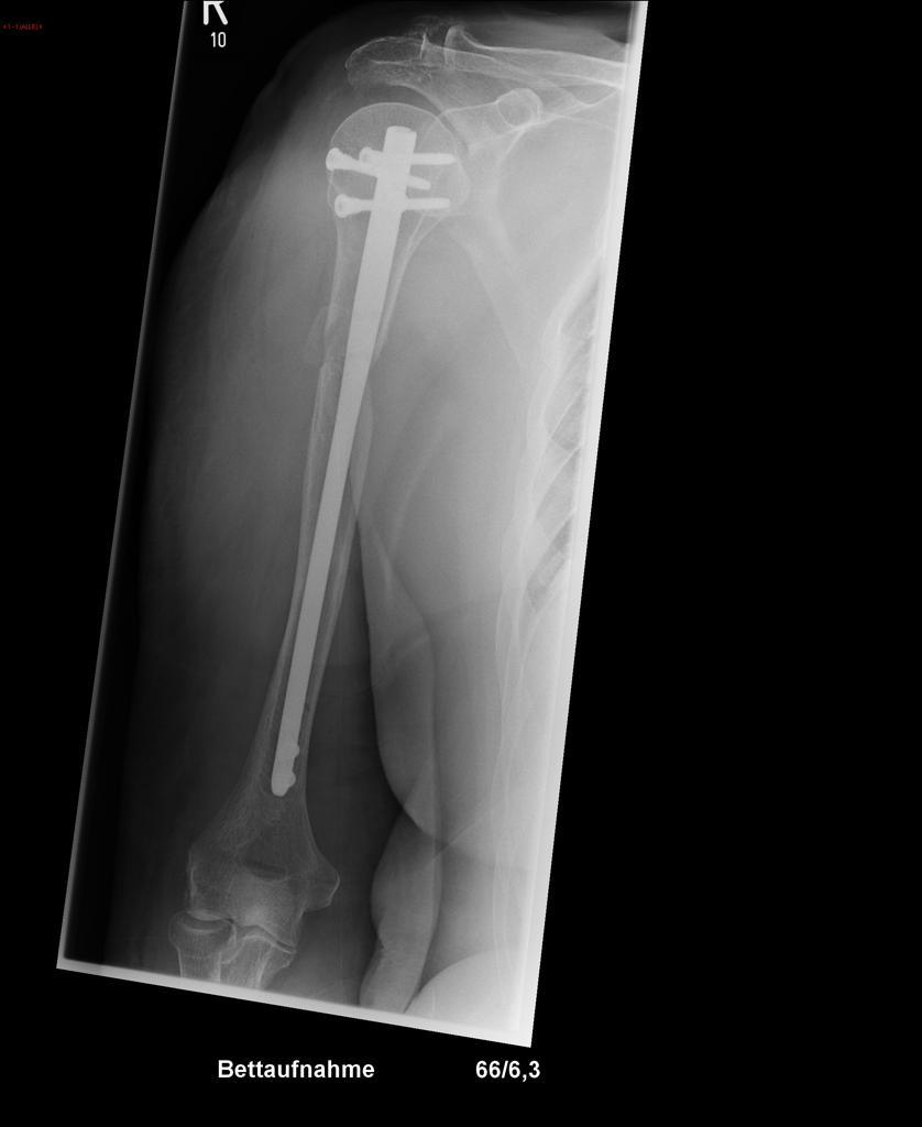
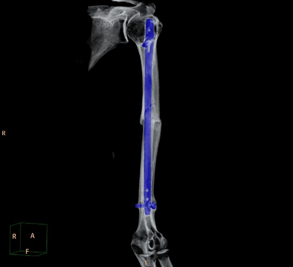
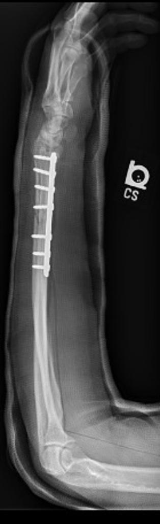
 Humeral-shaft-fracture-non-union-with-osteosynthesis (1).jpg DrMars
22:16, 16 April 2019
1,024 × 1,017; 47 KB
-
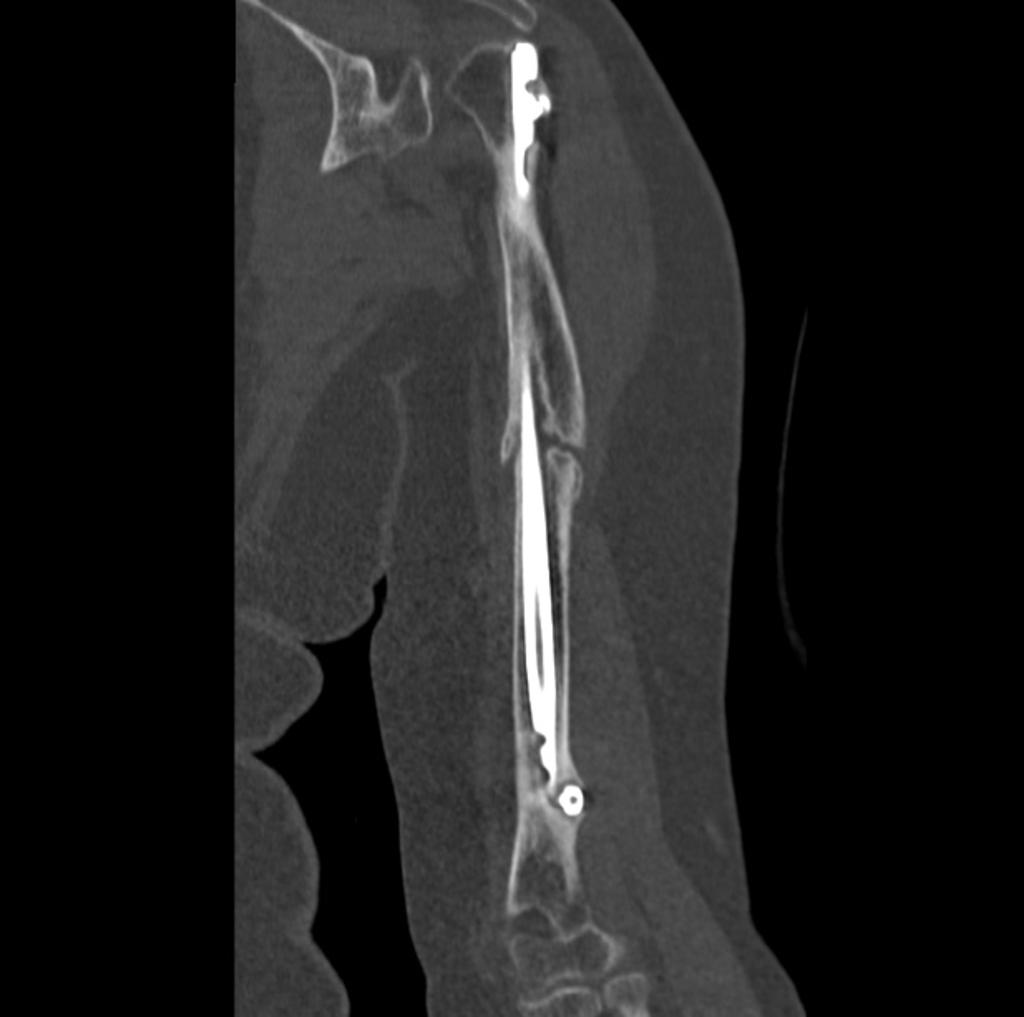
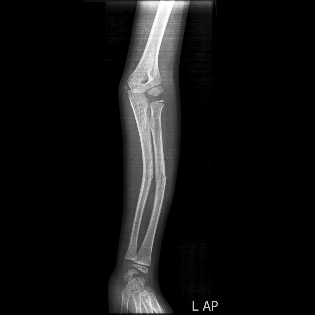
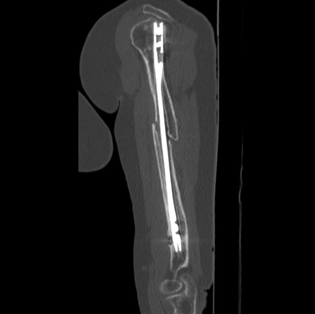 Humeral-shaft-fracture-non-union-with-osteosynthesis (2).jpg DrMars
22:16, 16 April 2019
1,024 × 1,020; 45 KB
-
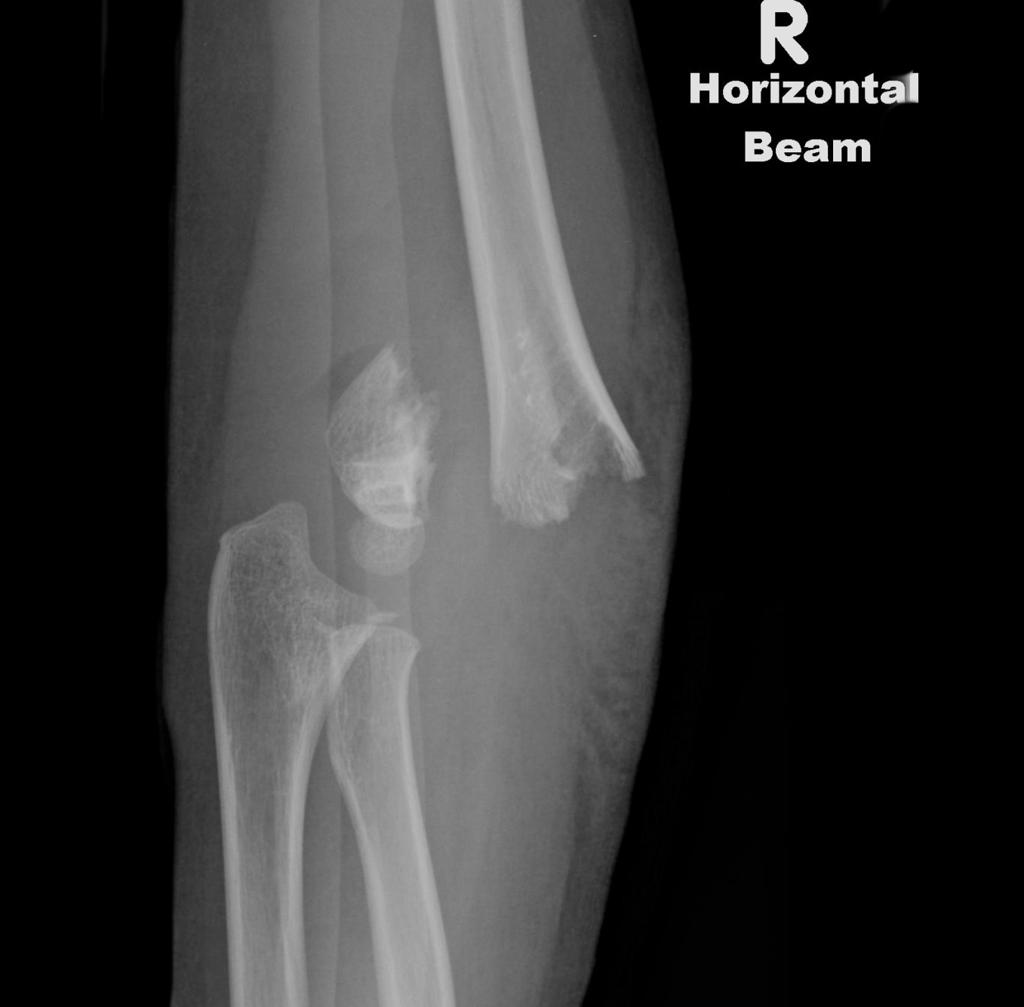
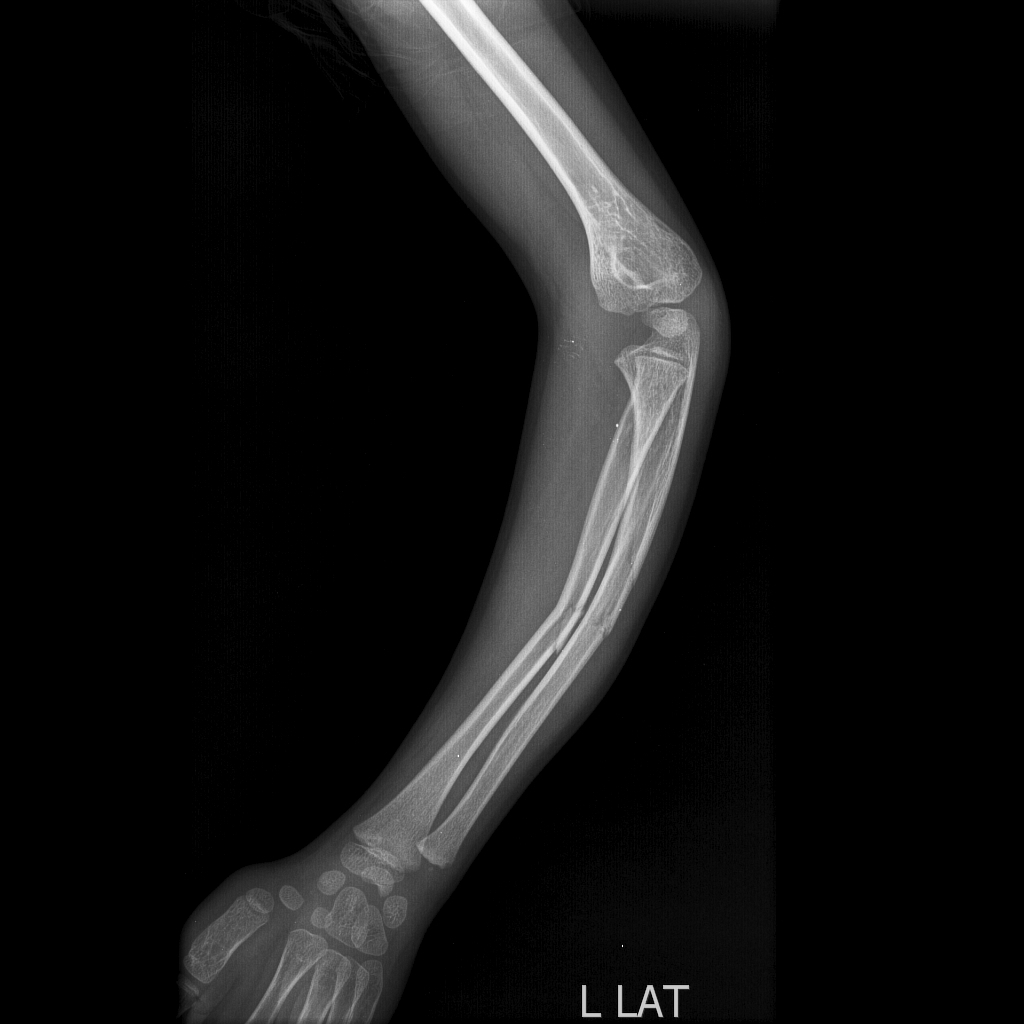
 Humeral-shaft-fracture-non-union-with-osteosynthesis.jpg DrMars
22:16, 16 April 2019
1,024 × 934; 33 KB
-
-
-
-
-
-
-
-
-
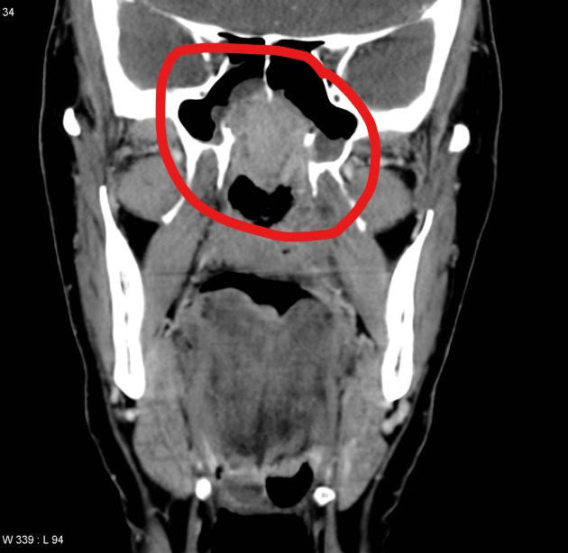
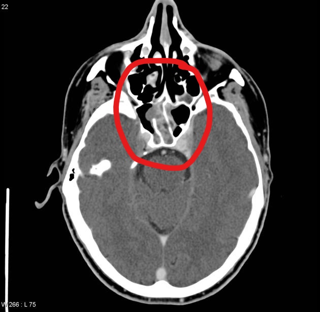
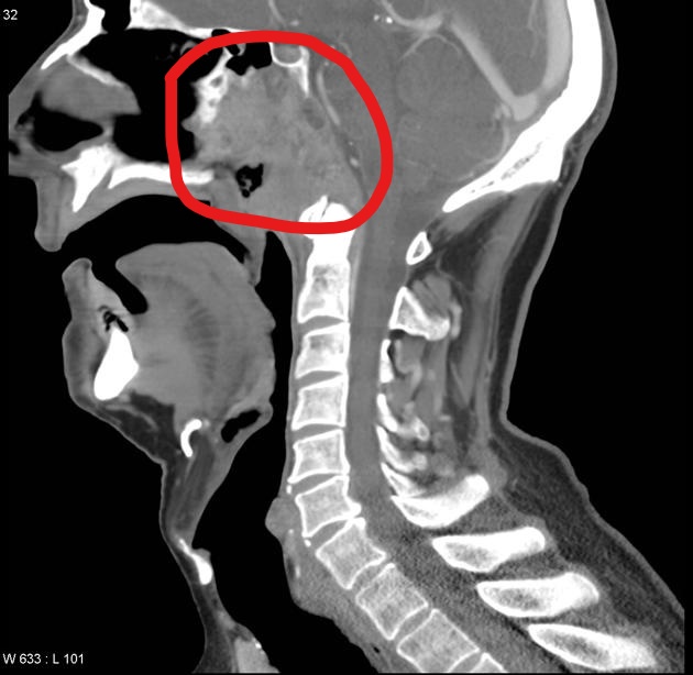
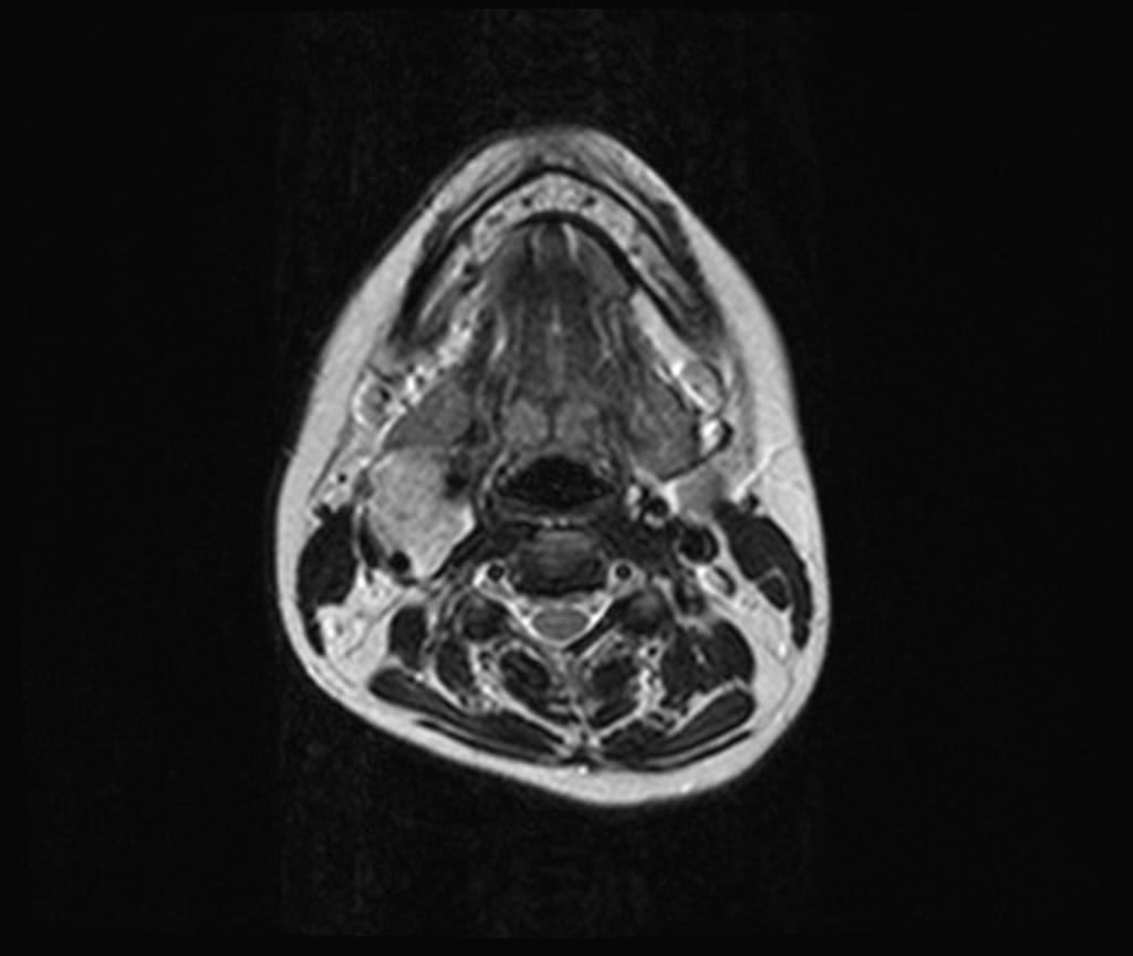
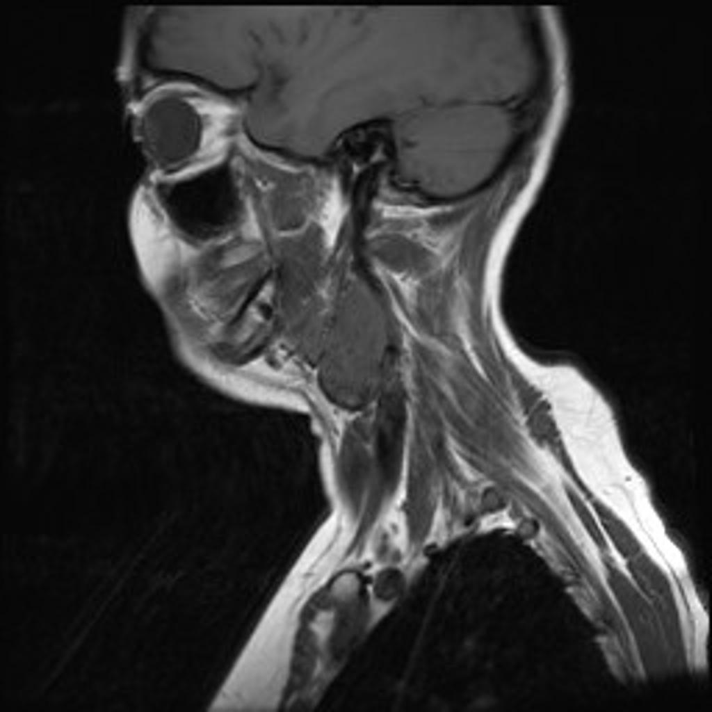
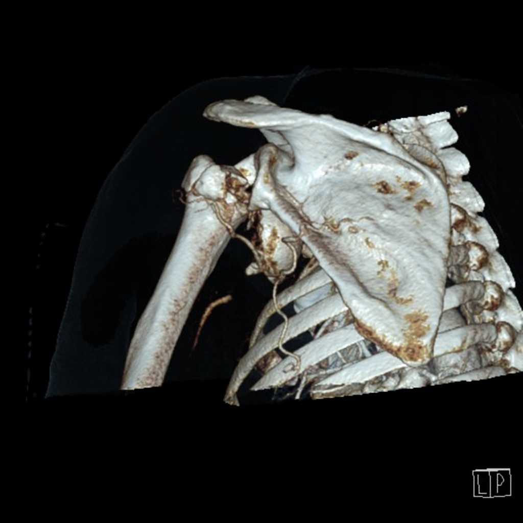
 Neck Paraganglioma CarotidBody HP CTR.jpg Sahar Memar Montazerin
17:28, 16 April 2019
2,048 × 1,536; 1.19 MB
-

-

-
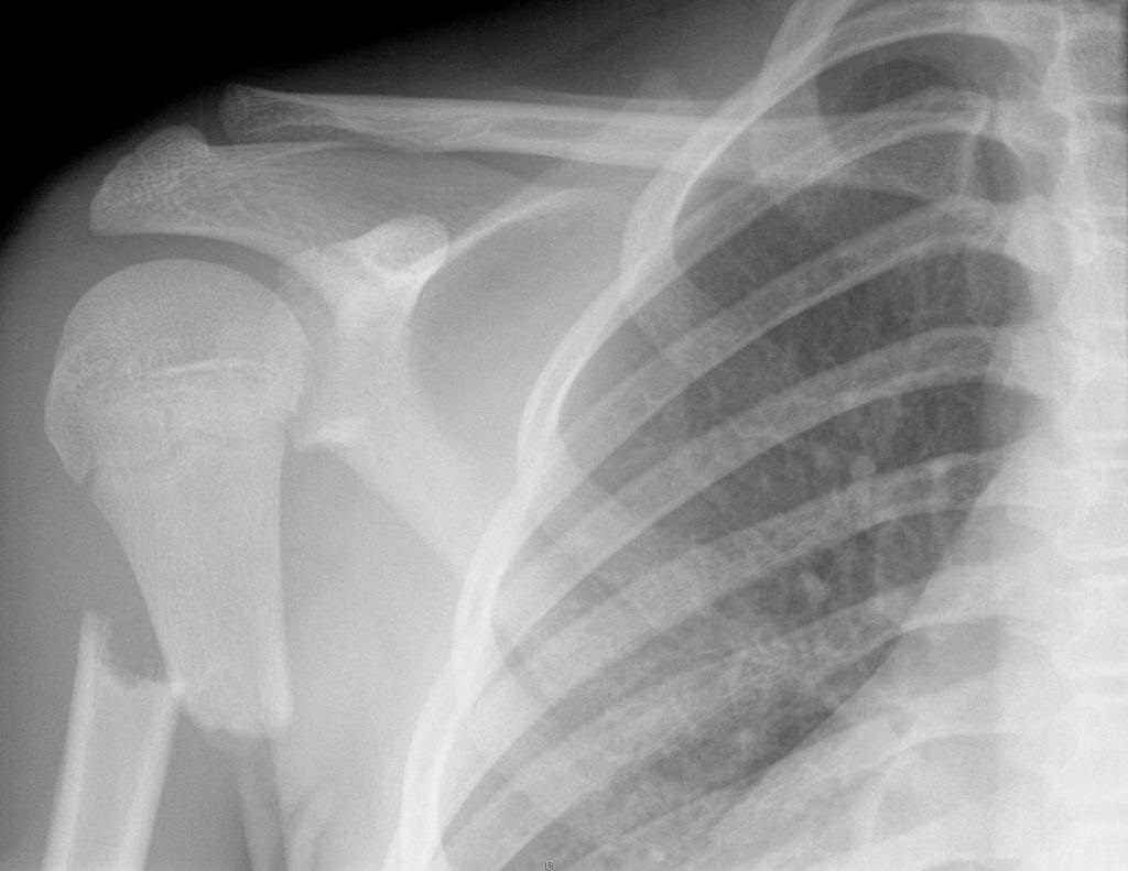
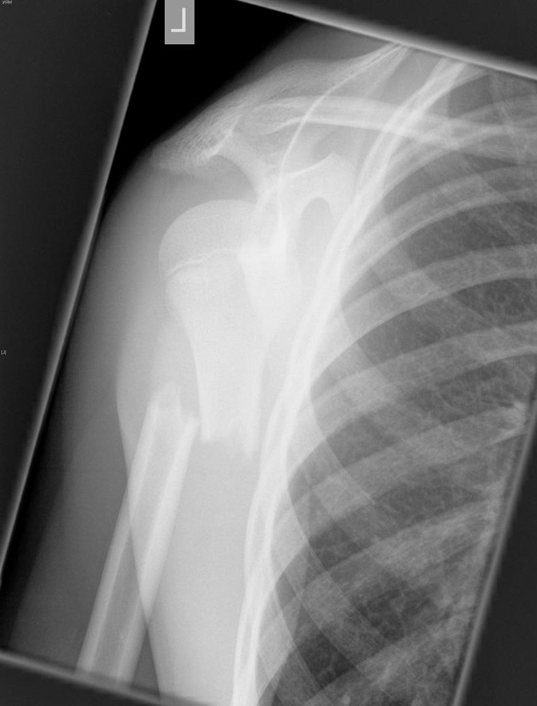
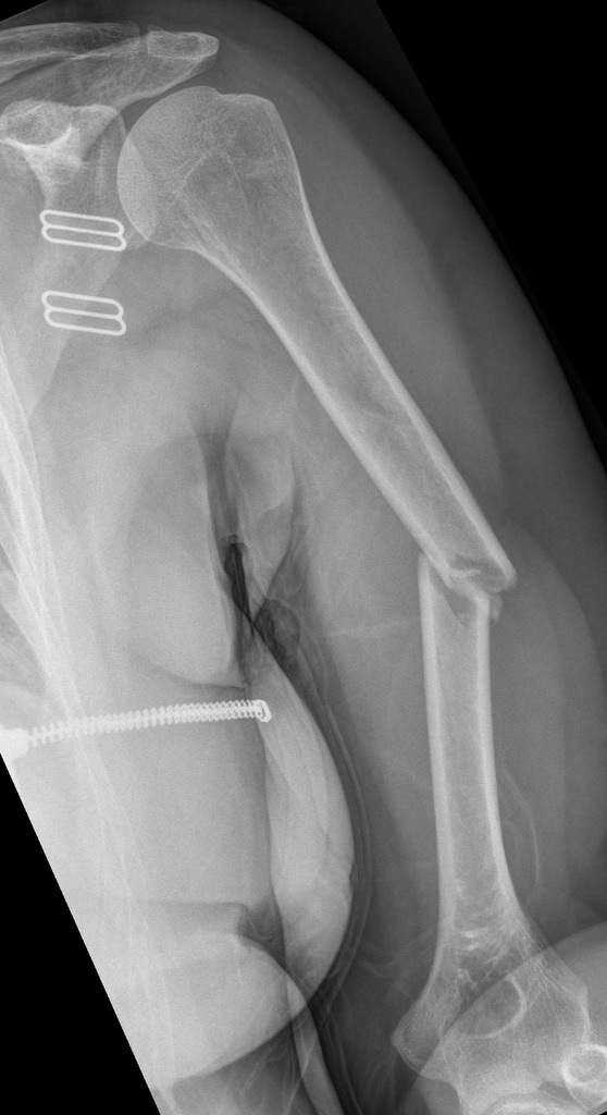
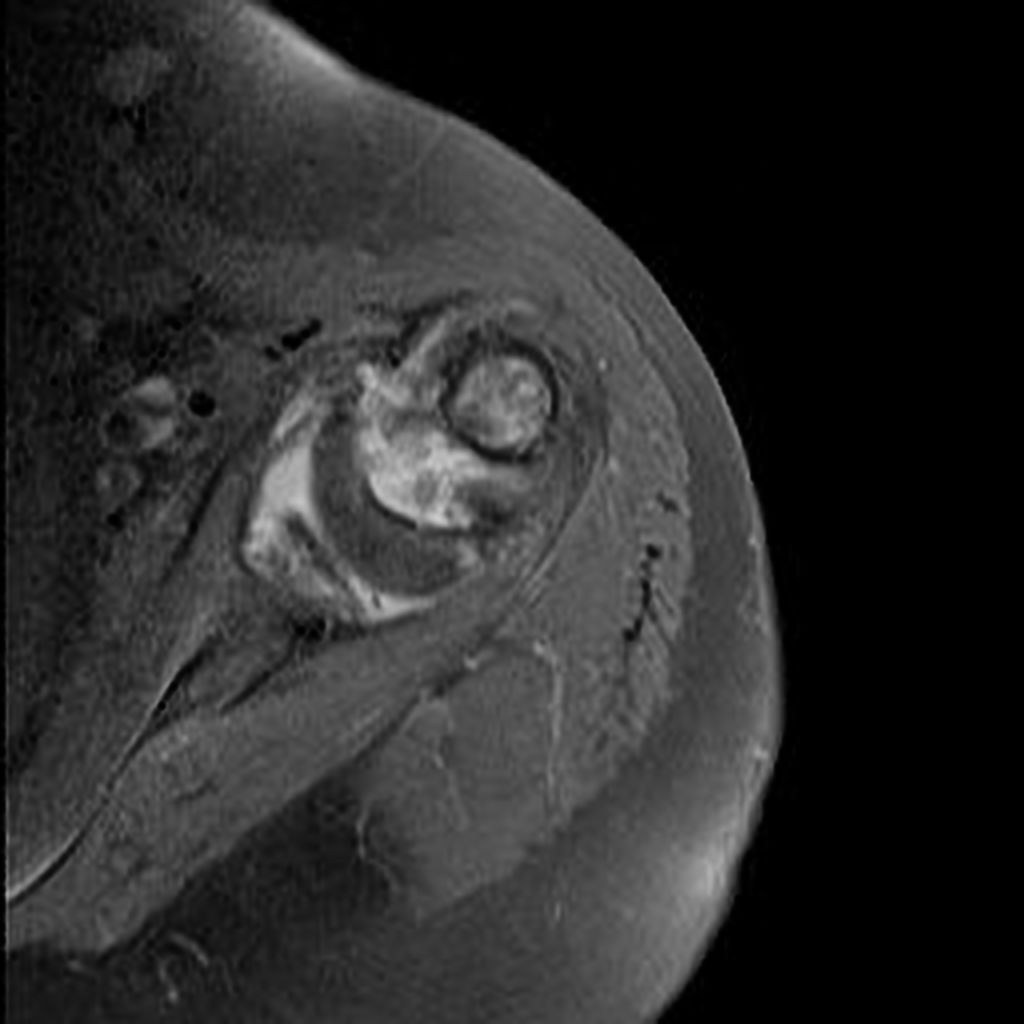
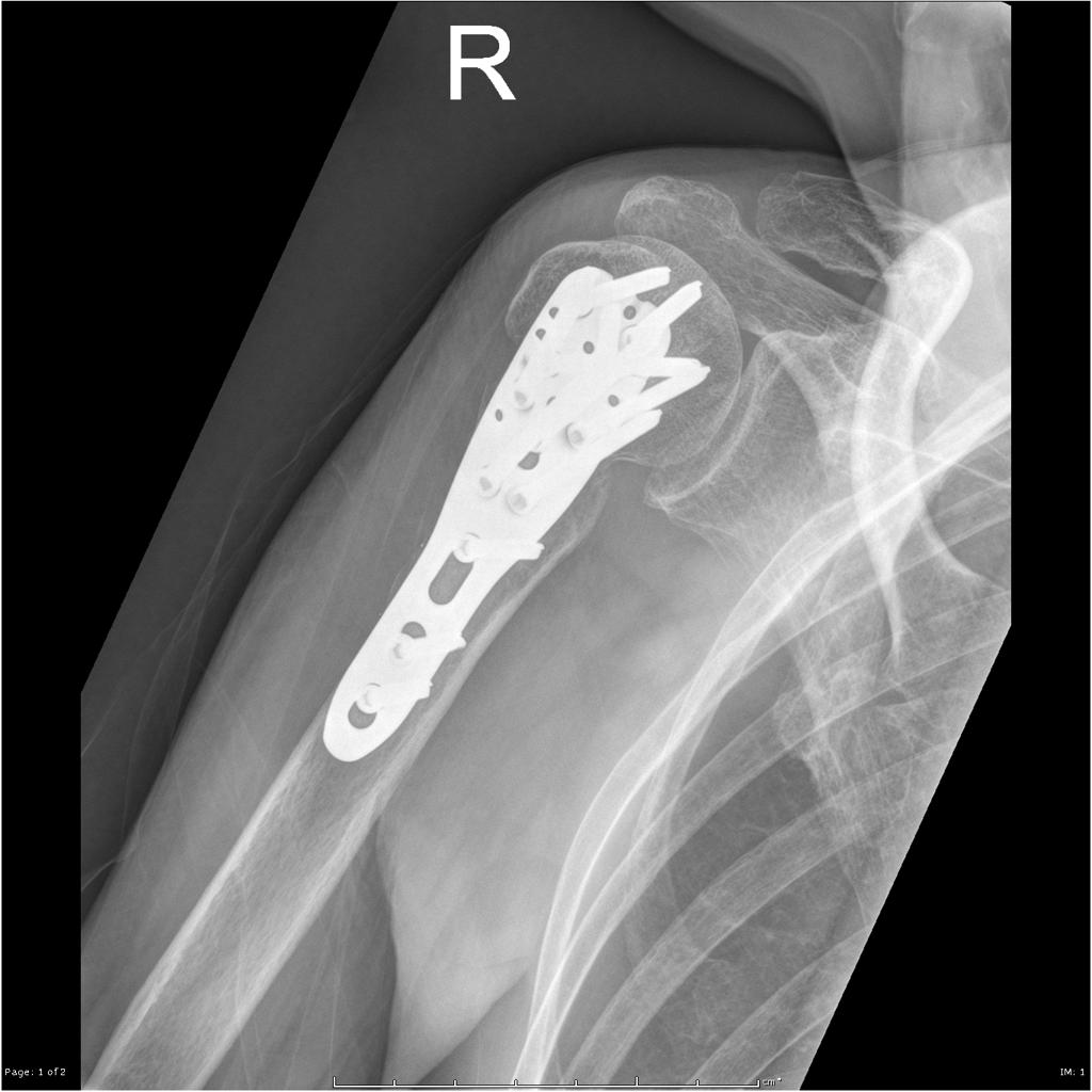
 Shoulder-fracture-dislocation-with-arterial-dissection2.jpg DrMars
21:32, 15 April 2019
1,024 × 1,024; 122 KB
-
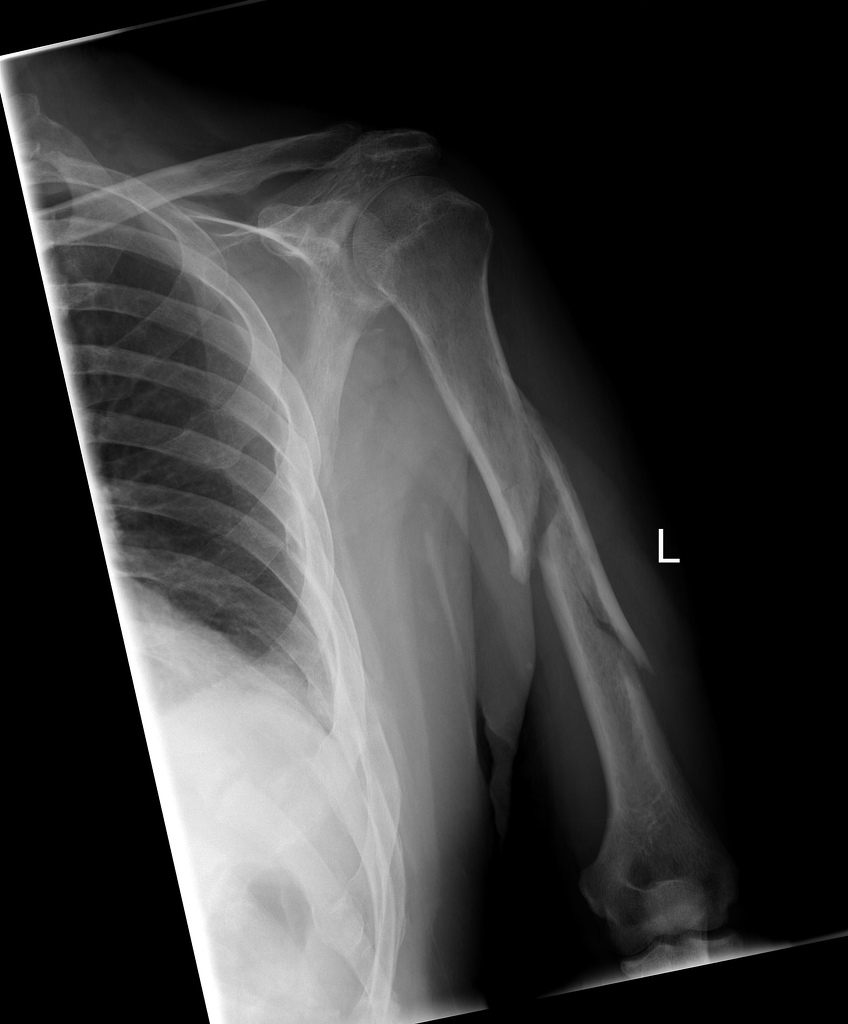
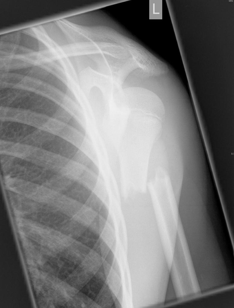
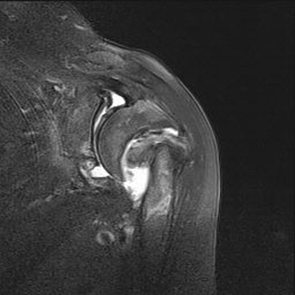
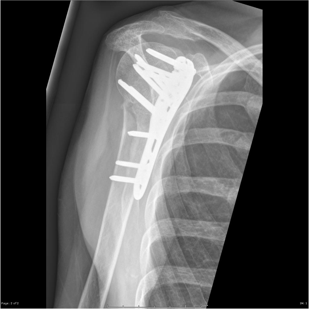
 Shoulder-fracture-dislocation-with-arterial-dissection1.jpg DrMars
21:32, 15 April 2019
1,024 × 1,024; 89 KB
-
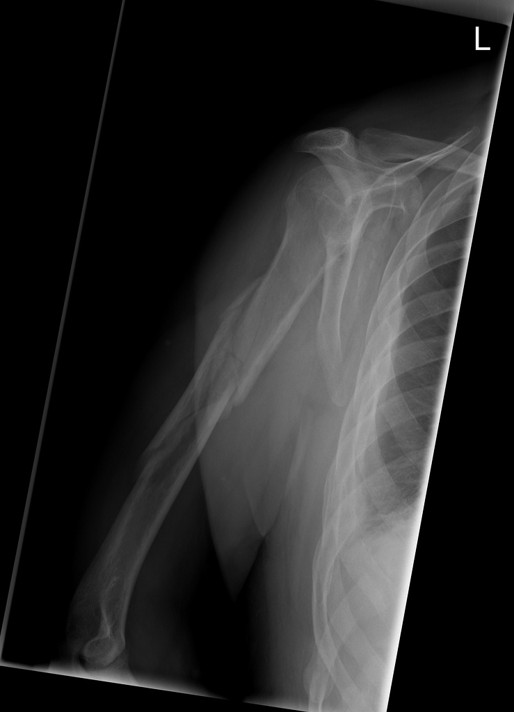
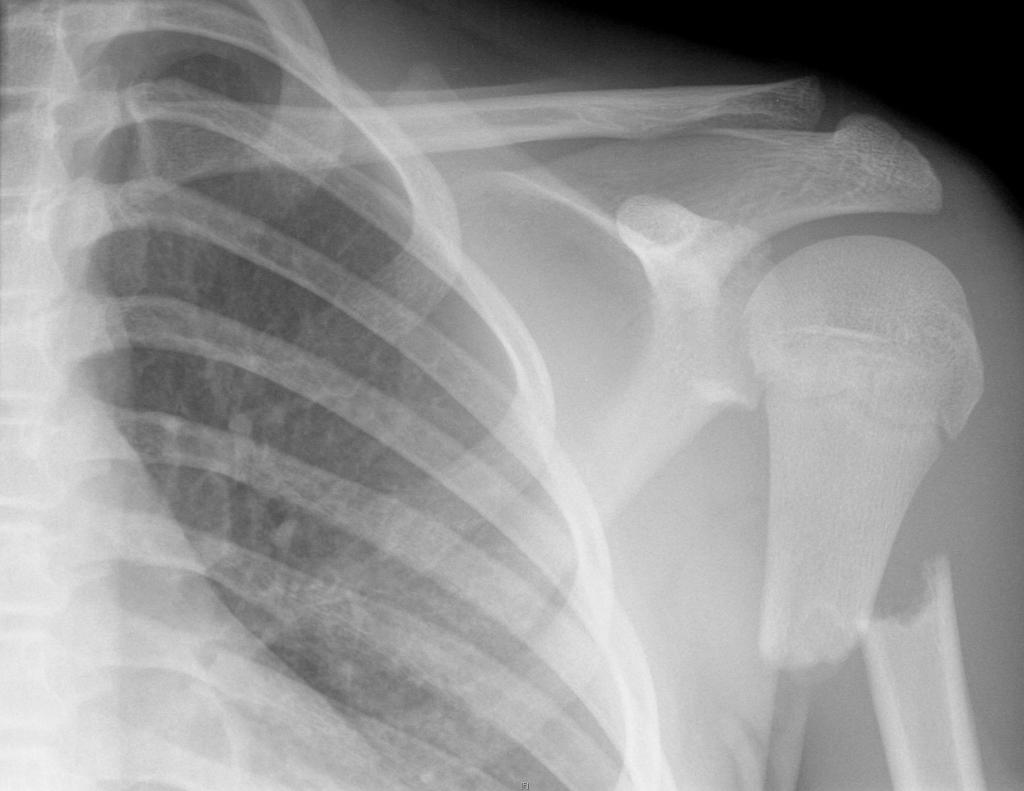
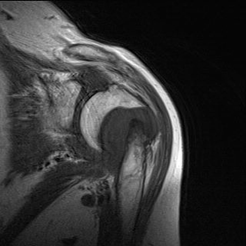
 Shoulder-fracture-dislocation-with-arterial-dissection.jpg DrMars
21:32, 15 April 2019
1,024 × 1,024; 72 KB
-

-

-
-
-
-
-
-
-

-

-

-
-
-
-
-
-
-
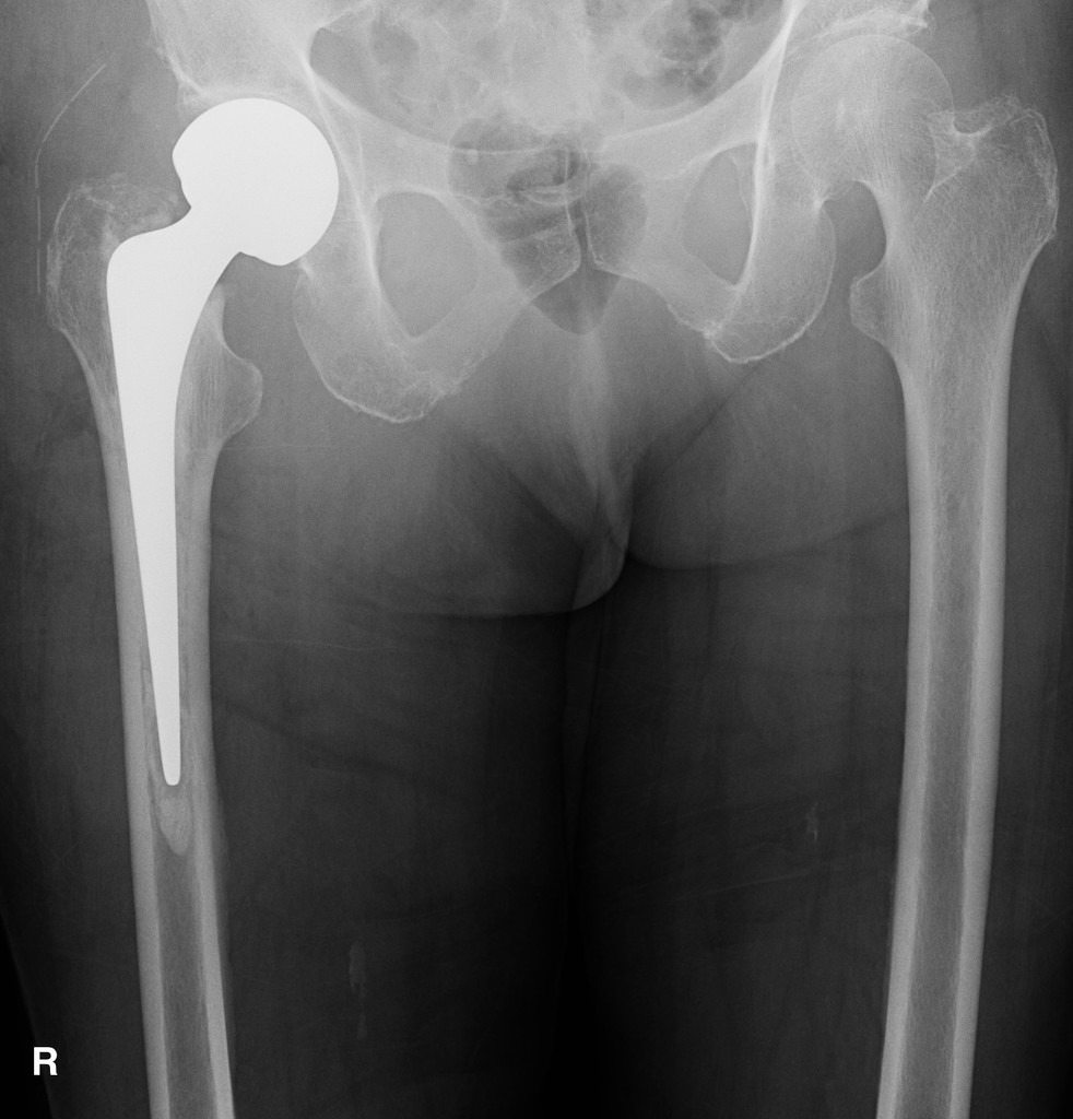
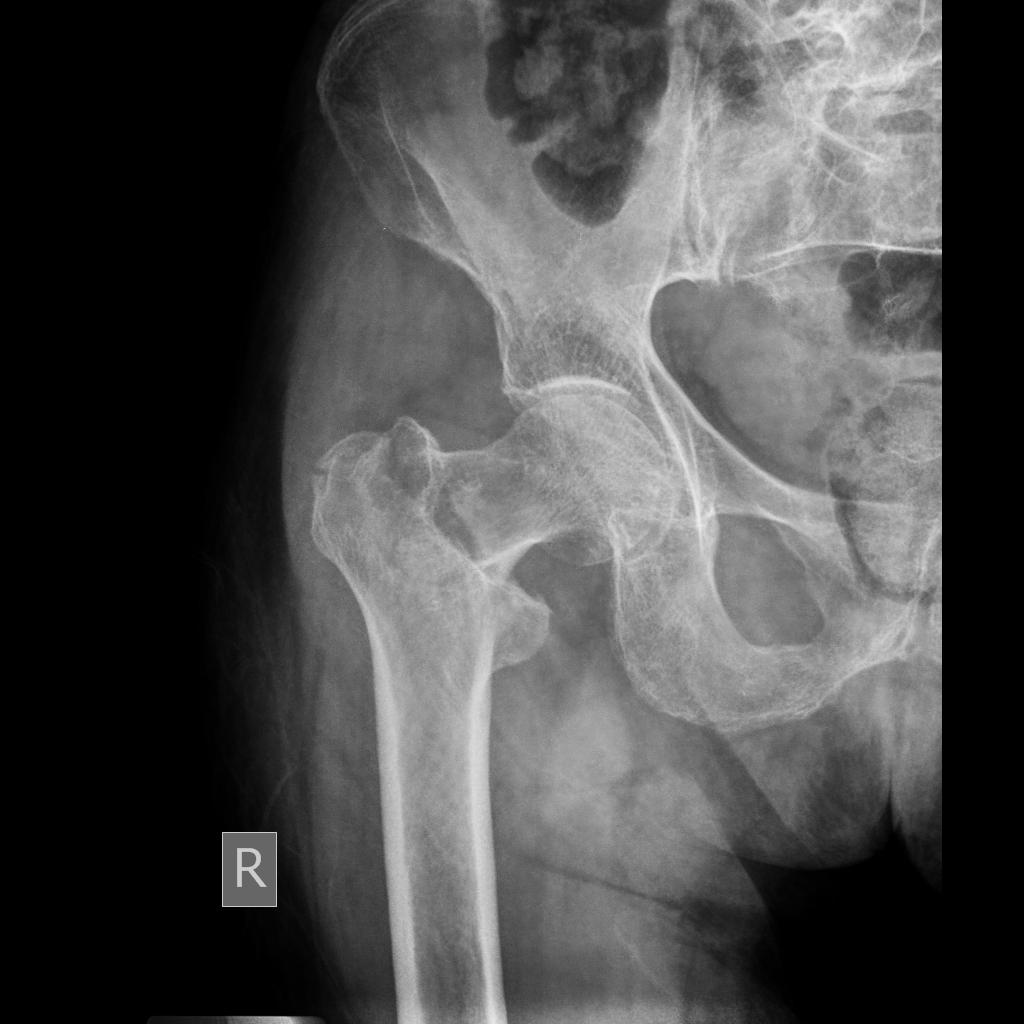
 Periprosthetic-femoral-fracture-vancouver-type-b2-1.jpg DrMars
14:19, 15 April 2019
1,024 × 919; 100 KB
-
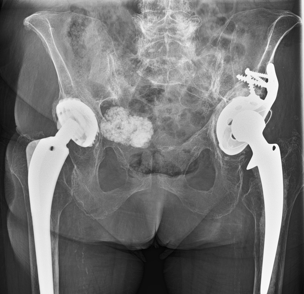
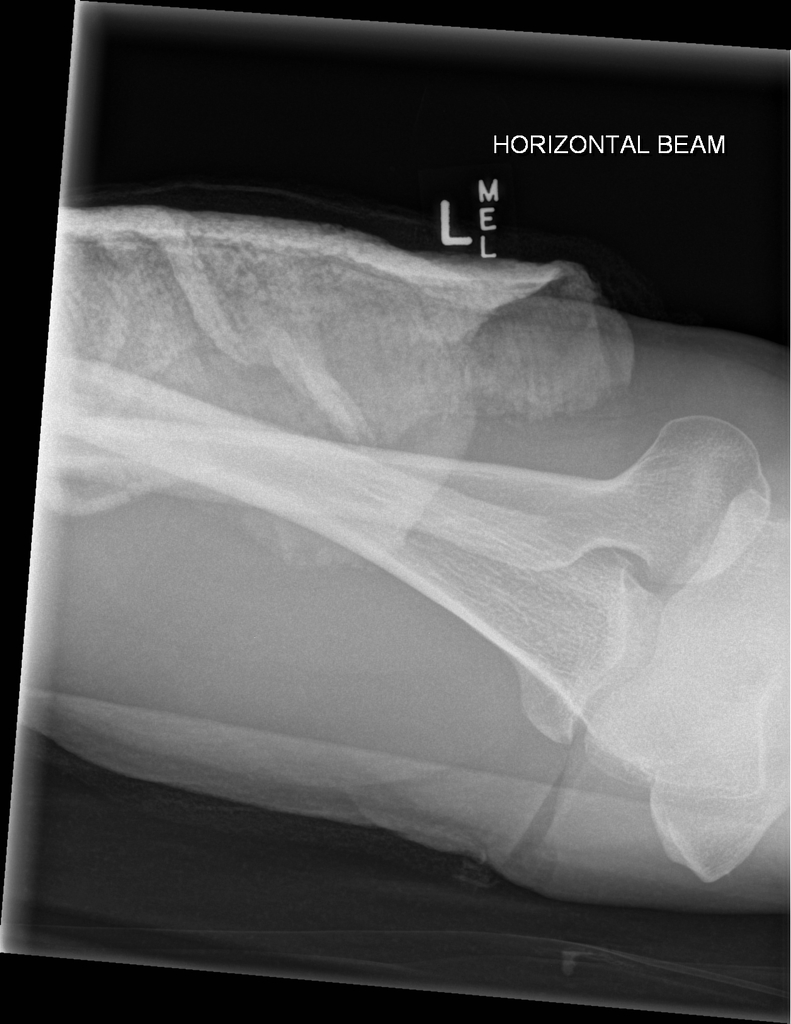
 Fractured-surgical-drain-after-right-hip-hemiarthroplasty.jpg DrMars
14:16, 15 April 2019
982 × 1,024; 132 KB
-
-
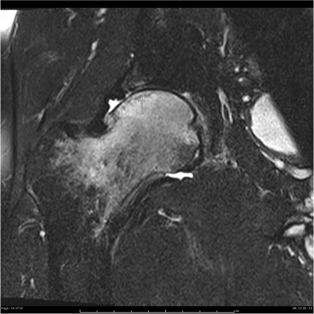
 Transient-osteoporosis-of-the-hip-underlying-subchondral-fracture (5).jpg DrMars
14:20, 14 April 2019
1,024 × 1,024; 89 KB
-
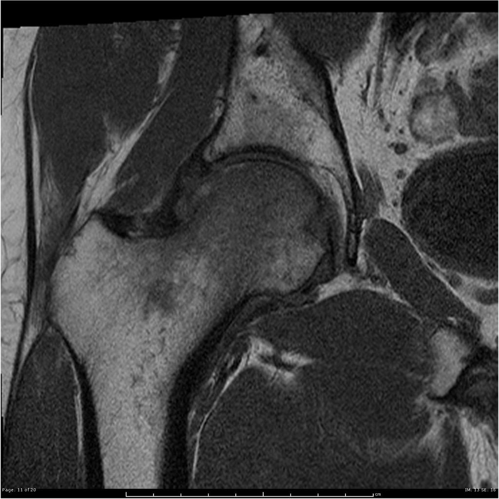
 Transient-osteoporosis-of-the-hip-underlying-subchondral-fracture (4).jpg DrMars
14:20, 14 April 2019
1,024 × 1,024; 92 KB
-
 Transient-osteoporosis-of-the-hip-underlying-subchondral-fracture (3).jpg DrMars
14:20, 14 April 2019
1,024 × 1,024; 95 KB
-
 Transient-osteoporosis-of-the-hip-underlying-subchondral-fracture (2).jpg DrMars
14:19, 14 April 2019
1,024 × 1,024; 119 KB
-
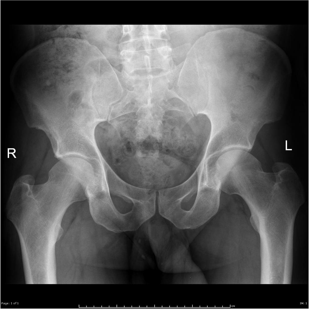 Transient-osteoporosis-of-the-hip-underlying-subchondral-fracture.jpg DrMars
14:16, 14 April 2019
1,024 × 1,024; 87 KB
-
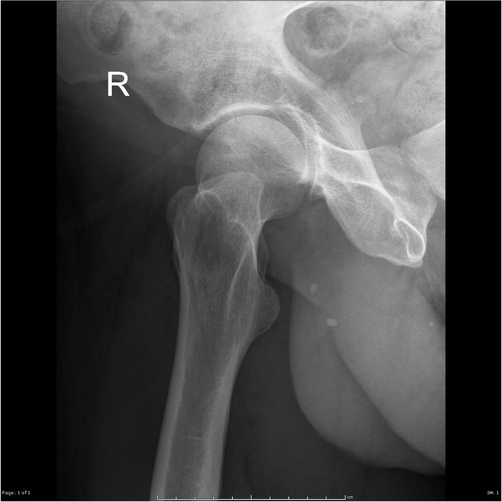 Transient-osteoporosis-of-the-hip-underlying-subchondral-fracture (1).jpg DrMars
14:16, 14 April 2019
1,024 × 1,024; 74 KB
-
-
-
-
-
-

-

-

-
-

-

-

-

-

-

-
-
-
-
-
-
-
-
-
-
-
-
-
-

-
 Epidermoid cyst of testis -- high mag.jpg Gertrude Djouka
19:47, 10 April 2019
2,848 × 4,272; 5.74 MB
-

-

-
 Intratubular germ cell neoplasia - 2 - very high mag.jpg Gertrude Djouka
16:07, 10 April 2019
4,272 × 2,848; 5 MB
-
 Mucinous Cystadenocarcinoma of the Ovary.jpg Qurrat-ul-ain Abid
15:54, 10 April 2019
1,022 × 900; 238 KB
-
 Intertubular seminoma -- intermed mag.jpg Gertrude Djouka
15:53, 10 April 2019
4,272 × 2,848; 5.2 MB
-

-

-
 Hemihypertrophy of the right chest, and a soft-tissue mass under the right crus.jpg Gunnam
17:49, 9 April 2019
628 × 511; 126 KB
-

-

-
 Mammography and scintimammography of carcinoma.jpg Soroush Seifirad
17:16, 9 April 2019
1,004 × 565; 159 KB
-

-

-
-
-
-
-
-

-

-

-

-
-

-
-

-

-

-
-
-
-
-
-
-
-
-

-

-

-
-
-
-
-
-

-

-

-

-
 Peau d’ orange Appearance in Breast cancer.jpg Soroush Seifirad
19:09, 2 April 2019
1,600 × 1,550; 339 KB
-
 Diagram showing stage 1A breast cancer CRUK 199.svg Soroush Seifirad
15:52, 2 April 2019
0 × 0; 42 KB
-

-

-

-

-

-
-

-
-
-
-
-
-
-
-
-
-
-
-
-
-
-

-

-

-

-
-
-
-

-

-

-
-

-

-
-
-

-

-

-

-
-
-

-

-

-

-
-
-
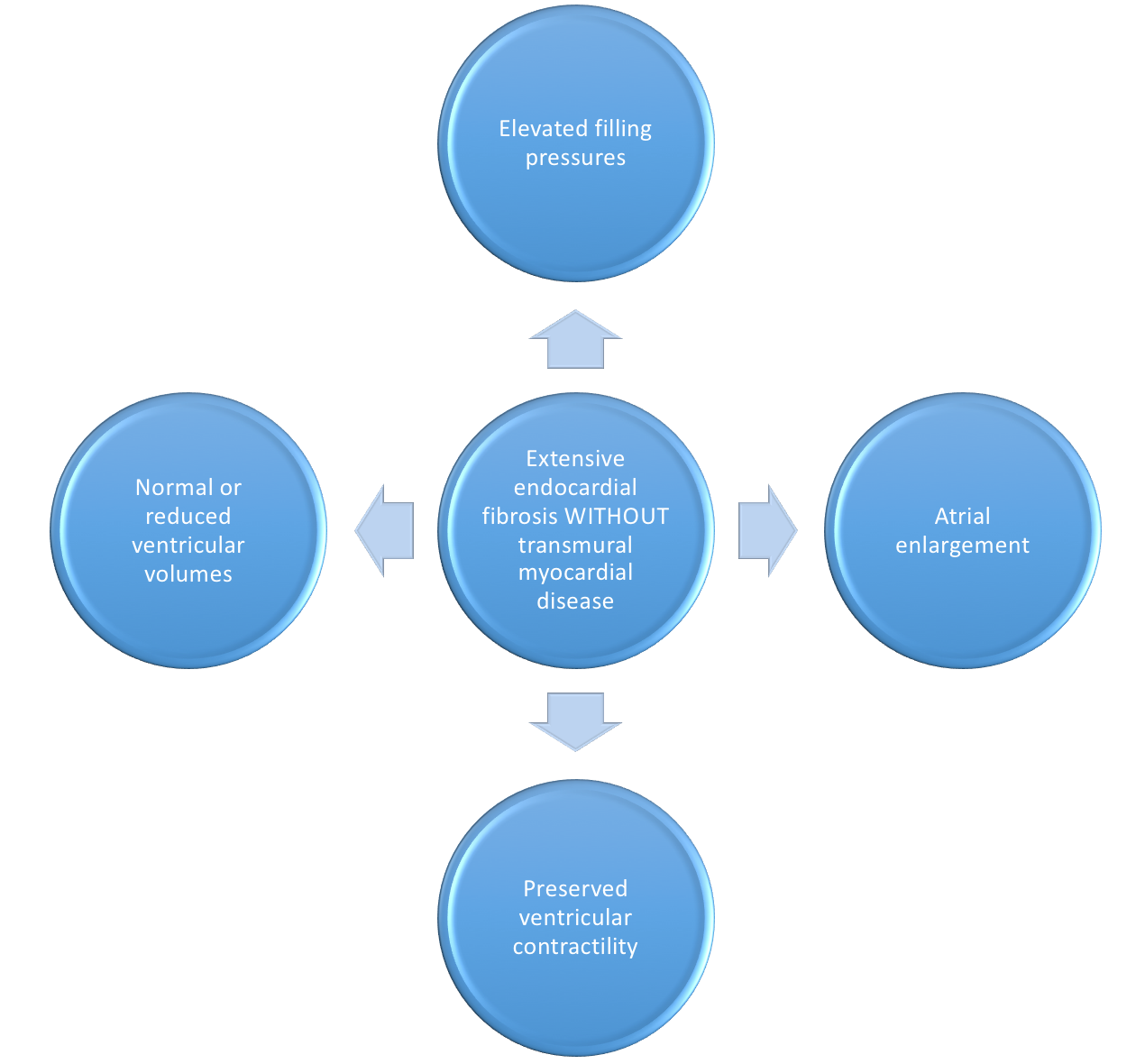
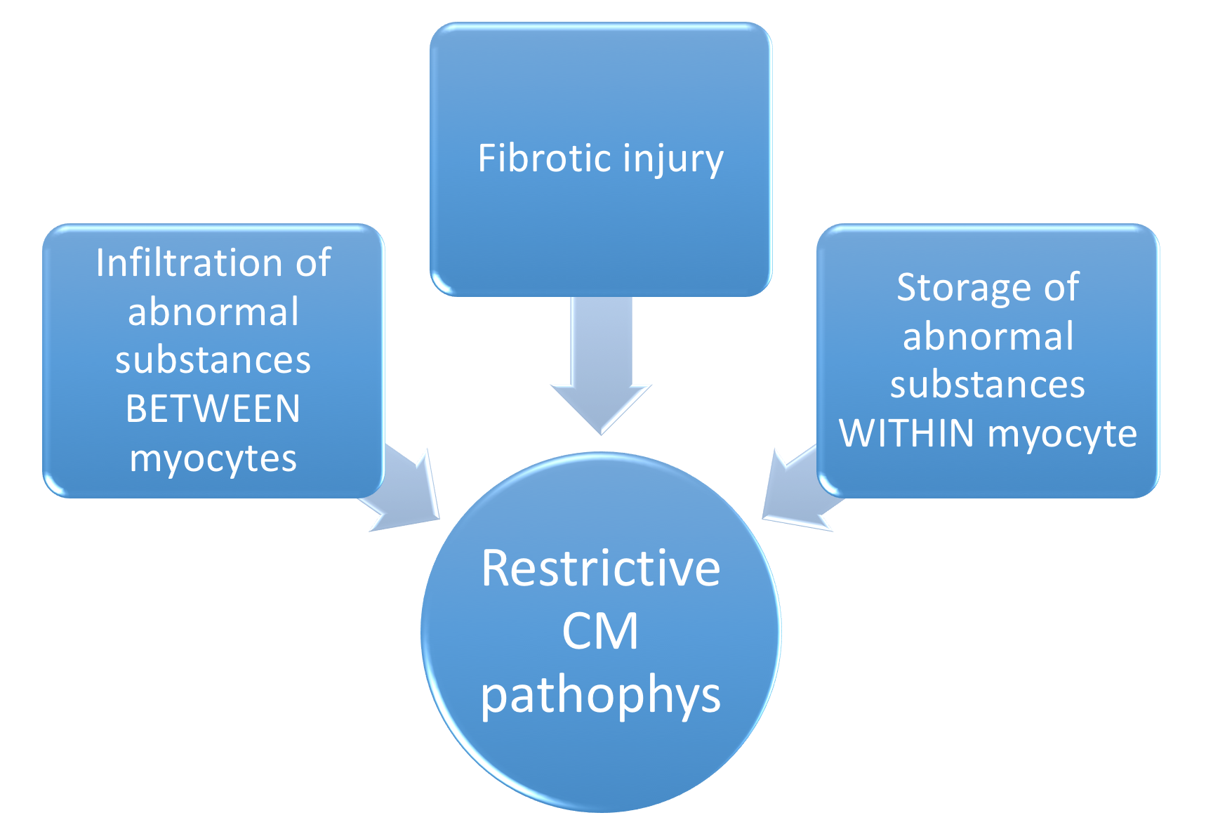
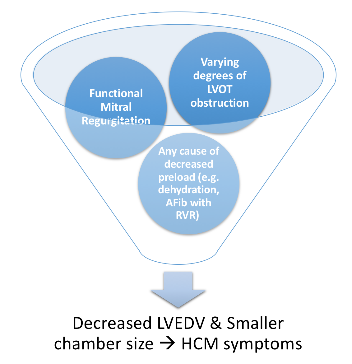
 Pathological cycle of LVOT obstruction in HCM.png Bade Fatunde
12:23, 26 March 2019
1,574 × 1,174; 438 KB
-


_image,_and_enhanced_depth_imaging_optical_coherence_tomographic_(EDI-_OCT).gif)


.jpg)


.png)


.jpg)


.jpg)
.jpg)


.jpg)


.jpeg)
.jpeg)


.jpeg)
.jpeg)

.jpg)
.jpg)

.jpg)
.jpg)
.jpg)


.jpg)

.jpg)


.jpg)
.jpg)
.jpg)


.jpg)

,_was_a_respected_Swiss_clinician_scientist._Source._Nationaal_Archief-_https---www.nationaalarchief.nl-onderzoeken-fotocollectie-detail-a9d4bf64-d0b4-102d-bcf8-003048976d84urce-.jpg)


.jpg)

.jpg)

.jpeg)

.jpg)


.jpg)
.jpg)

.gif)


_(1).jpg)
.jpg)


.jpg)

.jpg)
.jpg)
.jpg)
.jpg)
.jpg)
.jpg)
.jpg)
.jpg)
.jpg)
.jpg)
.jpg)

.jpg)

.jpg)
.jpg)
.jpg)


.jpg)


.jpg)
.jpg)
.jpg)


.jpg)
.jpg)
.jpg)


.jpg)

.jpg)
.jpg)
.jpg)


.jpg)
.jpg)


.jpg)
.jpg)
.jpg)
.jpg)


.jpg)


.JPEG)


.jpg)


.jpg)
.jpg)


.jpg)
.jpg)
.jpg)
.jpg)
.jpg)
.jpg)

.jpg)


.jpg)

.jpg)

.jpg)


.jpg)
.jpg)
.jpg)
.jpg)
.jpg)
.jpg)

.jpg)


.png)
.png)
.jpg)
.jpg)
.jpg)
.jpg)
.jpg)
.jpg)
.jpg)
.jpg)
.jpg)
.jpg)
.jpg)
.jpg)