Gallery of new files
Jump to navigation
Jump to search
This special page shows the last uploaded files.
-
-
-

-

-

-
-
-
-
-
-
-
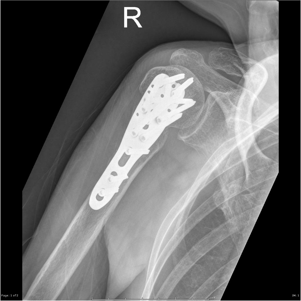
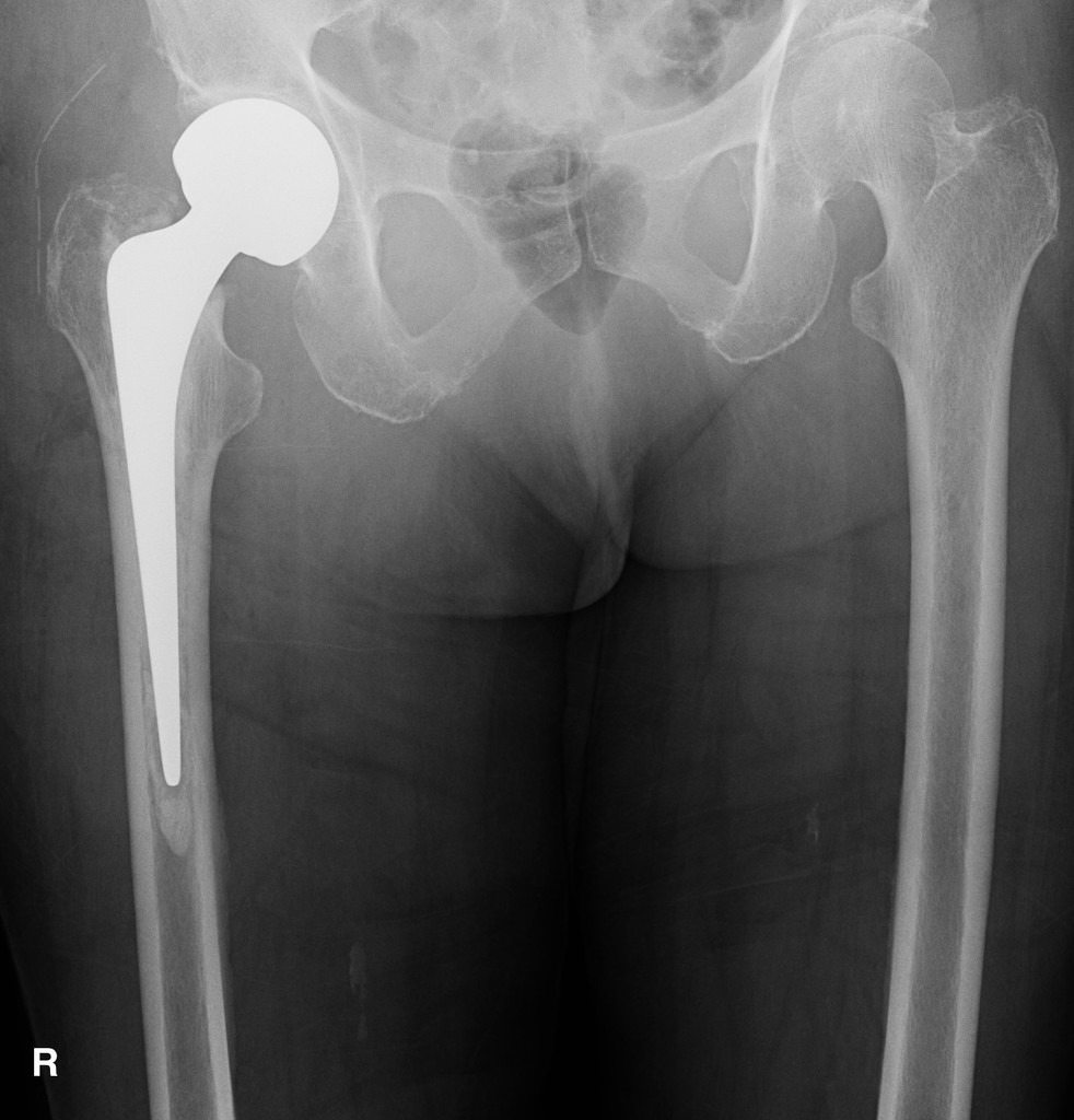
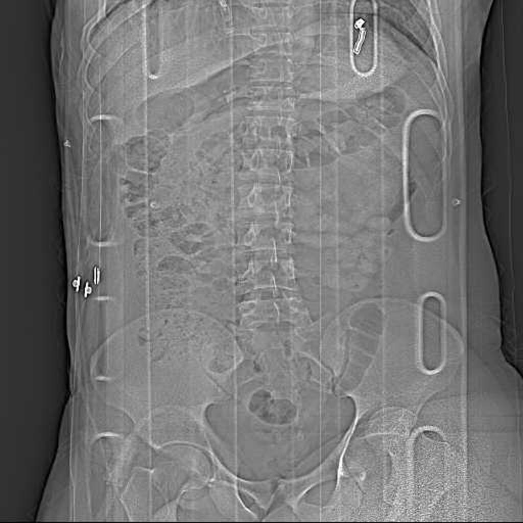
 Periprosthetic-femoral-fracture-vancouver-type-b2-1.jpg DrMars
14:19, 15 April 2019
1,024 × 919; 100 KB
-
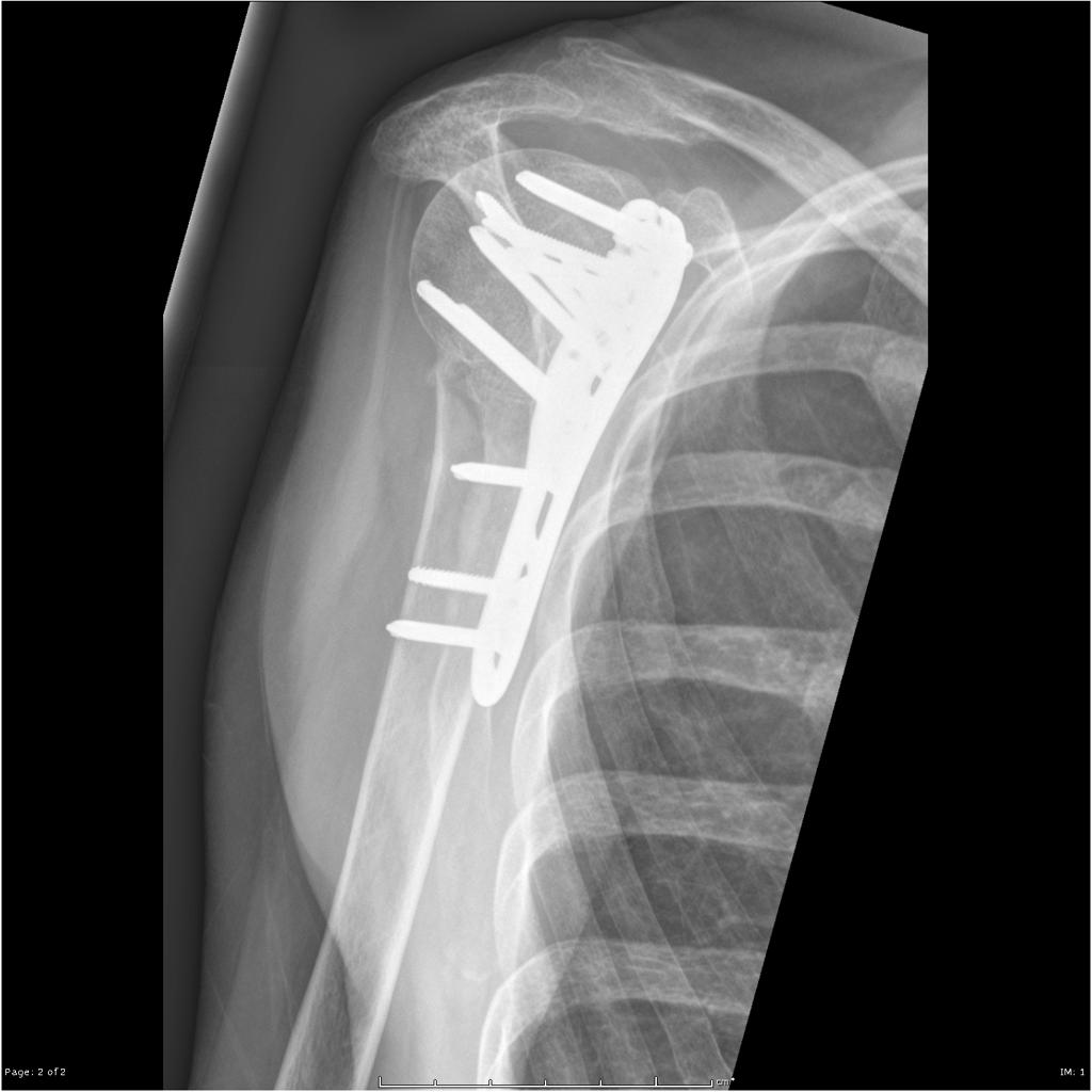
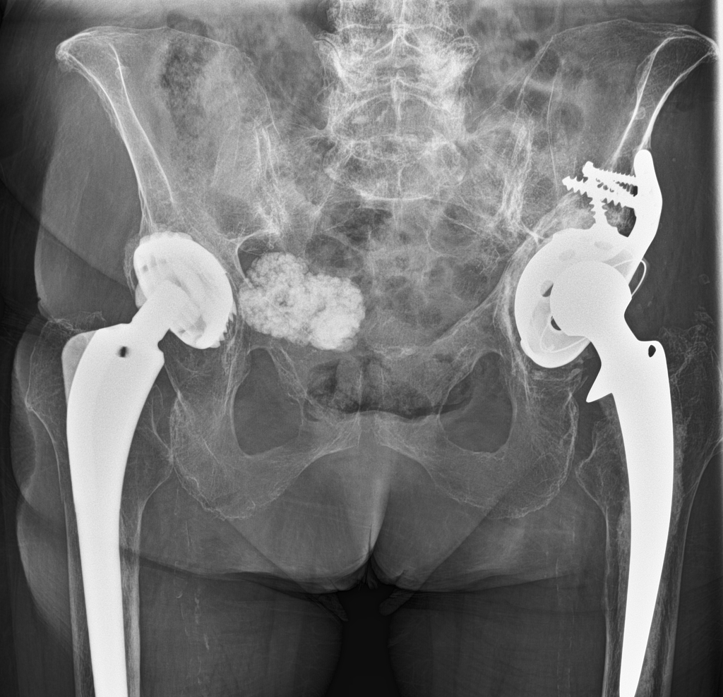
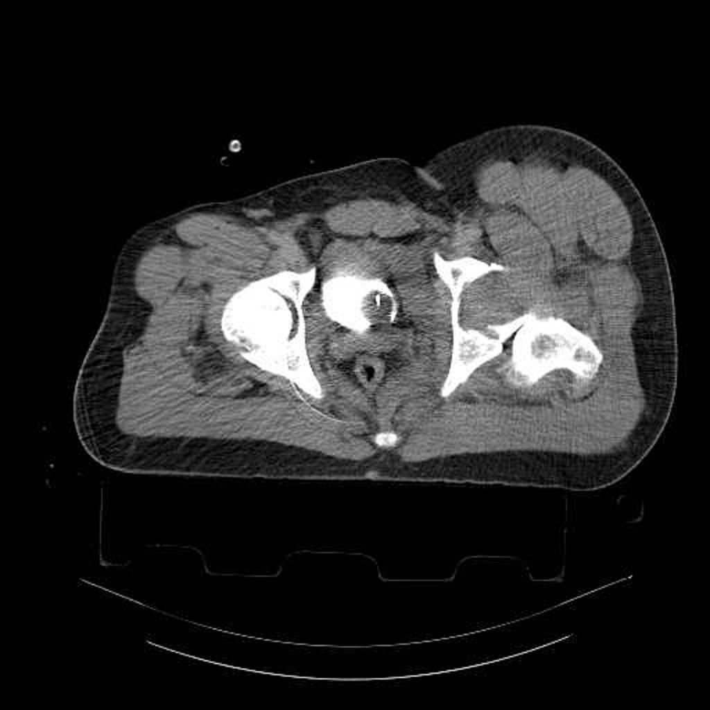
 Fractured-surgical-drain-after-right-hip-hemiarthroplasty.jpg DrMars
14:16, 15 April 2019
982 × 1,024; 132 KB
-
-
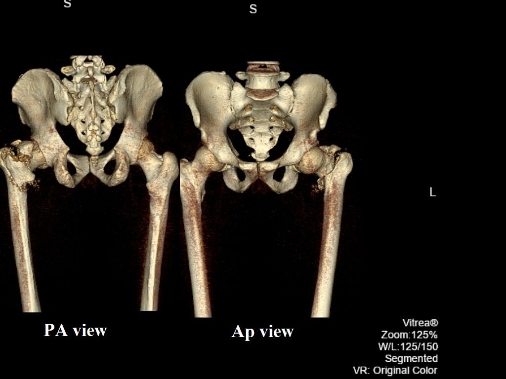
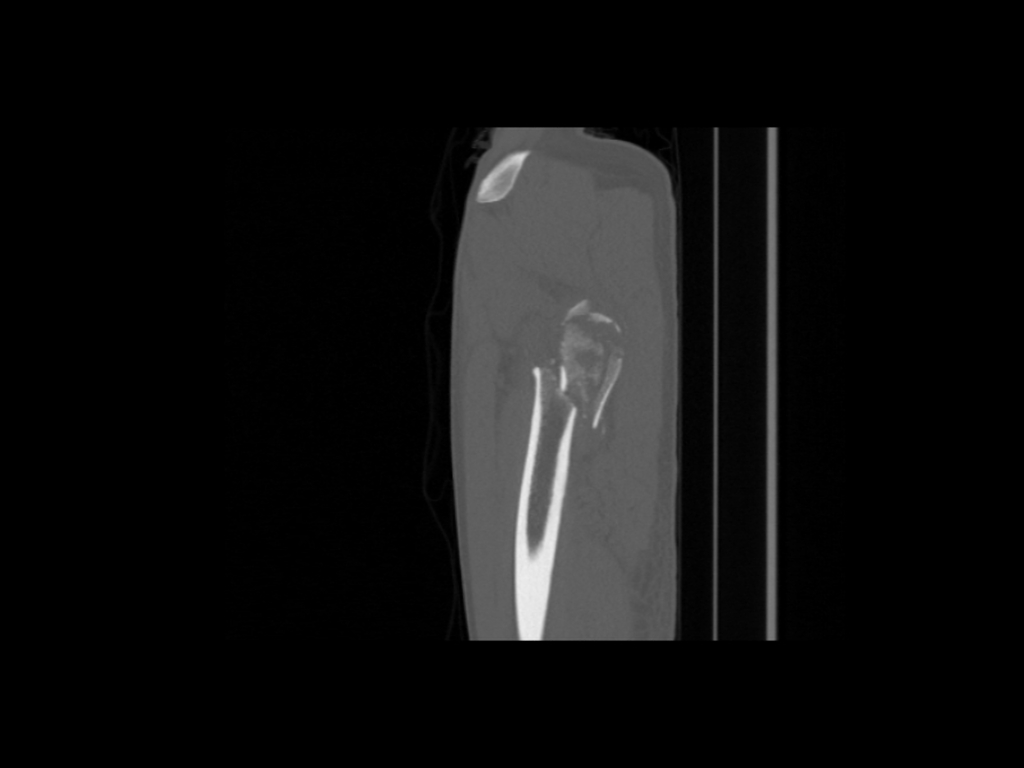
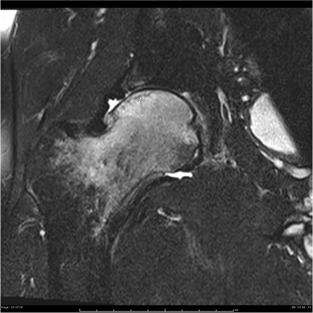
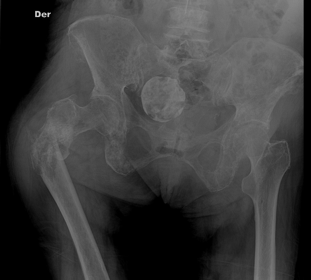
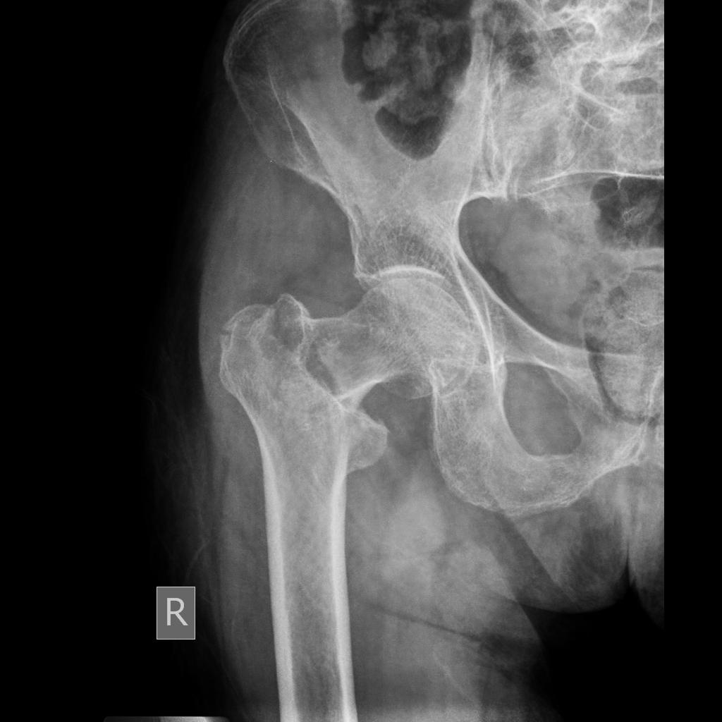
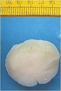
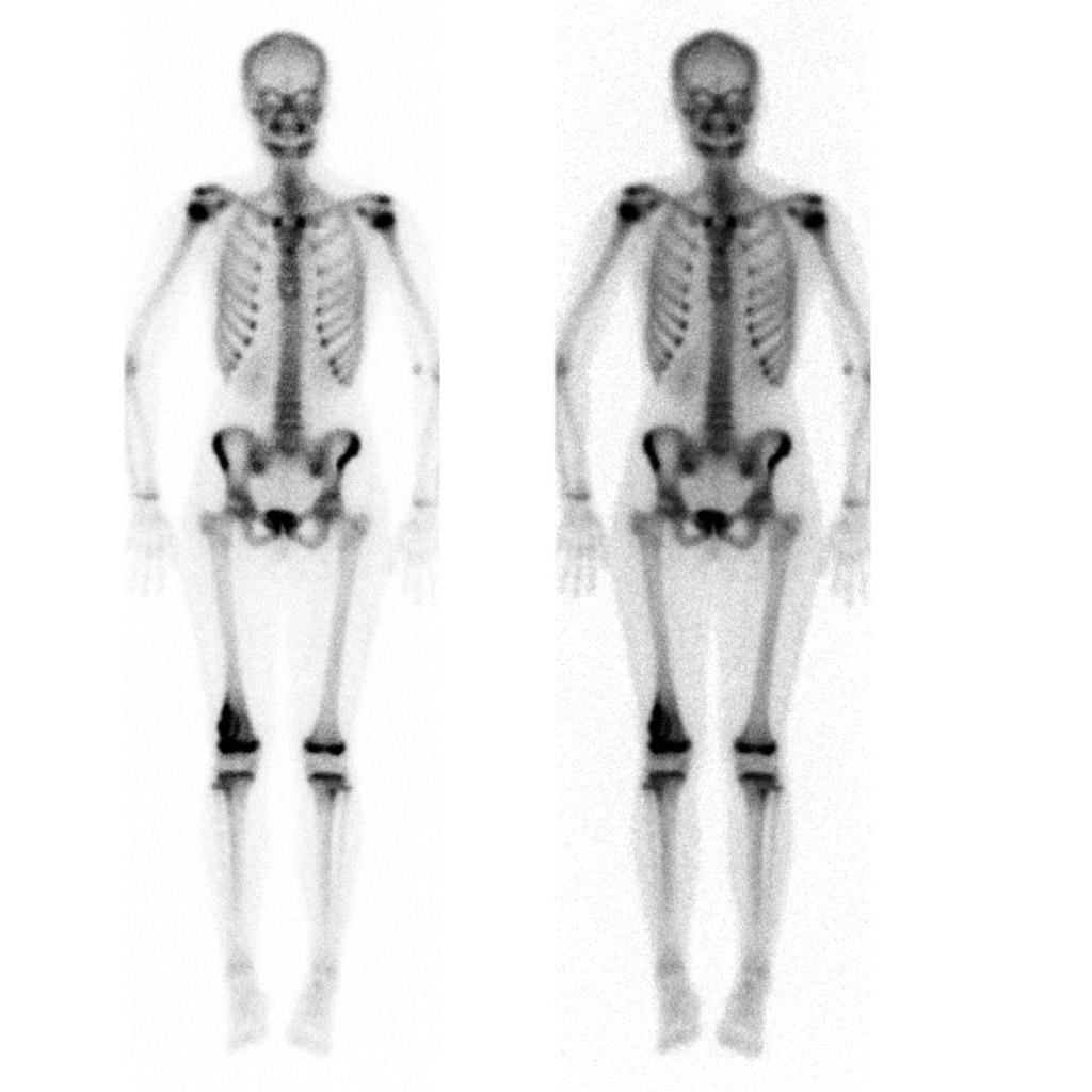
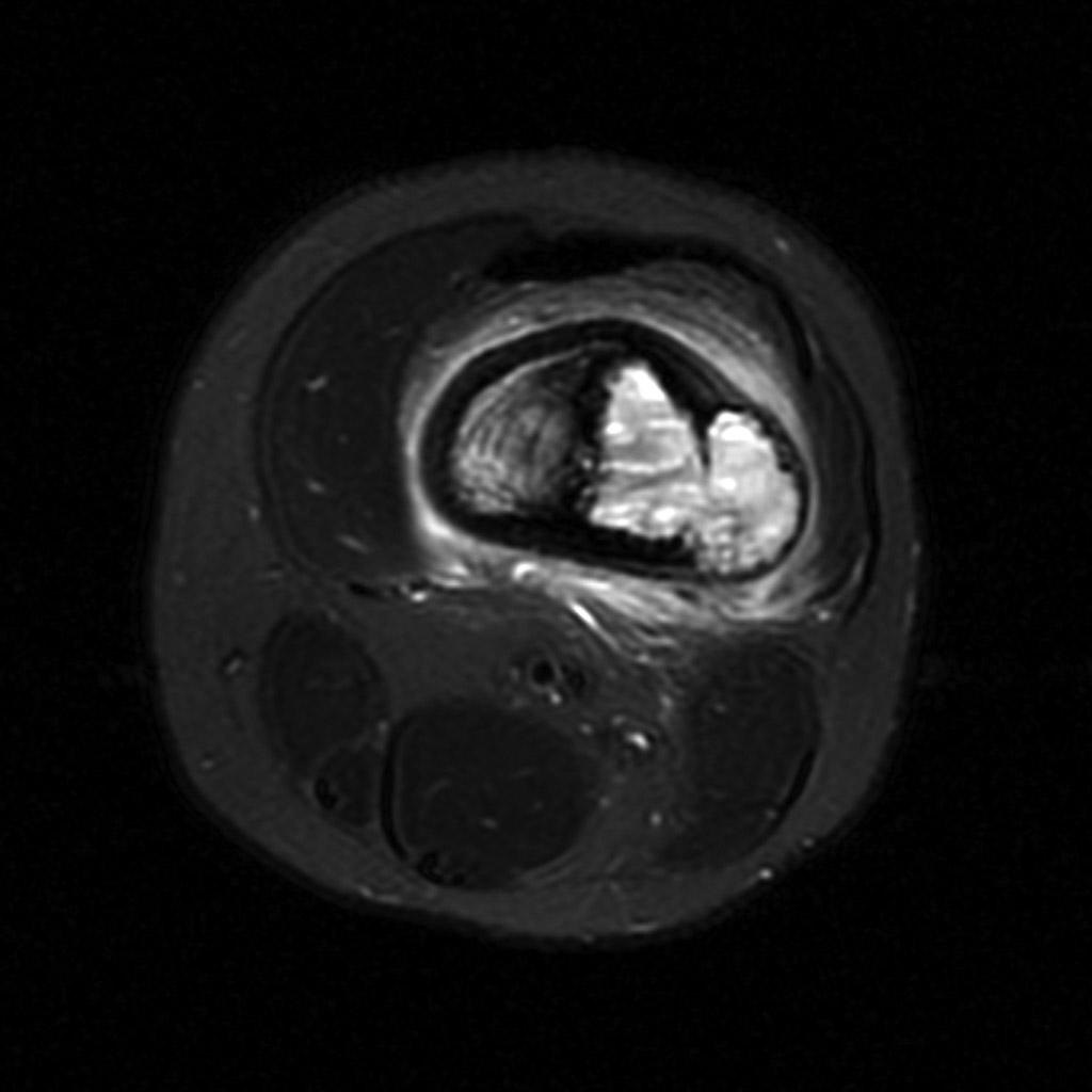
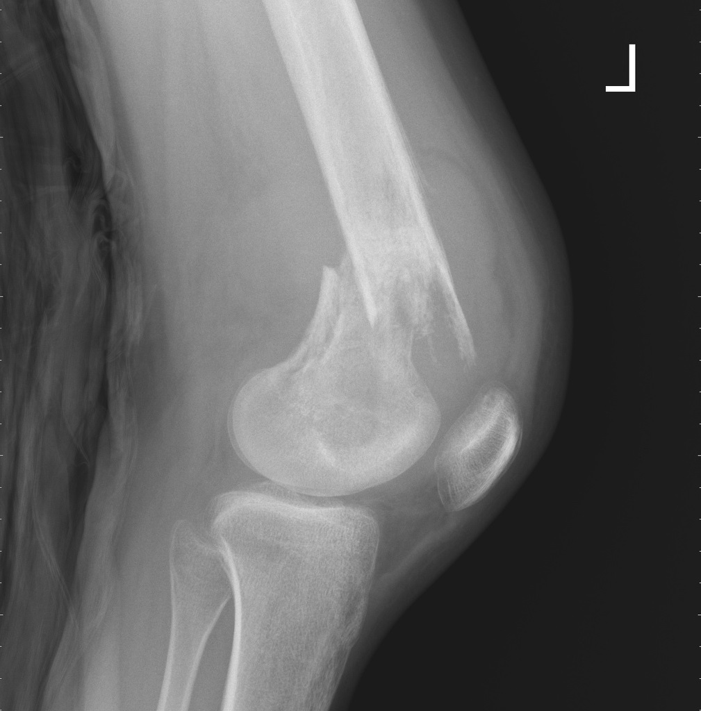
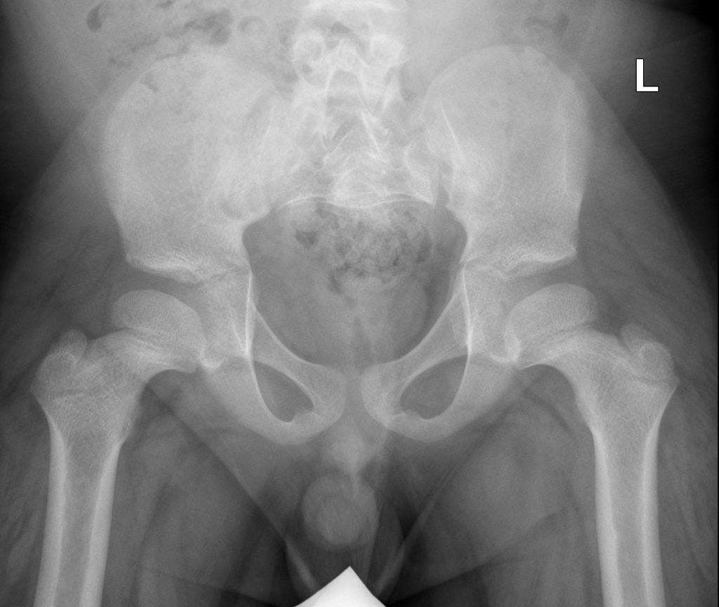
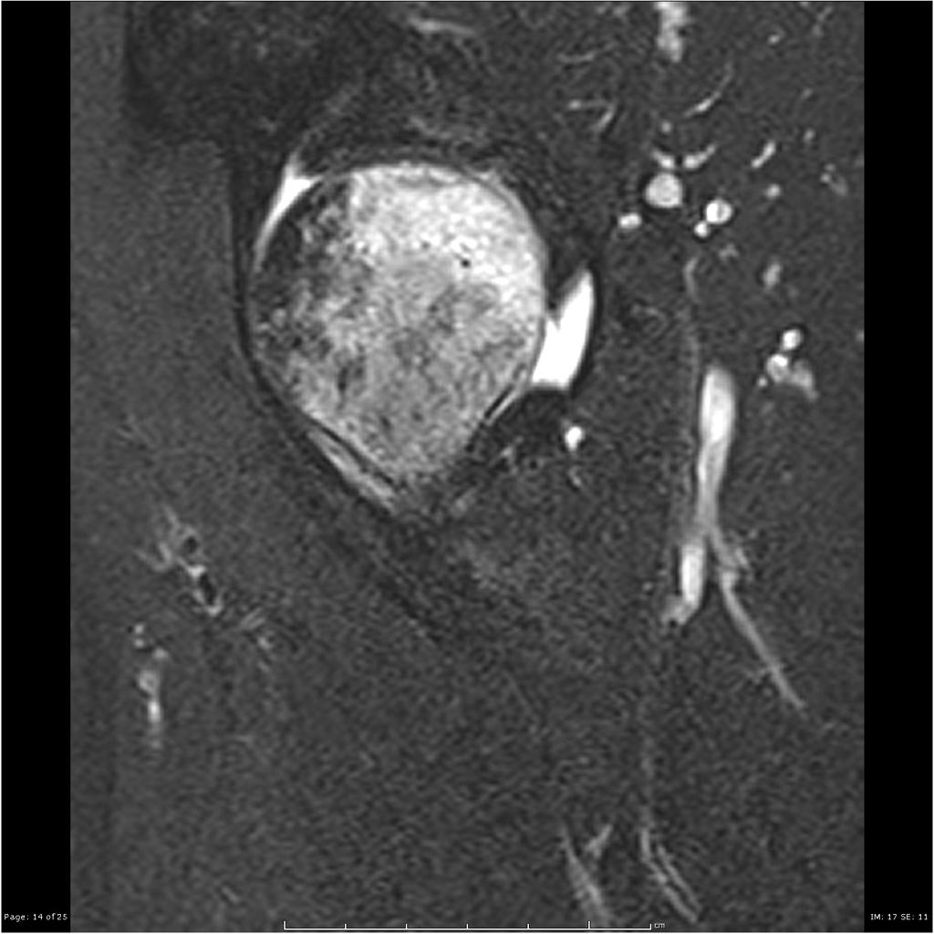 Transient-osteoporosis-of-the-hip-underlying-subchondral-fracture (5).jpg DrMars
14:20, 14 April 2019
1,024 × 1,024; 89 KB
-
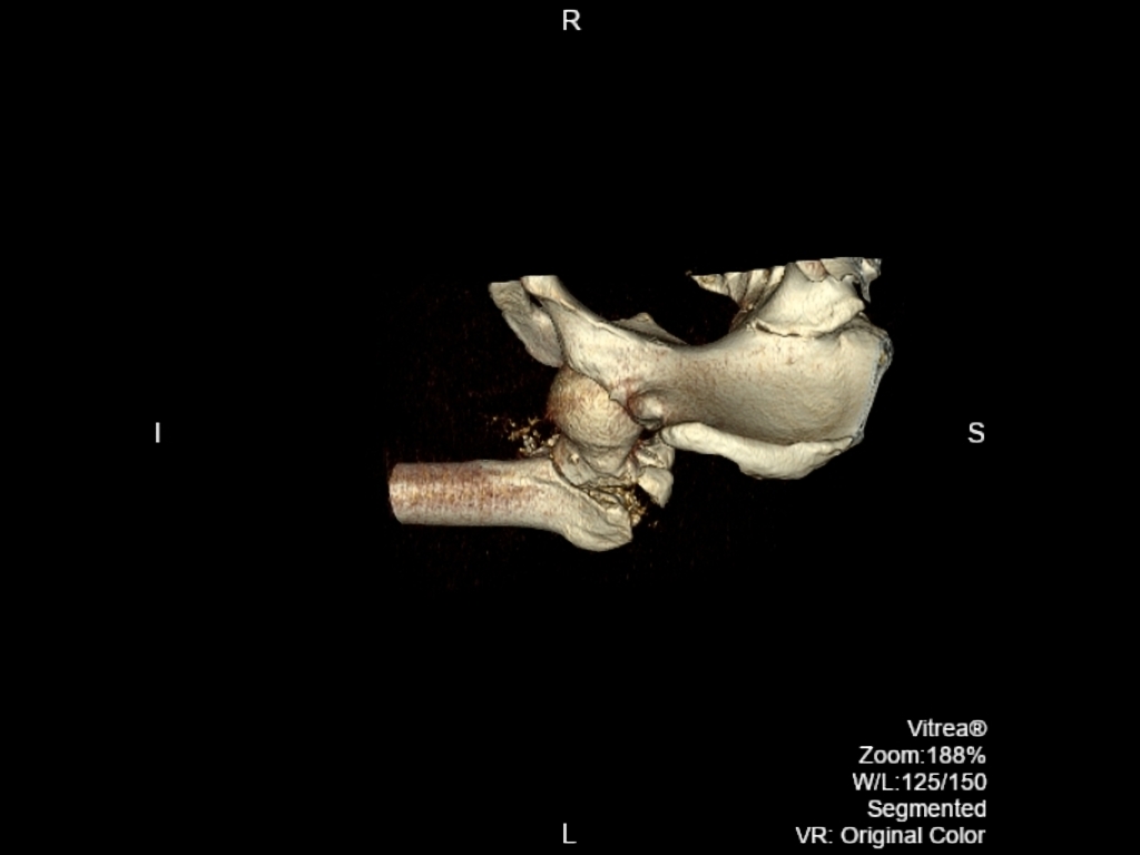
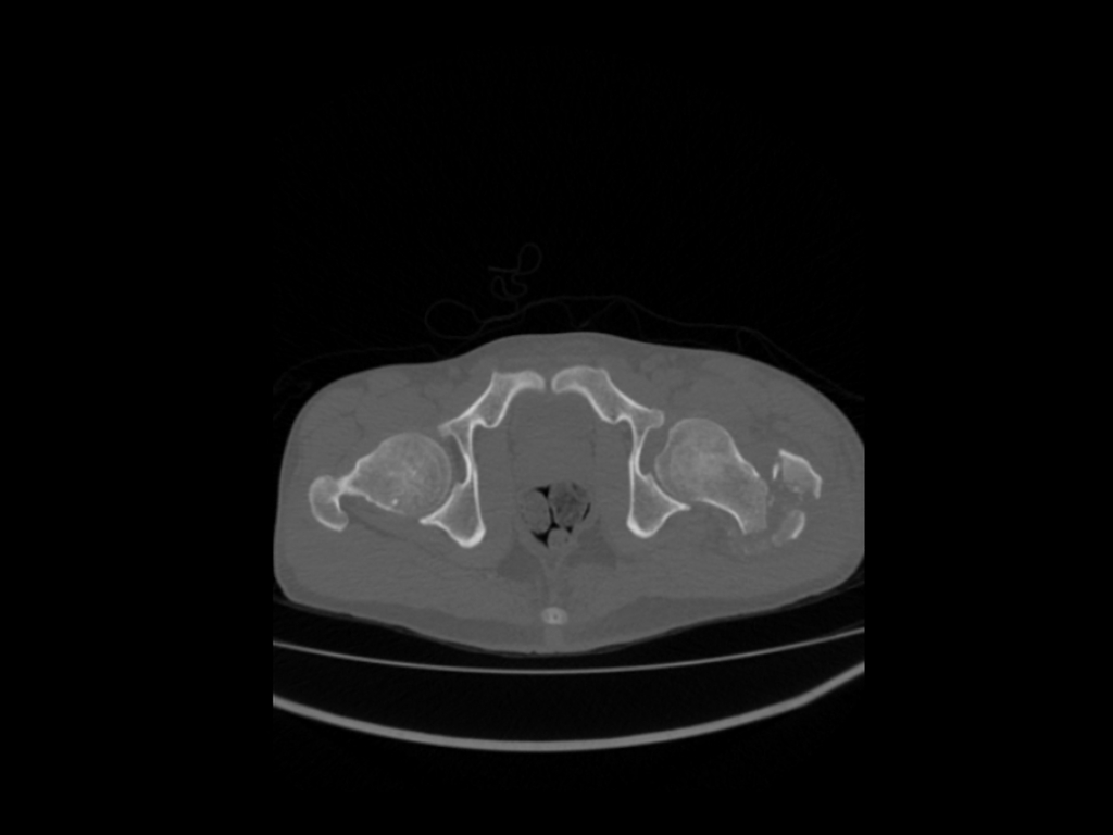
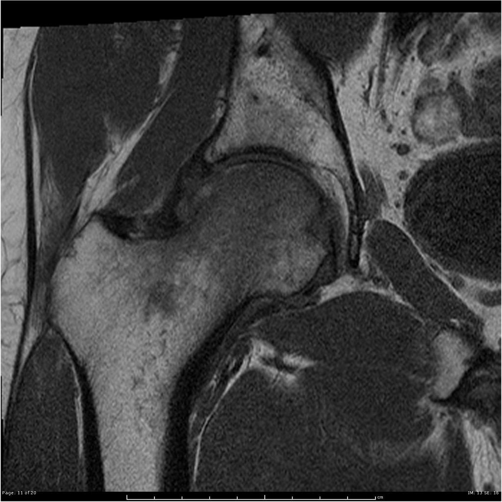
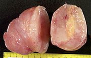
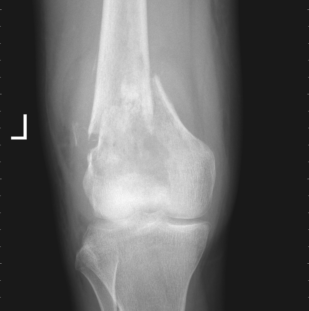
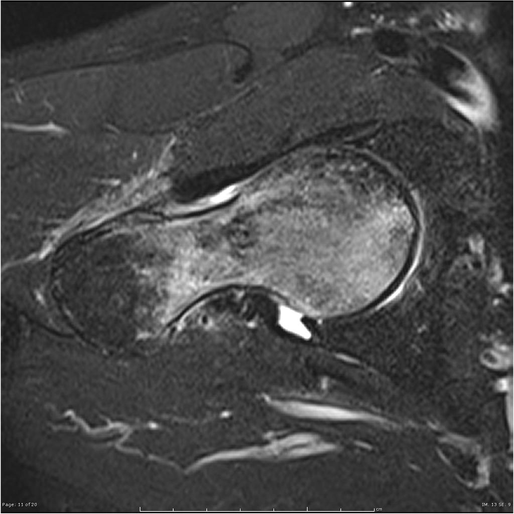 Transient-osteoporosis-of-the-hip-underlying-subchondral-fracture (4).jpg DrMars
14:20, 14 April 2019
1,024 × 1,024; 92 KB
-
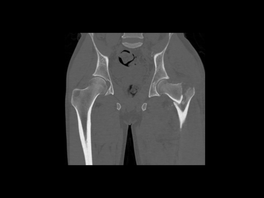
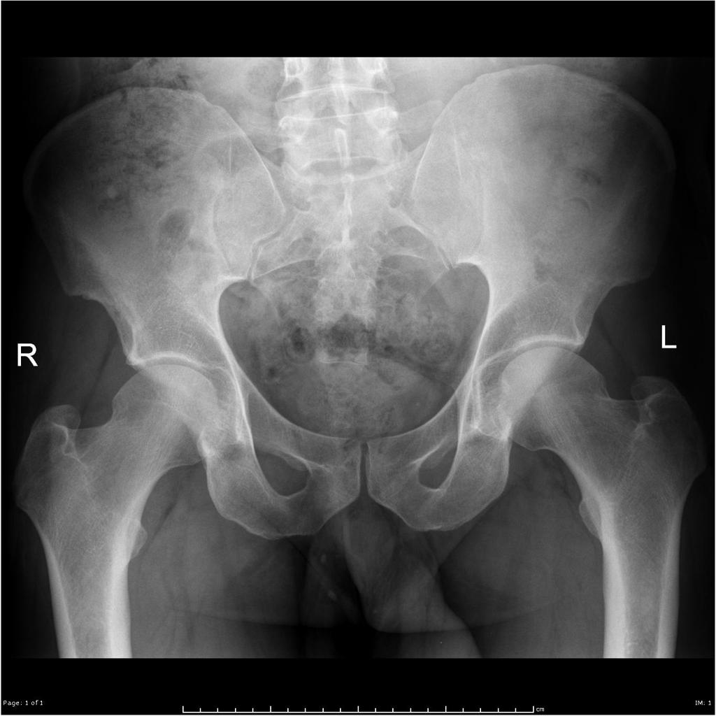
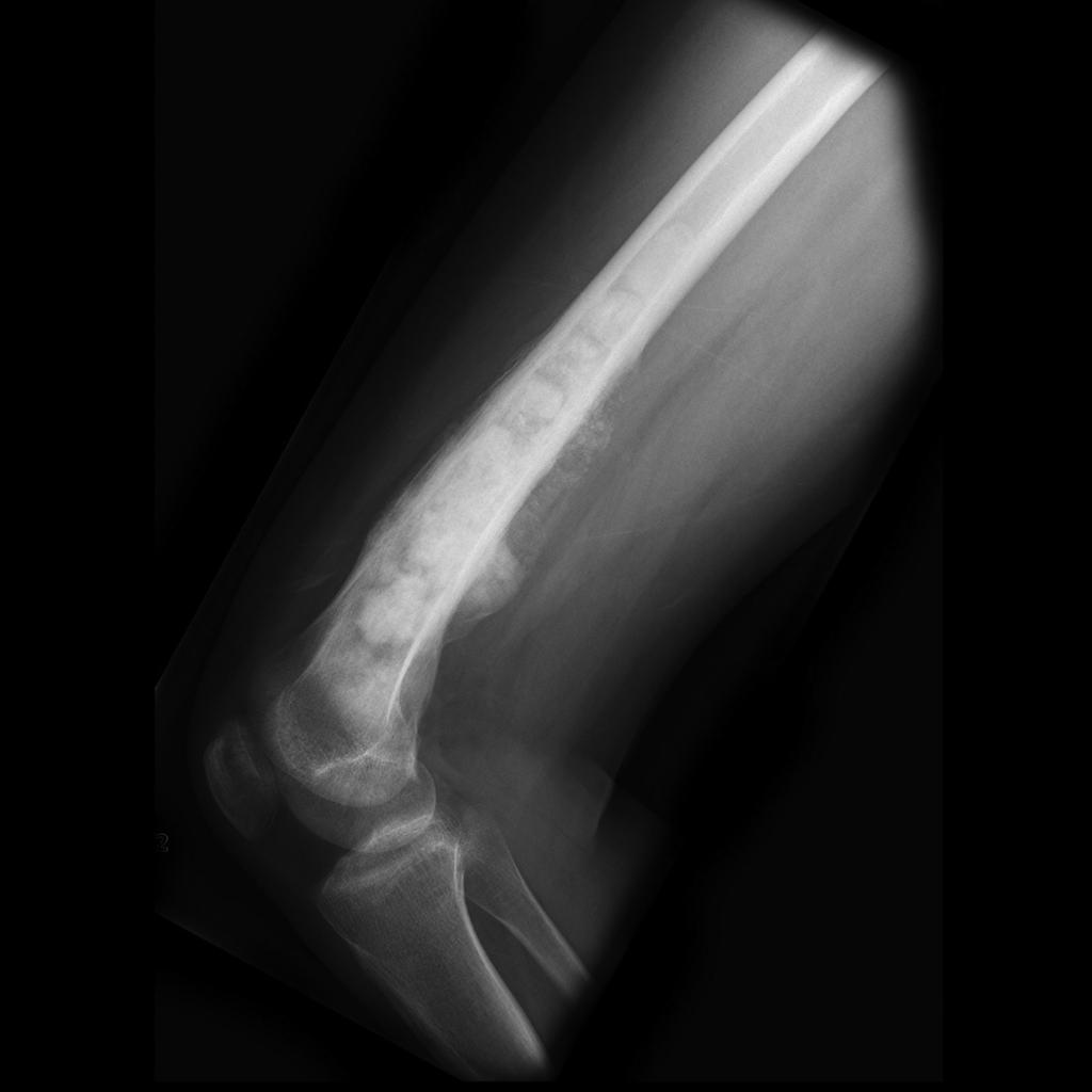
 Transient-osteoporosis-of-the-hip-underlying-subchondral-fracture (3).jpg DrMars
14:20, 14 April 2019
1,024 × 1,024; 95 KB
-
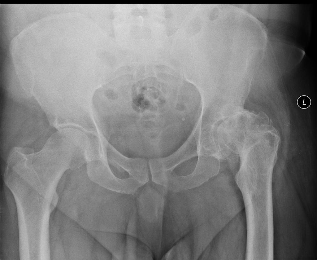
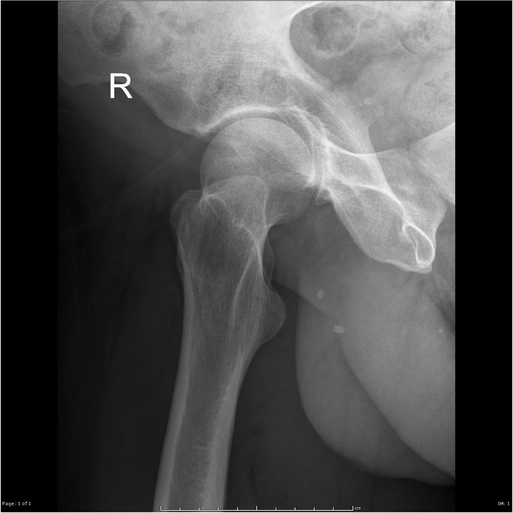
 Transient-osteoporosis-of-the-hip-underlying-subchondral-fracture (2).jpg DrMars
14:19, 14 April 2019
1,024 × 1,024; 119 KB
-
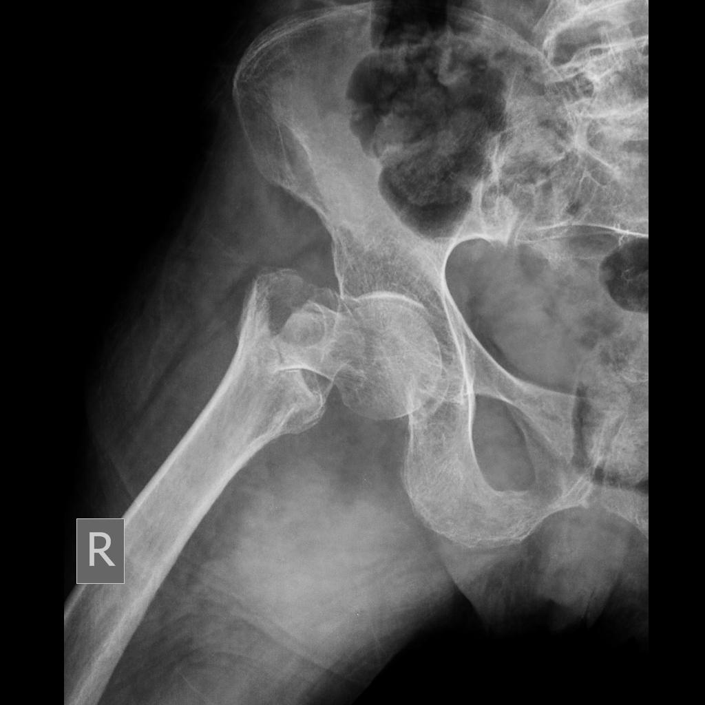
 Transient-osteoporosis-of-the-hip-underlying-subchondral-fracture.jpg DrMars
14:16, 14 April 2019
1,024 × 1,024; 87 KB
-
 Transient-osteoporosis-of-the-hip-underlying-subchondral-fracture (1).jpg DrMars
14:16, 14 April 2019
1,024 × 1,024; 74 KB
-
-
-
-
-
-

-

-

-
-

-

-

-

-

-

-
-
-
-
-
-
-
-
-
-
-
-
-
-

-
 Epidermoid cyst of testis -- high mag.jpg Gertrude Djouka
19:47, 10 April 2019
2,848 × 4,272; 5.74 MB
-

-

-
 Intratubular germ cell neoplasia - 2 - very high mag.jpg Gertrude Djouka
16:07, 10 April 2019
4,272 × 2,848; 5 MB
-
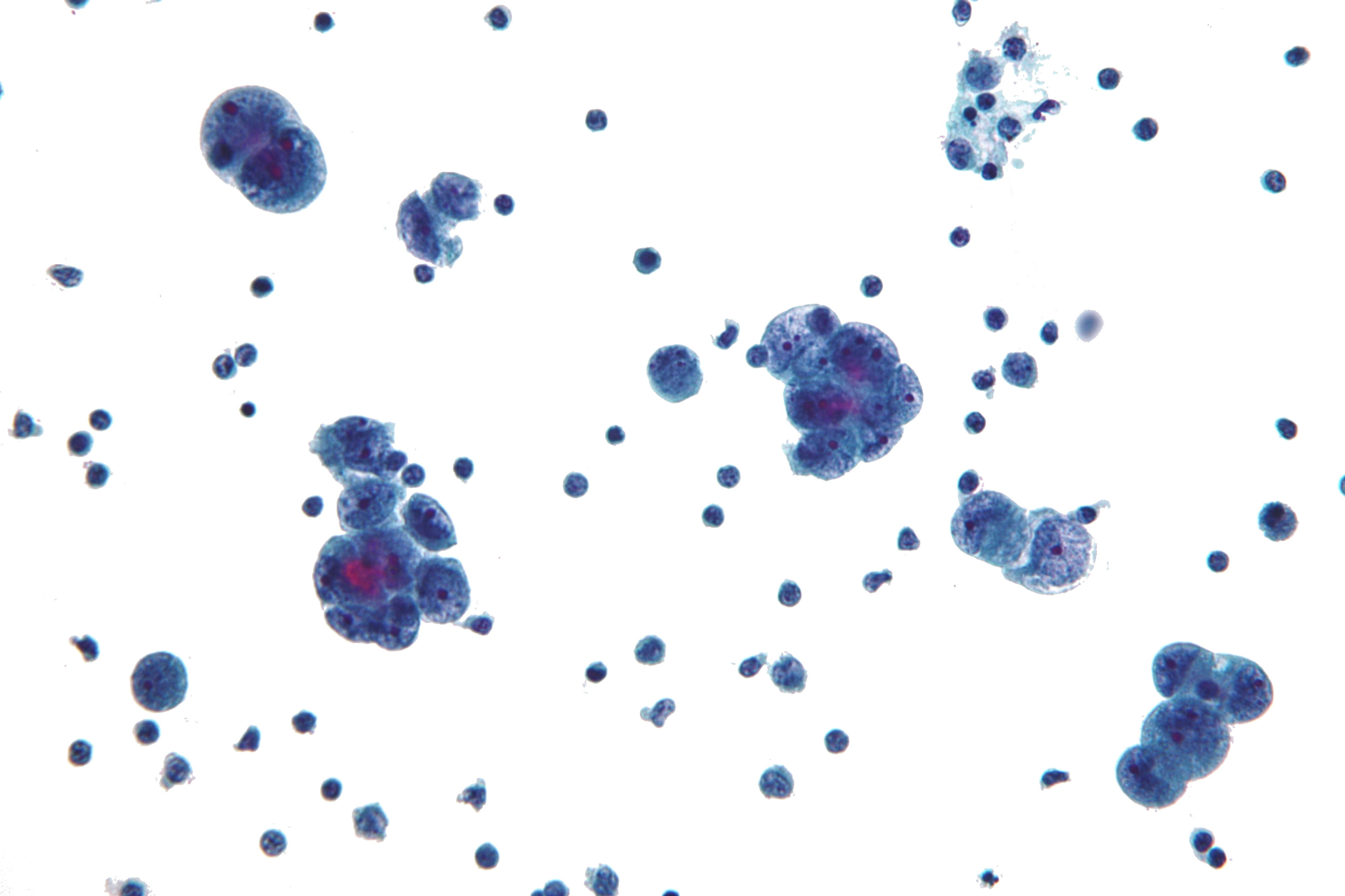
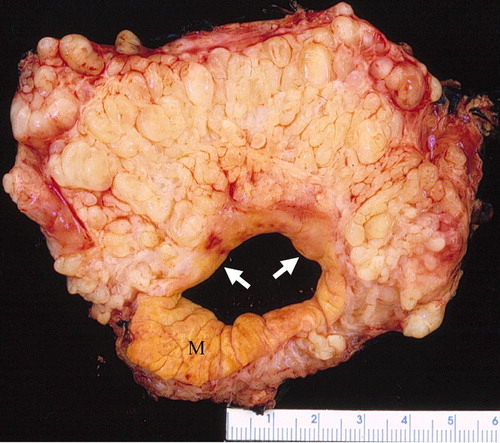
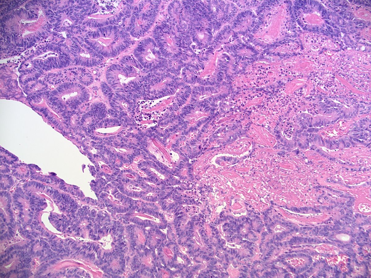
 Mucinous Cystadenocarcinoma of the Ovary.jpg Qurrat-ul-ain Abid
15:54, 10 April 2019
1,022 × 900; 238 KB
-
 Intertubular seminoma -- intermed mag.jpg Gertrude Djouka
15:53, 10 April 2019
4,272 × 2,848; 5.2 MB
-

-

-
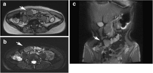
 Hemihypertrophy of the right chest, and a soft-tissue mass under the right crus.jpg Gunnam
17:49, 9 April 2019
628 × 511; 126 KB
-

-

-
 Mammography and scintimammography of carcinoma.jpg Soroush Seifirad
17:16, 9 April 2019
1,004 × 565; 159 KB
-

-

-
-
-
-
-
-

-

-

-

-
-

-
-

-

-

-
-
-
-
-
-
-
-
-

-

-

-
-
-
-
-
-

-

-

-

-
 Peau d’ orange Appearance in Breast cancer.jpg Soroush Seifirad
19:09, 2 April 2019
1,600 × 1,550; 339 KB
-
 Diagram showing stage 1A breast cancer CRUK 199.svg Soroush Seifirad
15:52, 2 April 2019
0 × 0; 42 KB
-

-

-

-

-

-
-

-
-
-
-
-
-
-
-
-
-
-
-
-
-
-

-

-

-

-
-
-
-

-

-

-
-

-

-
-
-

-

-

-

-
-
-

-

-

-

-
-
-
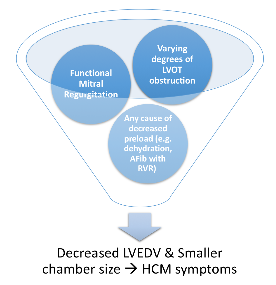
 Pathological cycle of LVOT obstruction in HCM.png Bade Fatunde
12:23, 26 March 2019
1,574 × 1,174; 438 KB
-
-
-
-
-
-

-
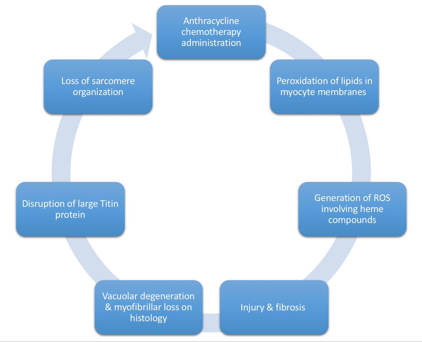 Mechanism of Anthracycline-induced dilated CM.png Bade Fatunde
10:14, 26 March 2019
1,462 × 1,184; 275 KB
-
-
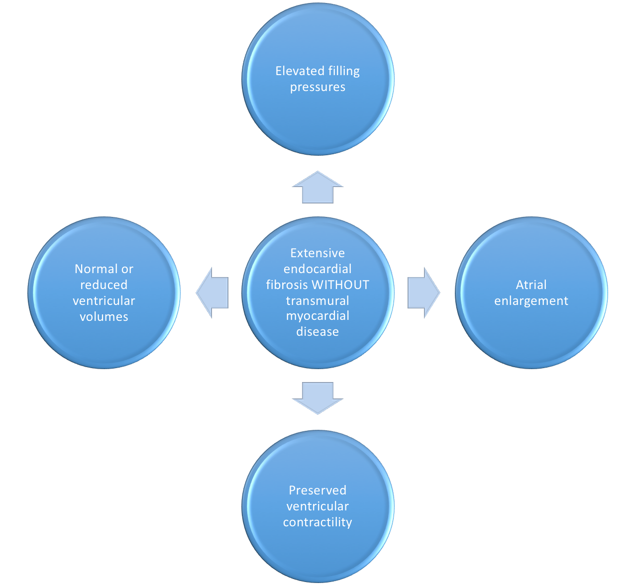
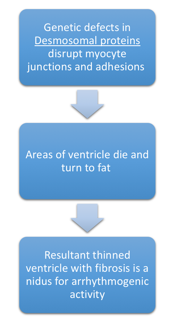
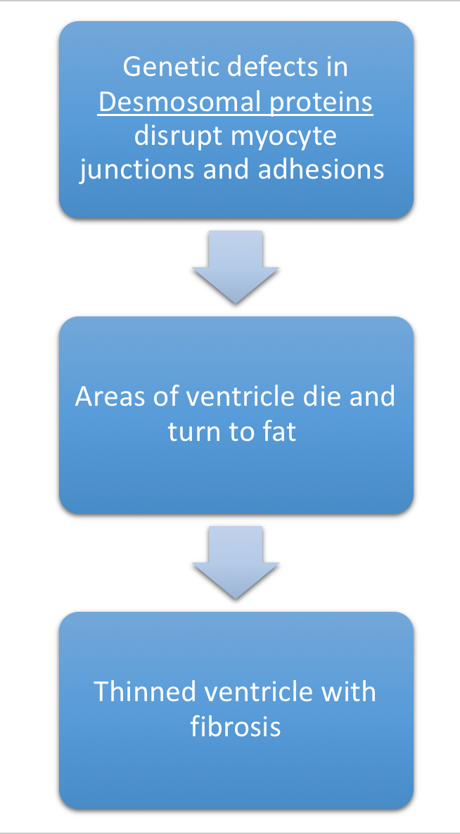
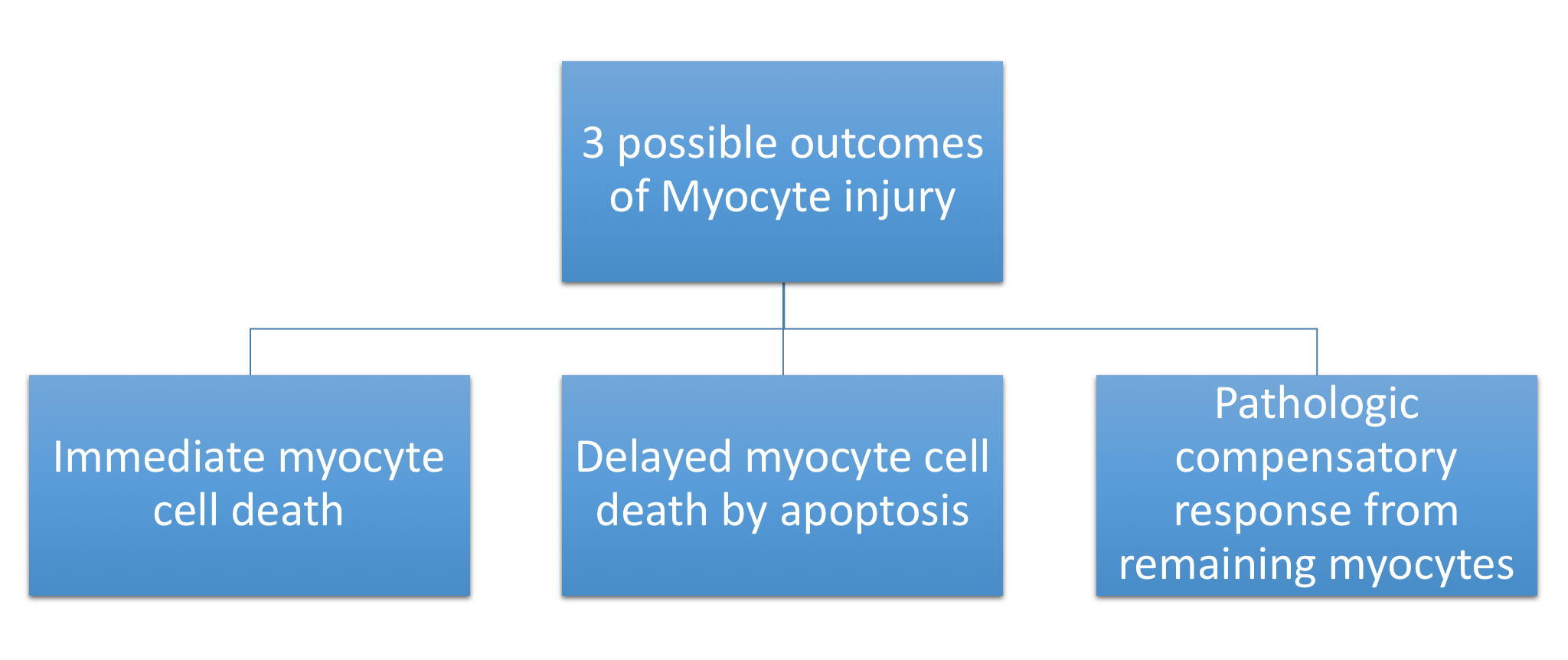
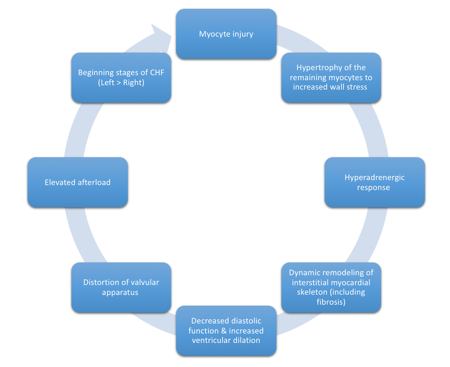 Pathologic compensation to cardiac injury.png Bade Fatunde
09:21, 26 March 2019
1,436 × 1,172; 266 KB
-
-

-
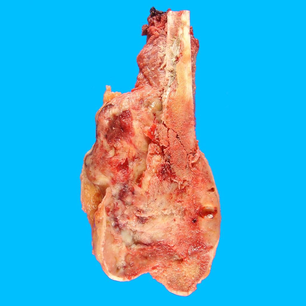
 Mature Cystic Teratoma of the Ovary Bone Tissue (4047143950).jpg Sahar Memar Montazerin
00:41, 26 March 2019
1,510 × 1,441; 1.18 MB
-

-

-
 Luteinised thecomas with sclerosing peritonitis.png Maneesha Nandimandalam
16:16, 25 March 2019
512 × 340; 496 KB
-
 1200px-Ovary SertoliLeydigCellTumor 4 PA.jpg Maneesha Nandimandalam
15:58, 25 March 2019
1,200 × 900; 307 KB
-
 1200px-Sex cord tumour with annular tubules - 2 - very high mag.jpg Maneesha Nandimandalam
15:46, 25 March 2019
1,200 × 800; 233 KB
-
 1200px-Juvenile granulosa cell tumour - very high mag.jpg Maneesha Nandimandalam
15:38, 25 March 2019
1,200 × 800; 250 KB
-
 450px-Juvenile granulosa cell tumour - very high mag.jpg Maneesha Nandimandalam
15:29, 25 March 2019
450 × 300; 57 KB
-

-

-

-

-
-

-

-

-
-
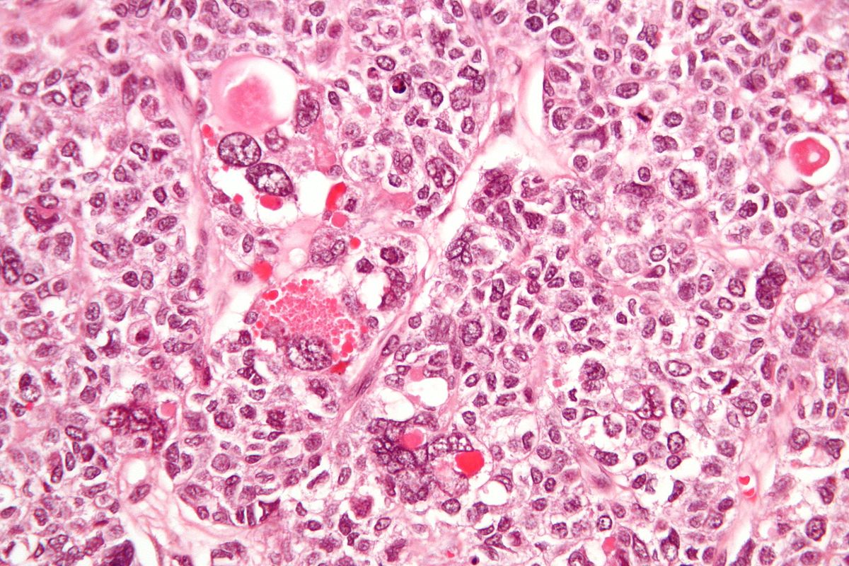
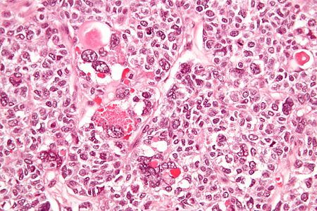
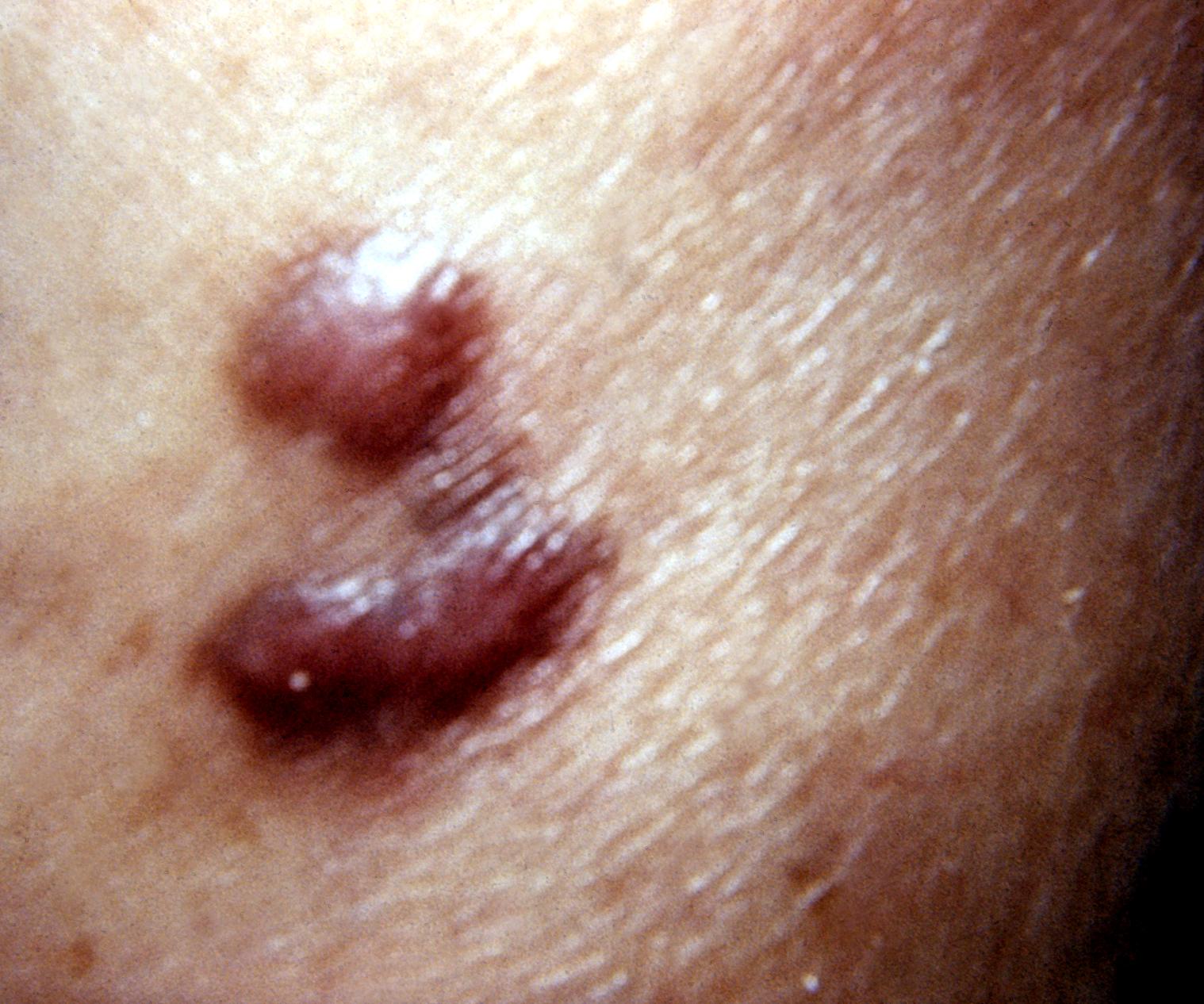
 Kaposi sarcoma new photo to help in diagnosis.jpg Amandeep Singh
20:55, 22 March 2019
800 × 1,066; 575 KB
-
-

-
-

-

-

-

-

-

-

-

-

-

-

-

-

-
-

-

-

-

-

-

-

-
 Diagram showing lobular carcinoma in situ (LCIS) CRUK 166.svg.png Soroush Seifirad
15:27, 21 March 2019
800 × 942; 206 KB
-

-
-
-

-

-

-
 Mature-cystic-ovarian-teratoma-6 (1).jpg Sahar Memar Montazerin
15:55, 15 March 2019
829 × 1,024; 116 KB
-

-

-
-
-

-

-
-
-
-
-

-
 Renal osteodystrophy microscopic pathology.jpg Parnian Jabbari
07:23, 9 March 2019
1,272 × 928; 517 KB
-

-

-

-

-

-
-
-
 Multiple papules may also be noticed on the lower labial mucosa in Cowden syndrome.gif Gunnam
15:48, 7 March 2019
600 × 461; 541 KB
-

-
-
-
-
-
-
-
-
-

-

-
-

-
-

-

-
-

-
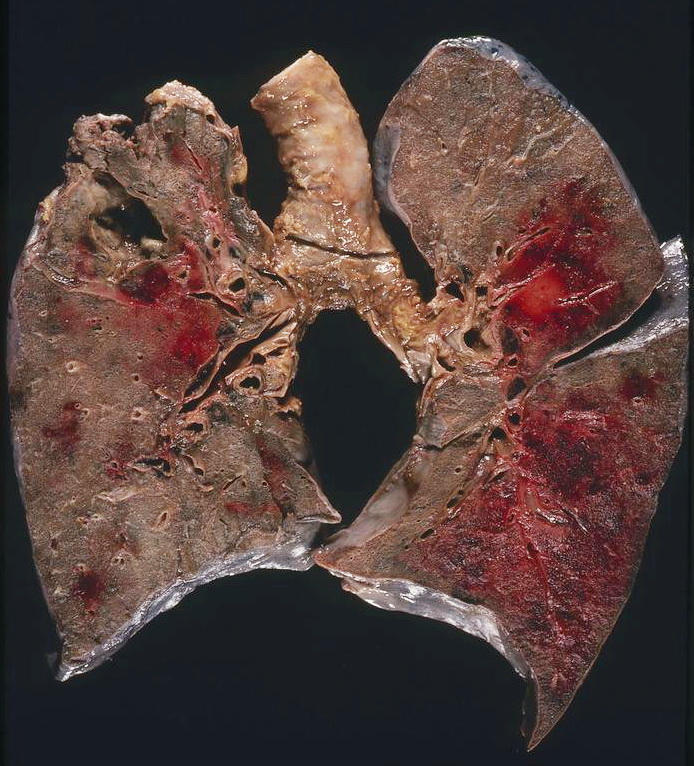
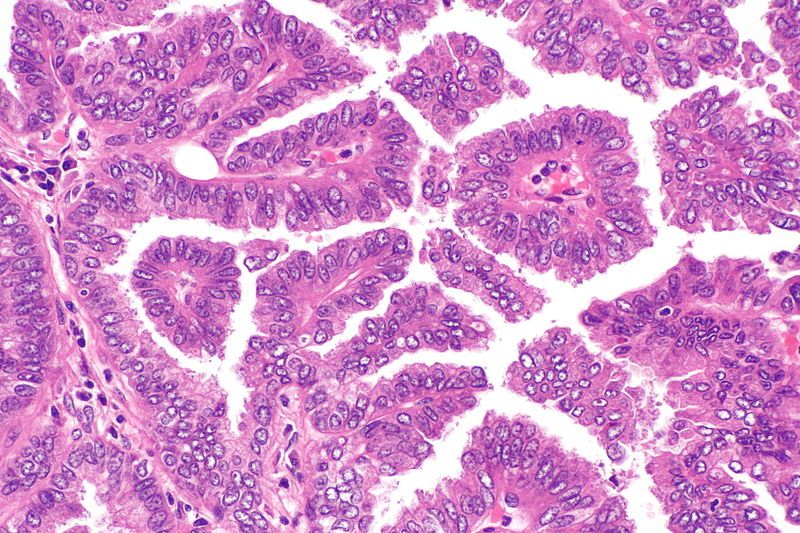 Papillary adenocarcinoma of the lung -- high mag.jpg Trushatank
17:40, 26 February 2019
800 × 533; 160 KB
-
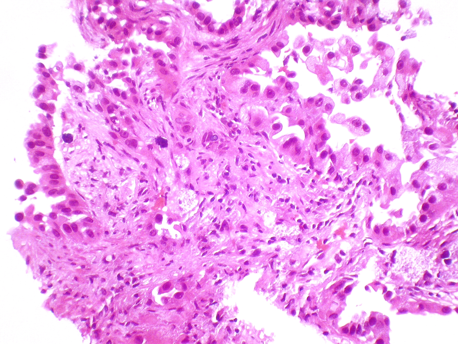 Adenocarcinoma with predominant lepidic growth.jpg Trushatank
17:36, 26 February 2019
1,600 × 1,200; 1.4 MB

.jpg)
.jpg)


.jpg)
.jpg)
.jpg)
.jpg)


.jpg)


.JPEG)


.jpg)


.jpg)
.jpg)


.jpg)
.jpg)
.jpg)
.jpg)
.jpg)
.jpg)

.jpg)


.jpg)

.jpg)

.jpg)


.jpg)
.jpg)
.jpg)
.jpg)
.jpg)
.jpg)

.jpg)


.jpg)


.jpg)


_CRUK_166.svg.png)


.jpg)


.jpg)
.jpg)
.jpg)
.jpg)
.jpg)
.jpg)
.jpg)
.jpg)
.jpg)
.jpg)
.jpg)
.jpg)